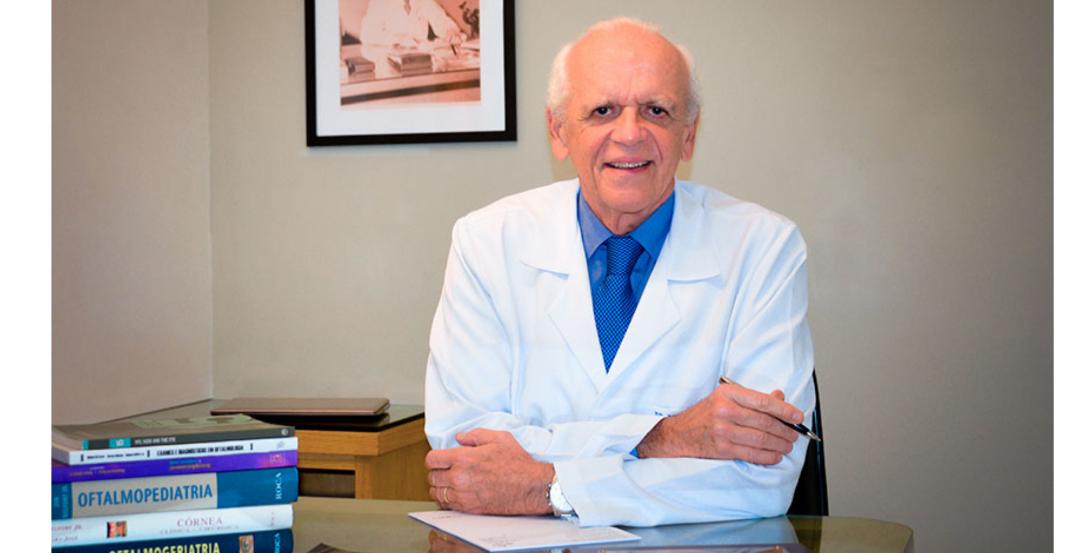
For one of the most renowned Ophthalmologists in Brazil, technology is fundamental in primary care because it allows the detection of eye diseases in a faster, simpler and cost-effective way.
For 20 years, Ophthalmologist Rubens Belfort Jr. has been looking for efficient ways to use telemedicine to diagnose ocular diseases. “Initially, the devices were very expensive and heavy, which made it very difficult, especially in Brazil, to use teleophthalmology,” recalls Belfort.
However, in the last few years, more modern, practical and cheaper models have appeared on the market. One of them is the smartdevice Phelcom Eyer, which is attached to a smartphone, performs fundus exams in a few minutes and without the need for pupil dilation. In addition, it allows the photograph to be sent and stored on an online platform for remote evaluation.
Through the scientific community, Belfort found out about the equipment still in the development phase. “I accompanied the whole process and helped in the improvement of this technology that revolutionizes the possibility of expanding the diagnosis of ophthalmological diseases. And not only in Brazil, but in all countries, since the lack of financial resources is widespread,” he says.
However, the doctor has never had any commercial relationship with the company that created the device, the startup Phelcom Technologies.
Belfort believes that the device has significant advantages over traditional retinal cameras, such as portability, easy handling, high image quality and extremely affordable price. “This type of technology is very important not only in Ophthalmology, but also for other specialties such as Endocrinology, Geriatrics, and Rheumatology. In addition, medical students need to learn how to use this model of device and retire the old one, which has existed for more than 150 years and is totally outdated”, he points out.
Vision Institute
Since 2019, the Eyer is used daily by Belfort’s team in primary care of SUS patients at the Paulista Institute for Studies and Research in Ophthalmology (IPEPO), popularly known as the Vision Institute, in São Paulo. The entity is a non-profit, philanthropic organization linked to the Ophthalmology Department of the Paulista School of Medicine, Federal University of São Paulo (Unifesp). Belfort has been a full professor in the Department of Ophthalmology since 1991.
The Vision Institute provides medical services through diagnosis, clinical and surgical treatments in assistance and didactic projects. Currently, it assists approximately 80 thousand SUS patients with the help of teleophthalmology. With quick training, the professionals of the area, such as technicians, nurses and students, under the supervision of doctors, perform the exams. So far, more than 80 thousand exams have been done to detect diseases such as glaucoma, cataract, diabetic retinopathy, maculopathies, toxoplasmosis, and others.
“Our experience shows that for every 1,000 patients, 850 do not have a disease that requires referral and treatment. In fact, 85% need glasses or have symptoms of dry eye or less dangerous problems. Therefore, through this technology, we are able to diagnose and already refer these 15% for the correct treatment. This provides significant savings in time and resources for the medical system and for the patient as well. Everybody wins,” points out Belfort.
In this sense, the Ophthalmologist believes that the use of Eyer in primary care is fundamental, because it empowers professionals to detect, in a faster and simpler way, ocular diseases. “That is the future. Even more: in a few years, the patient will be able to examine himself. This is the path that this technology is allowing”, he reflects.
Social actions and research
Belfort also uses Eyer in medical actions and research at Unifesp. “We use it in the Amazon Eye Oncology Center, in Manaus, and as of this year, we will also use it in our advanced campus in Rondônia, in partnership with the University of São Paulo,” he says.
In relation to research, the most recent one counted on Eyer in the detection of the new coronavirus (SARS-CoV-2) in retinal lesions. The discovery is unprecedented. The work was carried out in partnership with the Federal University of Rio de Janeiro (UFRJ) by scientist Wanderley de Souza and published in the journal JAMA Ophthalmology.
The researchers, led by Belfort, photographed the fundus of a patient’s eye with Eyer. “The image quality is very good. It’s certainly not inferior to other much more expensive models. It’s probably even better than these. The quality is so good that the photographs taken in the research are accepted in the best ophthalmology journals in the world,” says Belfort.
Have you heard of blockchain in health? This technology is an incorruptible, decentralized and transparent data security system. It has a robust encryption that hides the IDs both from the users and owners of the information. In addition, it can only be validated by consensus and cannot be changed by a single person, without authorization of the others.
One of the key benefits of this industry is superior protection of the large amount of data. It grows stronger with the General Data Protection Regulation (GDPR). Learn more about blockchain and its promising possibilities of use in the field of health.
What blockchain is
Blockchain emerged approximately 10 years ago as an ultra secure and flawless security tool for conducting financial transactions with cryptocurrencies (digital currency) such as Bitcoin. Currently, it is evaluated as the technology with the greatest impact on the digital revolution since the appearance and popularization of the internet.
The decoding tool is based on a data structure known as a “blockchain”. Each of them has transactions with a certain number of information which only authorized people can access, change or view. Information is validated only by consensus.
It is completely private, as it hides the identification of the user and the owners of the information through complex codes due to powerful encryption. This blocks the access of unauthorized people and makes the information almost impossible to leak.
This, therefore, is the great difference of blockchain in relation to other data security systems.
It is widely used in security software and companies in various industries are already using its advantages for other purposes, such as: certification and authenticity of documents, registration of contracts and intellectual property.
In healthcare services, healthtechs have already used blockchain in solutions such as cloud data storage, where it is possible to share everything from medical research to patient information.
Blockchain in Health: key advantages
Undoubtedly, the main advantage of using blockchain in healthcare is the superior data security compared to other systems. For example, each person or institution involved in the process has an access key that decodes the information. Only the patient can allow access to his/her data. This way, he/she becomes the “guardian” of his/her own medical information. All this in an agile and non-bureaucratic way.
It enables access to the complete history of the patient and provide care in a more assertive way. For example, get more accurate diagnoses and direct to more effective treatments.
Professionals can also obtain clinical trials and new research more simply and quickly. For example, decentralized studies, done in several countries and still in secrecy, can be the data entered into a tool with blockchain and shared only with people of interest.
Other possible benefits are to control and track hospital and pharmaceutical supplies, even more so with legislation that imposes several transport and storage rules. It can also decrease problems with false drug supply.
This technology is useful to monitor diseases and predict possible epidemics.
Finally, it can be applied to pricing and payments in healthcare services. It can improve identity management, provide smart contracts, and streamline receipts with immediate transfers. For example, there are systems that allow applications to be processed in a matter of seconds instead of weeks or months.
Blockchain in health: promising tool
In fact, we can state that using blockchain in healthcare is quite promising by ensuring extreme data security, storing the patient’s medical information, and expanding access to the patient’s history, clinical trials, and still-confidential new research results.
With this, the tool can transform the industry by reorganizing operations, generating new business models, and integrating patient medical records.
Reviewed by Paulo Schor, ophthalmologist, free professor and director of innovation of the Federal University of São Paulo (Unifesp) and collaborator of the Faculty of Medicine of the Albert Einstein Hospital.
Follow the Phelcom blog and stay on top of the main news about office management!
Attracting patients is one of the most important issues on the agenda of office and clinic managers. After all, any business needs customers to exist. Including the healthcare services.
In recent years, the rise of internet has strongly changed the consumer-business relationship. Nowadays, it focuses on the needs of the human being. Therefore, it is critical to invest in the relationship with patients to meet their demands and create deeper connections.
You need to create well-segmented strategies to attract your target audience. More than that, you need to keep and build customer loyalty with the existent one. We picked 5 practical tips on how to attract patients and increase credibility and revenue of your business.
1. Register your office to Google My Business
Have you ever searched for a company on Google and received all its essential information on screen, such as phone number and opening hours? That’s Google My Business.
More than a profile with your clinic information, the app allows you to connect with customers through Google Search engine and also Google Maps, all for free.
First step: register your office with the basic data, such as address, phone number, opening hours and website. Then you can add photos of your workplace.
Patients can also make reviews and leave messages on your profile. You receive instant notifications via e-mail when they do it. Be attentive not to answer late.
Another interesting feature is the possibility to know followers and and how often they interact with the profile. For example, you can know where they are from and how many have used the registered phone to schedule the service.
Google is the largest search engine on the internet nowadays. Many people use it to search for doctors. After finding a professional of interest, they search more thoroughly, to evaluate online recommendations before setting an appointment.
2. Invest in digital marketing for doctors
We are currently experiencing the post-digital era without clear boundaries between offline and online world. We use technology all the time for research, entertainment, social interaction, virtual shopping and much more.
That’s why it’s essential to be present online, to build up and keep both relevance and authority in the market. And, thus, attract and retain more and more patients. Especially in social networks, as they offer a great potential for interaction and relationship with the public. The greater the interactions with quality, the greater the visibility of the professional.
So, it is necessary to invest in producing relevant content for each type of platform. In fact, there is no need to all the platforms, which are too many. But there is more doctor-focused social media. To help you plan your main content strategy, we’ve developed a series of articles with practical tips for Facebook, Instagram, LinkedIn, WhatsApp and Telegram.
3. Build the ideal patient journey
Do you know the patient journey? Basically, it is the path the patient travels towards your office. And this involves many phases, such as noticing symptoms, deciding to make an appointment, choosing doctors, receiving healthcare service and the evaluating the whole process.
Undoubtedly, this path can be easy and fast in some cases. However, most of the time, there is a long journey until one feels confident and chooses a specialist. Several aspects influence the final decision, such as complexity of the disease, quality of the professional, evaluation from other patients on the internet or close people, concern about health, inter alia.
Thus, it is essential to understand and map the journey to increase the opportunities of how to attract patients and retain existing ones for your office.
To improve it, you can:
Offer online appointment scheduling;
Have a comfortable office;
Avoid delays;
Invest in technologies;
Set a quality standard for services;
Humanize service;
Reduce waiting time gaps between the journey stages.
Check which patients made only the first appointment and did not return anymore. Or returned and never came back. It could surely be because he/she no longer needed the service or does not remember the office name and address. But, there is a chance that the patient did not like the service.
In this case, it is worth contacting the patient, by phone or e-mail, to find out the real reason and try to redeem the patient.
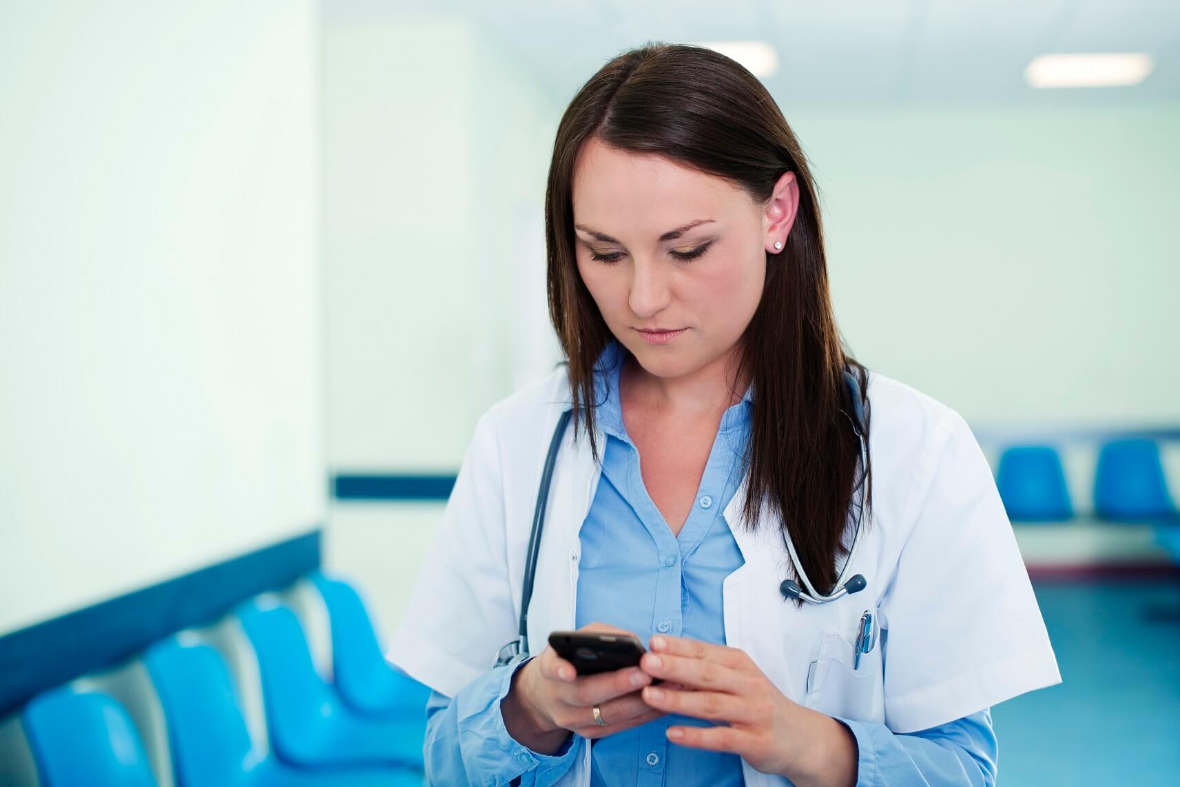
5. Keep a relationship with the patient
Young female doctor texting
To keep in touch with the patient, it is worth to invest in an aftercare process. For example, carry out satisfaction surveys regularly to identify bottlenecks in care and implement improvements.
Another suggestion is, the day after the appointment, send an SMS to the patient saying that you are happy to provide him/her your services, and that you are available. You can also remind him/her of the return visit, congratulate the birthday and wish a happy holiday.
In fact, SMS is just one of the ways to approach. You can also use email and WhatsApp.
By the way, e-mail allows you to send longer contents. For example, you can talk about news in the health area that interest you, give prevention tips and announce innovations of the office, inter alia.
It demonstrates your attention and continuous care for your patients. By this,they will notice your concern for their well-being, which can directly reflect the evolution of treatment.
However, ask the patient if he/she wants to receive content from the clinic and the formats he/she accepts.
And, of course, speed up the process by automated messaging systems. They are faster and ensure information security.
Reviewed by Paulo Schor, ophthalmologist, free professor and director of innovation of the Federal University of São Paulo (Unifesp) and collaborator of the Faculty of Medicine of the Albert Einstein Hospital.
Follow the Phelcom blog and stay on top of the main news about office management!
With the pandemic and the need for social isolation, children around the world began to stay longer at home. A daily life with longer time in front of screens and less outdoor activities in leisure moments. The “new normal”, experienced a year and a half ago, is already paying its price: the growth of myopia among children aged 6 to 8 years in China.
Learn about the research, the data raised and what are the recommendations of experts to slow the growth of the disease among young people.
The research
Researchers examined 123 thousand children and teenagers, from 6 to 13 years old, in schools in Feicheng, China, in 2020. The evaluation technique used was photoscreening, a camera that analyzes the eyes and does not require pupil dilation.
Children aged 6 years were the ones who suffered the most from the increase in myopia: from 5.7%, between 2015 and 2019, to 21.5% in 2020. The 7-year-olds, in the same period, showed a raise from 16.2% to 26.2% and the 8-year-olds, from 27.7% to 37.2%. The increased degree of myopia also drew attention: 1.5-2 degrees.
In the 9 – 13-year group, there was no significant evolution.
Another interesting result is that girls developed myopia earlier than boys.
With this, researchers concluded that the social isolation caused by the new coronavirus pandemic can influence myopia in children. Especially among those aged six to eight years because they are at a stage more sensitive to the problem.
Does increased myopia also occur here as overseas?
In Brazil, there are no concrete data on the increase in myopia in children and teenagers during the pandemic. But in a recent survey conducted by the Brazilian Council of Ophthalmology (CBO), 72% of ophthalmologists reported an increase in diagnoses in patients from zero to 19 years old.
295 ophthalmologists, specialized in various areas, such as retina, cornea, glaucoma and pediatrics, were heard between April and June this year. 76% of doctors believe excessive exposure to electronic devices may directly relate to the explosion of cases. 22% believe only smartphones and tablets are to blame. On the other hand, a small percentage of experts believe there is no link between the two events.
Less screen, more outdoor action
The increase of myopia in young people during the pandemic is influenced by genetic and environmental factors. The disease can be hereditary, passing from parents to sons. In relation to external conditions, the problem lies in the longer period focused on objects very close to the eyes, not resting nr being exposed to sunlight.
Looking at things too closely, less than 33 centimeters from the eyes, without intervals, causes the release of chemical agents inside the eye, which can grow the eyeball larger and increase myopia.
Another aggravating factor is the progression to severe myopia, which seriously affects vision. Currently, this untreated disease is the leading cause of mild and moderate visual impairment and the second largest cause of blindness in the world, according to the World Health Organization (WHO). Besides this, it can cause more serious problems in the future, such as glaucoma, cataracts and retinal detachment.
The Brazilian Society of Pediatrics (SBP) has recommendations on the use of screens by children and teenagers. One of the main is not exposing children up to two years to screens, even if passively. From two to five years, only one hour a day. From six to ten years, two hours a day. Other guidelines are to avoid screens during meals and two hours before bedtime. And, when using, take periodic breaks every 30 minutes or 1 hour in a row.
At the same time, it is critical to increase outdoor activities so that cases decrease. Sunlight releases neurotransmitters that reduce eye enlargement.
Myopia: the epidemic of the century
It has been a few years since the WHO warns of a worldwide myopia epidemic. The entity estimates that the disease currently affects 35% of the population and may reach more than half (52%) by 2050. Only in Brazil, the organization believes that there are 59 million short-sighted people.
Regular visits to the ophthalmologist
How to slow the increase in myopia among children and teenagers taking other actions than reducing close focus without intervals and having more outdoor activities? It is advisable for parents or legal guardians not to only take youngsters to the ophthalmologist after a visual issue. It is essential to keep a routine of visits to the specialist, mainly because at this age it is possible to prevent and early diagnose eye disorders.
Reviewed by Paulo Schor, ophthalmologist, free professor and director of innovation of the Federal University of São Paulo (Unifesp) and collaborator of the Faculty of Medicine of the Albert Einstein Hospital.
Follow Phelcom’s blog and stay on top of the main news about coronavirus and the eyes.
The use of robotics in health has grown increasingly in recent years. By 2025, global investment in this sector is expected to increase approximately 20%, according to a report from Zion Market Research.
The technology generates several advantages for doctors and health institutions, such as more accurate and reproducible procedures. In fact, doctors’ knowledge allied to robotics make the procedure safer, faster and reduces pain and trauma to the patient.
The technology is employed in robot surgeons and even medical software. Learn about the health benefits of robotics and how it works in areas such as surgery, healthcare and even office management.
Robots X doctors
First, robotics does not replace doctors in the healthcare field. In fact, robots only work when guided by humans.
Equipments work as an extension of the surgeon’s hand, for example. This way, they follow commands and avoid the natural tremor of a person, which is a great benefit to delicate surgeries that demand millimeter movements, mainly.
Another approach is planning of the entire procedure and monitoring the execution. Therefore, if an unforeseen event occurs, the professional can adjust at the same time.
1. Robotics in surgery
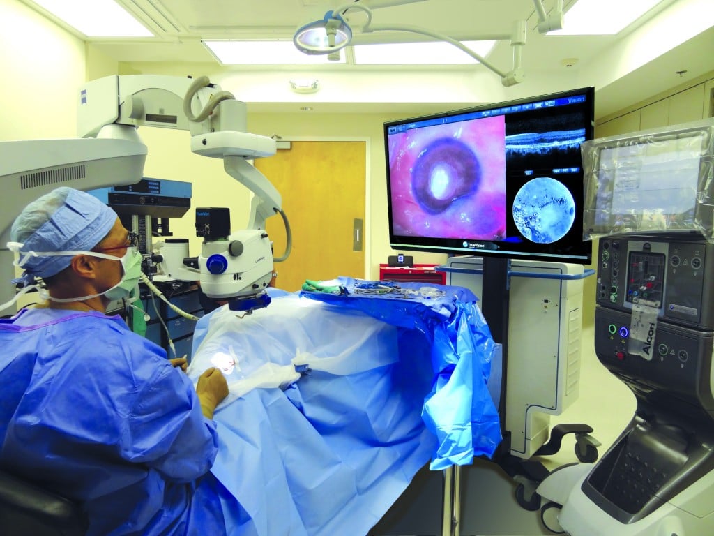
It seems like something new, but the health field started employing robotics many years ago. First surgeries with such equipments occurred in 1988, in Paris.
Ophthalmology is one of the areas in which the use of robotics has been done for the longest time. One of the first uses was treating diabetic retinopathy with laser. The technology measures the duration, size and power of each pulse. For this, the device is pre-programmed so that each laser pulse is the same as the previous one.
Nowadays, the tool is also used in refractive surgeries to correct myopia. Robots guide all the programming of the procedure of laser pulsing in the patient’s, always monitored by a specialist.
The robotics system NGENUITY uses 3D technology in high definition that greatly increases the surgeon’s view of the eye. It turns the ophthalmological surgeries more accurate. The doctor operates by looking at a high-definition 3D screen, which allows a more proper posture and reduces fatigue during the procedure.
Another ease is using digital filters to customize visualization during the procedure, increasing the image of eye structures and tissue layers.
There are also robots to carry out cataract surgery and corneal transplantation, such as the Ziemer and LensX. However, they are less used in Brazil.
Da Vinci
Several technologies have this goal nowadays. Undoubtedly, the most known is Da Vinci, the robot surgeon most used in the world. Currently, more than 5 million patients are treated. Even in Brazil.
These systems carry out minimally invasive surgeries in different procedures. One of its tools is a console – inspired by flight simulators – in which doctors visualize the high-definition 3D images and make the operative movements with their own hands, through a joystick, which are transmitted to the robot.
The doctor can move the axes for 360 degrees, reaching angles that human hands would only reach with great difficulty.
It also applies to train and requalify students, residents and specialists in attendance or within the surgical center. Thus, it is possible to analyze ethnographically the performance of the professional – who is alone during the procedure – by capturing images from a local camera.
The technology can also be operated remotely via telemedicine.
2. Robotics in healthcare
Several robots already improve medical care. Such as robot Laura, which identifies potentially dangerous generalized infections in patients and informs the medical staff. For this, the robot is connected to electronic medical records and monitors the health reports and clinical information of each patient. When it identifies any worsening or abnormality, it generates an alert. It also uses the principle of machine learning for this purpose.
Since its invention in 2016, it has helped reduce the overall mortality rate by 25%, saving 18 patients a day in institutions that work with the technology.
In ophthalmology, one of the proposals is the Adam robot, which can assist in checking primary visual acuity. This is because it identifies visual difficulty levels. In up to five minutes, it can detect diseases such as myopia, astigmatism, hyperopia and presbyopia.
Even for procedures that involve needle placement, such as taking blood and biopsy, there are robotic systems. This is the case of Veebot. It uses a pouch similar to blood pressure gauges, making the veins more visible. It then uses an infrared light and a camera to find the best vein through an image analysis software.
The depth with which the needle is pricked is pre-calculated and the whole process takes about a minute. The system succeeds in choosing the vein in 83% of cases. In addition, the procedure is less painful to the patient.
3. Robots in office management
Imagine receiving your patient with the help of a robot? Invented in Japan, Pepper is used in several countries for the reception of shops, exhibitions, public places and even in medical offices.
The robot has a human appearance: giant eyes, child’s face, arms, hands, 1.20 meters high, 30 kilos and a screen attached to the chest. One of its main features is the ability to analyze people’s emotions through facial expression and tone of voice.
It uses voice recognition technology, cameras and sensors and evaluates all this data in a system based on artificial neural networks. It is able to modify the voice, the color of the eyes, move the arms and show images on the screen according to the emotions of the person.
In fact, neither Pepper nor any other robot can replace professionals in the field. However, it helps greatly to improve care, reduce costs, carry out more precise and less invasive procedures, increasing agility and reducing pain in the patient.
Reviewed by Paulo Schor, ophthalmologist, free professor and director of innovation of the Federal University of São Paulo (Unifesp) and collaborator of the Faculty of Medicine of the Albert Einstein Hospital.
Follow Phelcom’s blog and stay on top of the main news about coronavirus and the eyes.
Ocular syphilis is a manifestation of syphilis that can arise when the disease is not treated properly. This stage occurs years after infection and has a challenging diagnosis. Despite directing lesions, we call the treponema palidum (etiological agent of the disease) “the great copycat”. The agent can simulate several different manifestations. At this stage, the problem can even cause blindness.
But, a new study pointed out that Optical Coherence Tomography (OCT), A common ophthalmological examination in SUS, can help in the early identification of ocular syphilis. The University of São Paulo (USP) carried out the research and published it recently in the journal Ocular Immunology and Inflammation.
Learn about the research, results and what should be the next steps for the use of OCT to diagnose the disease.
The research
Researchers from the Faculty of Medicine of Ribeirão Preto (FMRP), from USP, evaluated one of the eyes of 54 patients with ocular syphilis admitted to the FMRP clinical hospital (HCFMRP). After part of them received the treatment, scientists still analyzed 31 eyes.
Through Optical Coherence Tomography (OCT), researchers found retinal lesions that may aid in early diagnosis of the disease.
Results
The ophthalmological exam identified round spots, irregularities, elevations and detachment in the retinas studied. According to the authors of the work, it is the first time that OCT checks for frequent changes in the retina in a large series of cases of ocular syphilis. These modifications are imperceptible on clinical exams.
Undoubtedly, the findings of OCT have diagnostic value in ocular syphilis, but do not predict the prognosis. However, the examination – common both in the Brazilian Unified Health System (SUS) and private clinics – can help visualize signs of the disease even in early stages. After confirming the diagnosis with serology and referring to the indicated treatment, the patient has a good chance of not having permanent sequelae in vision.
Photo: Eduardo Paulino Eye Institute.
Reviewed by Paulo Schor, ophthalmologist, free professor and director of innovation of the Federal University of São Paulo (Unifesp) and collaborator of the Faculty of Medicine of the Albert Einstein Hospital.
Follow Phelcom’s blog and stay on top of the main news about coronavirus and the eyes.