Google is the most used search engine on the internet. There are 5.5 billion searches a day worldwide. In fact, the platform brings answers to countless and unimaginable types of questions.
In addition, it also takes the internet user wherever he/she wants. With Google My Business, companies can provide full address, opening hours, contacts and photos, among other information.
With a single click, your potential patients can find your clinic, know the services you offer and schedule an appointment. All this at no cost to you.
So how about attracting more customers using Google My Business for doctors? Learn more about the tool, how to register and the advantages for your business.
Google My Business for doctors – what is it?
Google My Business for doctors is a free and easy-to-use tool that enables offices, clinics, hospitals, laboratories and other healthcare institutions to manage their online presence on Google, including Search and Maps.
To do this, you can create a profile of your business with all the essential data. That way, when people search directly by your name or the office or by keywords related to your specialty, Google will show your professional profile.
Regarding keywords, the results presented by Google can be from the nearest locations or even from every city and in municipalities in the region.
Undoubtedly, having one-click access to all the information of your clinic is quite useful to patients. Will you make an appointment? Just “Google” and find the phone and service times. Is it the consultation date? Click on the address to open Maps right away.
In addition, it is possible to lure more customers. For example, when the internet user searches for “ophthalmologist in Vila Madalena”, Google will immediately show your clinic profile.
Another advantage is to increase interaction with your audience. The patient can call the registered number directly, send a message, leave comments or evaluate your office. And you can answer him/her, solving doubts or thanking the reviews.
By the way, evaluations help increasing your clinic reliability and increasingly rise your position on the search engine page. That is, the higher the chances of appearing in the first results on a keyword search. So, the better the “grades”, the more positive comments and interactions with the audience, the more your authority in the area grows.
This is because a good review and five stars, for example, helps to demonstrate your service excellence to potential customers. This influences their decision to schedule an appointment.
Reports are another benefit of Google My Business to doctors. The platform provides metrics such as number of followers and interactions on your profile through phone calls, visits to the registered site or route requests.
This way, you can know how customers research your business and where they come from, essential data for a more efficient digital marketing strategy.
Registering your office or medical clinic on Google My Business is easy. Read the step-by-step guide:
Create an email in Gmail;
Access Google website. At the top right, go to “Google Apps” and click ” My Business”;
To make sure there is no record of your business already, enter your name or that of the clinic. Then click ” Include your company on Google”;
Put the name and choose the business category;
Then allow the address to be included in Google Maps and direct searches;
Fill in the address fields;
Inform if you operate outside your workplace and the area you serve;
Include contact forms such as phone, website and email;
Upload images of your clinic. According to Google, Profiles with photos and videos receive 42% more route requests on Google Maps and 35% more clicks to access the site;
Confirm all data.
Finally, Google will send a code, by letter, to the address provided to confirm the veracity of the information registered. When you receive the letter, enter the code in the virtual area of Google My Business.
Ready! Your profile is up and visible to thousands of people who use Google.
Reviewed by Paulo Schor, ophthalmologist, associate professor and director of innovation of the Federal University of São Paulo (Unifesp) and collaborator of the Faculty of Medicine of the Albert Einstein Hospital.
The World Health Organization (WHO) has long warned about the danger of diabetes. The disease grows year by year around the world, and in the past 40 years the number of cases has quadrupled.
According to the 10th edition of the Diabetes Atlas, published by the International Diabetes Federation (IDF) and recently released, 537 million people aged 20 to 79 have diabetes worldwide. A growth of 16% compared to 2019.
This equals one diabetic out of ten people. The scenario gets even worse: almost half (44.7%) do not even imagine that they face the disease. The projection for next years are 643 million diabetics in 2030 and 784 million in 2045.
Lifestyle, lack of access to healthcare in developing countries and the present pandemics – which increased sedentary lifestyles, poor diets and postponed medical care – are the main factors for these numbers. Learn more about preliminary data IDF presented.
Diabetes around the world
According to the survey, done every two years, 10.5% of the world’s population have diabetes. Thus, the number proportionally exceeds the global population growth. Until then, one person out of 11 was diabetic.
More than that, 44.7% don’t even know they are sick. That can greatly aggravate diabetes, since people only seek help when symptoms arise. Undoubtedly, figures are worrying. Lack of control can lead to other serious problems, such as blindness, kidney damage, changes in the heart and even death.
The disease is also one of the most deadly: 6.7 million people have lost their lives due to diabetes. That is, every five seconds a person dies from this condition. This account does not yet include deaths resulting from complications of other diseases that have been aggravated due to diabetes, such as covid-19.
The presence of the disease is much higher in developing countries: 81% of sick adults live in these localities. That is, 4 out of 5 diabetics. According to atlas, 32 million diabetics are from Latin America and Central America.
So many sick people cost a lot of money: USD 966 billion were spent worldwide with healthcare, a rise of 316% in the last 15 years, according to IDF.
Diabetes in Brazil
In the 2019 edition, there were 16.8 million diabetics in Brazil. In the world ranking, we are in 5th place behind only China, India, the United States and Pakistan.
Among Brazilian Capitals, Rio de Janeiro stands out with the highest rate of diagnoses in the country: 11.2%. Then there is Maceió (11%) and Porto Alegre (10%). The disease also affects more women (9%) than men (7.3%) here.
Data are from the research “Vigilância de Fatores de Risco e Proteção para Doenças Crônicas por Inquérito Telefônico” (Vigitel), 2020, a telephone survey research of the Ministry of Health.
With regards to the costs invested in the treatment of Brazilian diabetics aged 20 to 79, Atlas estimates USD 52.3 billion per year. This equals to USD 3 thousand per adult.
More data on Brazil should be published in the full edition, with a release preview for December 6.
Causes
Experts claim that diabetes is increasingly out of control and that there is a lack of information and awareness for prevention. Current lifestyle is one of the main factors for the increasingly high number of the disease cases. Sedentary lifestyle and poor diets, rich in fats and carbohydrates, have brought problems such as hypercholesterolemia, hypertension, overweight, obesity and pre-diabetes, among others.
In low-and middle-income countries, which have the largest number of diabetics, there is a lack of access to healthcare, delaying diagnoses, treatments and even guidance for a balanced diet.
Diabetes and the eye
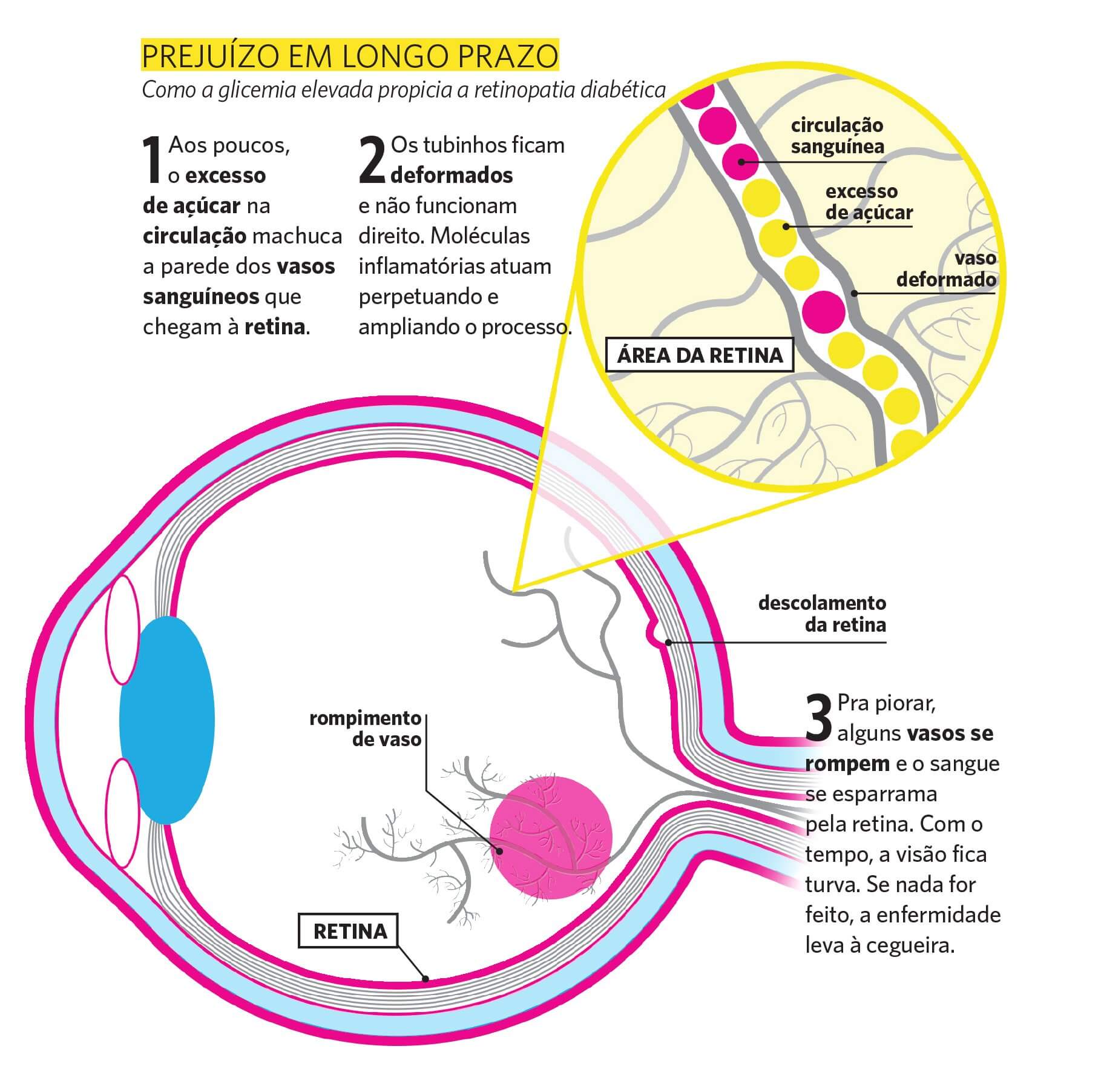
One of the possible complications of diabetes is in the eyes. According to a study by the Brazilian Society of Ophthalmology (SBO), 40% of people who suffer from diabetes present ophthalmic changes.
Diabetic retinopathy figures among them. Currently, about 40% of the 4 million Brazilians diagnosed with retinopathy have diabetes. In fact, the duration of diabetes and the uncontrolled blood glucose have a direct relationship with retinopathy.
Source: Infographic on diabetic retinopathy – Saúde magazine
This disease subdivides into two types: pre-proliferative, which does not require laser treatment, and proliferative, in which neovases occur and also demand therapy.
For the diagnosis of the correct type, one must evaluate the fundus with examinations.
The disease can also cause glaucoma. The Ministry of Health estimates that people with diabetes are 40% more likely to develop the problem.
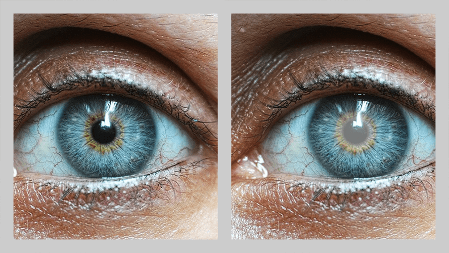
In addition, diabetics are 60% more likely to have cataract earlier. In this situation, the problem appears earlier and progresses faster than senile cataracts. Therefore, it is the main cause of vision loss in diabetic patients. However, it is reversible through surgery.
Comparison between a healthy eye and one with cataracts.
It is surely essential for diabetics to be attentive to any changes in their vision. Therefore, even if everything is fine, it is important to undergo periodic examinations with an ophthalmologist.
Prevention and early diagnosis are the keys to avoid serious damage, such as severe visual impairment and even blindness, in case of complications from diabetes.
Reviewed by Paulo Schor, ophthalmologist, associate professor and director of innovation of the Federal University of São Paulo (Unifesp) and collaborator of the Faculty of Medicine of the Albert Einstein Hospital.
Follow Phelcom blog and stay on top of the main health news.
Managing medical clinics can, indeed, be a real challenge for doctors. Office management involves various simultaneous responsibilities, such as organizing data, financial control, personnel administration, marketing and patient experience, inter alia.
So, many assignments may incur in procedure failures, which can cause diverse losses. One of them is spending more than necessary. In fact, clinics may spend more money from not knowing nor measuring resources involved in all areas through assertive cost and investment plans.
That may also prevent healthy, responsible and profitable business growth. For this reason, we have selected 6 practical tips to reduce costs in the office. Check them out!
1. Organize and analyze billing and expenses
Undoubtedly, good financial management is one of the main requisites to avoid losses and decrease clinic costs. This requires developing an assertive planning with incomes and expenses.
In this sense, it is important to recognize and measure resources involved in all areas of the clinic. Therefore, you will have available values in hands to decide what direction to take and how to apply each one of them efficiently.
For example, checking all cash outflows allows identifying which processes spend a greater amount of financial resources.
Some tips to help you on this goal follow below:
Have a proper control of your cash flow;
Do not mix up personal and clinic accounts;
Automate internal processes;
Have a sound financial planning;
Follow expenses and incomes;
Use a good medical clinic management system.
Those are many data – and free time available is not a strength in the medical area. Therefore a medical software is essential to organize all them.
Nowadays, medical softwares help automating all the clinic internal processes. Their main functionalities include medical record storage, online calendar, control of inventory inputs and outputs, as well as financial, administrative and human resource – as doctors and employees – information.
For example, a consultation schedule through an online calendar is instantly shared in the system, as all the information that follow: doctor, payment method or healthcare plan, examinations, etc. This reduces the time a team dedicates to such tasks and increases productivity, focused in more complex activities. That is, increased operational efficiency.
It improves and speeds up financial management by providing concrete data that allow a wider view on the business. Having efficient indicators at hand – such as appointment/miss/cancellation rate, average ticket, new patient search, user retention and billing – allow an in-depth analysis of your business.
Thus, you can see exactly how to reduce office costs assertively. On the other hand, a medical software only for online calendar, without data access, may not pay off.
In this case, it is more economical to stick with the good old paper agenda.
3. Decrease patients that miss scheduled appointments
Undoubtedly, patients who miss the consultation – without prior notice or cancellation – are one of the most frequent difficulties in medical offices and clinics. This fact clearly impacts clinic incomes, disrupts the routine and wastes time.
Therefore, it is essential to adopt strategies to decrease absence and urgent cancellations rates. Learn 8 steps to reduce clinic costs with lacking patients:
Offer online scheduling;
Confirm the scheduled consultations through electronic tools;
Clearly provide address and communication channels;
Confirm with considerable advance;
Ensure a well managed calendar;
Ask for a previous payment of a percentage of the consultation;
Manage the patient journey;
Do not be late for the consultation.
How to put all this into practice? We made a step-by-step guide in this article.
4. Reduce use of office supplies
By opting for technological systems, daily use of paper in the office reduces significantly. For example, diaries and medical records become electronic. You also save money because you do not need do rent or buy printers, ink, maintenance and team workforce to fill out the papers.
More than reducing office costs and helping the environment, information also become centralized, which is important to have a 360º-view of your business.
5. Negotiate with suppliers.
By knowing the exact amount of supplies and materials at the exact time the office needs, you can negotiate more attractive values and prices with suppliers. Your purchases will be predictable and also an advantage to your partners.
It is undoubtedly essential to control stock inputs and outputs, know which supplies are most used and which are the periods when each item is used the most. This reduces office costs.
The first step is to standardize processes. Register each material with a code and detailed description. When the product flows out, it is essential write them off.
Also keep an eye on products expiration dates and their proper storage not to lose any item.
Another tip: make an inventory. This will allow you to know all the stored products and their usage profile from inputs and outputs. In addition, it helps calculating stock costs with maintenance, losses and waste of materials.
To make a good stock control, you can chose for management systems and softwares. The entire catalog is safely stored in the cloud, easily accessible. You can track financial flow and costs with periodic reports and spreadsheets. Thus, such data-based management turns decisions more assertive.
For example, some tools allow you to view inputs by vendor and outputs by procedure types. By this, you know which materials are more used and are able to negotiate better values and payment methods for your business.
Undoubtedly, medical management systems help reduce costs in the office by automating processes, increasing productivity, centralizing data, and providing complete, intuitive and easily accessible reports for more assertive decision-making.
Reviewed by Paulo Schor, ophthalmologist, associate professor and director of innovation of the Federal University of São Paulo (Unifesp) and collaborator of the Faculty of Medicine of the Albert Einstein Hospital.
Follow Phelcom blog and see tips on how to improve the management of medical offices and clinics.
Business Intelligence (BI) is a concept aimed to assisting business management from collection, management and analysis of data. Even in the healthcare area.
More and more offices, clinics, hospitals and other institutions in the sector have joined the system. Its main advantages include: information evaluated more assertively, increased productivity, optimized processes and identification of possible improvements and opportunities.
Learn how the system works, its benefits and how to install BI in healthcare.
BI in healthcare: how it works
BI collects all data from the clinic and safely stores it in a single location. This function itself is a great advantage, mainly due to the new General Data Protection Regulation (GDPR). Information analysis and decision-making – such as new actions to improve processes or correct bottlenecks – turn faster and more effective with that functionality.
In fact, the ease of integration of numerous data and the variety of specialized BI systems on the market allow more intelligence to the medical offices. This may range end-to-end: from patient consultation to daily financial balance, when integrated with already implemented medical management software.
Simple charts and reports allow widening the strategic view on the business as a whole. In BI, health can show different points of view on your management model.
You can also use BI in equipments while managing data from each patient. It allows to learn deeply about the potentialities of each device.
BI in healthcare: advantages
Learn the main benefits of using BI in healthcare:
1. Deep data analysis
Every day, your clinic generates large amounts of data, such as appointments, patient medical records, inventory entry and exit, expenses, incomes, etc. It is practically impossible to evaluate all that data manually. That’s why more and more healthcare businesses are opting for BI tools.
The system makes the data available in reports and graphs that are simple to interpret and more agile. For example, you can measure your average number of patients per day, consultation length, absences, follow-up appointment rates and patients’ healthcare plans.
The information at hand enables troubleshooting and improving processes. For example, you can identify the number of monthly absences and take actions to decrease them, such as sending appointment reminders the day before. You can also set the approximate lifetime of disposable material and request for replacements in advance.
It surely increases quality of daily activities and assures a more profitable business.
3. Preventing risks and identifying opportunities
Process optimization links directly to risk reduction. As BI applied to healthcare helps identifying business bottlenecks, you can anticipate problems that can bring serious harm and headaches. For example, the number of patients cared for decreases at certain times of the year. You can prepare your budget for that phase by predicting this scenario.
More than predicting risks, the tool also allows to identify growth opportunities. For instance, is there a constant demand for services from your clinic and a long waiting list? It may be an indication that it is time to expand business. All depends on what the other data says.
4. Higher quality management
All this naturally leads to improve the management. By showing the real situation of your business, BI in health allows you to correct errors, implement new processes, diagnose more assertively, organize routine, improve productivity, increase profitability and reduce costs. And, of course, improve the quality of patient care.
To use the tool, you need to resort to medical management systems for input data to the tool. If you still do not have it or seek for new, more complete options, you need to consider the ease of access and the organization of the information provided. Also evaluate if it provides state-of-the-art technology, cloud storage with full data security and high-quality support.
Reviewed by Paulo Schor, ophthalmologist, associate professor and director of innovation of the Federal University of São Paulo (Unifesp) and collaborator of the Faculty of Medicine of the Albert Einstein Hospital.
Follow Phelcom blog and stay on top of the main news on office management!
For some time now, researchers have been investigating whether eye diseases can be a risk factor to manifest other health problems. For example, scientists at Sun Yat-sen University in China found that one in four people with eye disorders also develops depression.
Now, a new study links age-related macular degeneration (AMD), cataract and eye disorders brought on by diabetes to the increased risk of dementia. The researchwas recently published in the British Journal of Ophthalmology.
Understand the work, its results and how eye diseases can be directly linked to dementia cases.
Research and results
Researchers at the Guangdong Academy of Medical Sciences, in China, evaluated data from 12,364 adults with AMD, cataract or glaucoma, aged 55 to 73, from 2006 to 2010. Participants had follow-up until 2021.
The risk of cognitive decline was 26% higher in patients with AMD, 11% higher in those with cataracts and 61% more in diabetics compared to those who did not have eye diseases at the beginning of the study. Glaucoma was not considered one of the risk factors.
The scientists also looked at eye and systemic diseases alongside the incidence of dementia. Patients with cataract and a systemic condition were 1.19 to 2.29 times more likely to develop dementia compared to those without these problems. Regarding eye diseases related to diabetes and systemic diseases, such as diabetic retinopathy, this figure was 1.50 to 3.24 higher.
From the beginning, the study detected that diabetes, heart diseases, strokes and depression associate with increased risk of dementia. Hypertension joined the list until the research ended. All mediated the association of cataract and incipient dementia, as well as other eye diseases related to diabetes incipent dementia.
Despite the expressive results, it is worth noting that the research is observational. However, the scientists state in the article that “AMD, cataract and diabetes-related eye diseases associate with increased risk of dementia. Individuals with ophthalmic and systemic diseases have an even greater risk.”
Reviewed by Paulo Schor, ophthalmologist, associate professor and director of innovation of the Federal University of São Paulo (Unifesp) and collaborator of the Faculty of Medicine of the Albert Einstein Hospital.
Follow Phelcom blog and stay on top of the main news on health research!
Have you heard about the advantages of Clubhouse for doctors? One of the latest social networks has stood out due to the quality content shared by professionals who are references in the health area.
One of the great features in the new platform is the exclusive use of audio. Everything happens in real-time, in rooms the users themselves create, so it offers plenty of rich contents only found there.
Learn more about Clubhouse, its benefits and 5 must-see clubs for doctors.
Clubhouse-how it works
Clubhouse is a social network based on voice chats. Conversations take place in rooms – the clubs – created by members according to their common interests, more informally, even on important matters.
Chats are always live. The “speakers” mediate the discussions: they are users who can speak during the chat, and the” listeners” can only listen to the conversation. You can ask the group admins for speaking. They may accept the request or not.
At the end of the chat, clubs are no longer accessible. Any member can create new rooms, private or public, and invite participants. You can also create an event, providing date, time and content description.
The audience of each room is measured according to the number of members, participation of renowned experts, spontaneous interactions and exchange of genuine experiences. That is, content is king to attract members, even more so when there are no features like photos and videos.
This way, Clubhouse offers a set of audio discussions, podcasts and live calls. Created rooms are suggested to members according to the interests they select by creating their profiles.
However, it is only possible to join the social network through an invitation, which makes the application even more exclusive. Another “stumbling block” until a few months ago was the exclusive availability for iPhone (iOS). Since May, Android users may also download the app.
Clubhouse for doctors – advantages
In fact, Clubhouse offers advantages not only for doctors, but professionals from various fields. For example, live and audio-only content make communication more natural, with spontaneous and assertive responses. It is also a big plus for those who do not like to record videos.
One of the benefits pointed out by several doctors is to be in contact and exchange information with college friends, colleagues of the area and renowned specialists. Another advantage is the possibility to share knowledge and become a reference in your area of activity, strengthening your authority and credibility not only among fellow professionals, but also potential clients.
This is because Clubhouse for doctors can be used to create and strengthen professional relationships and also for medical marketing.
Reviewed by Paulo Schor, ophthalmologist, associate professor and director of innovation of the Federal University of São Paulo (Unifesp) and collaborator of the Faculty of Medicine of the Albert Einstein Hospital.
Follow Phelcom blog and stay on top of the main news about social networks for doctors!


