Clear vision: How ophthalmologist Simone Brandão uses Eyer2 for anterior segment exams

Clear vision: How ophthalmologist Simone Brandão uses Eyer2 for anterior segment exams

Dr. Simone Brandão, an ophthalmologist and professor at the Federal University of São Carlos (UFSCar), has been using the Eyer fundus camera for over two years. Since then, she has conducted over 5,000 exams at her clinic, in classrooms, at the São Carlos Teaching Hospital, and as part of a social project developed by the Academic League of Ophthalmology at UFSCar.

Due to her extensive use of the device, Phelcom invited Dr. Brandão to test Eyer2, a comprehensive visual examination platform that captures high-quality images of both the posterior and anterior segments.

The increase in the field of view (FOV) of retinal images from 45° to 55° now allows for the photo documentation of the ocular and periocular surface in a single image, reducing the need for multiple shots. Additionally, the device’s flash is more patient-friendly, enhancing the overall examination experience,” explains Brandão, a specialist in oculoplastic surgery.

With new embedded tools and improved features, the new equipment makes it possible to detect various diseases and conditions of the anterior segment of the eye, such as blepharitis and other eyelash alterations, meibomian gland dysfunction, styes, conjunctival tumors, eyelid tumors, advanced cataracts, foreign bodies, burns, corneal lesions and general keratitis caused by dry eye, contact lenses, infections and ulcers, among others.

Cases

Brandão recently encountered a patient with a welding burn. “I took the photo with the Eyer2 and showed the patient how his eye looked when he didn’t wear the goggles during welding,” she says.

In another case, a patient reported that she had hit her eye with a leaf, but had not hurt herself. Much to the patient’s surprise, the ophthalmologist carried out an examination and detected an eye injury. In another situation, a patient developed an ulcer after his son threw water in his eyes while playing in the pool.

Img Olho Ulcera

 Eyer2 examination of a patient with an eye ulcer after playing in the pool with his son.

Dr. Brandão uses the portable fundus camera during all her appointments, allowing patients to view their own fundus in real-time during the examination. “Real-time imaging not only educates but also motivates patients, helping them better understand their condition which enhances their commitment to treatment and recovery,” Dr. Brandão explains.

The exams are documented instantly on EyerCloud, a cloud platform. At subsequent appointments, the doctor reviews the images and shows the before and after of the treatment. “After an operation, a patient said that the suture in her eye was imperceptible. I took the picture and we could perfectly see the 10-0 thread, which is thinner than a strand of hair,” she recalls. 

For Brandão, the platform speeds up the process of issuing reports and allows exams to be accessed and shared quickly, facilitating communication with other health professionals, students and the patient themselves. “On one occasion, a colleague requested tests for a patient hospitalized at the São Carlos Teaching Hospital. I accessed EyerCloud remotely, issued the report, and shared it instantly.”

Before adopting Eyer, Dr. Brandão would often search for images on Google to explain conditions to her patients. Now, she accesses EyerCloud to display real, relevant images that depict both healthy eyes and those with specific diseases.

New features

The doctor also highlights the practicality of Eyer2’s magnetized connection module, which makes it easier to change modules during exams and does not detach from the equipment, making the process more efficient and precise.

Infrared light and cobalt light are also new features in the Eyer2 portable fundus camera, which the ophthalmologist has already used. “For example, sometimes in a blepharoplasty post-surgery, complications such as keratitis can occur. I apply the fluorescein and take the photo with the cobalt light so that the patient understands the picture. What’s more, this combination of techniques allows for a more precise and detailed assessment of eye conditions, helping with diagnosis, treatment and patient follow-up,” she adds.

Img Olho Ulcera Exame Luz Cobalto

Cobalt light examination with the Eyer2 of a patient with an eye ulcer after playing in the pool with his son.

Infrared light helps to detect changes in the choroid as it penetrates deeper into the structures in the back of the eye, and to perform meibography. This test, which assesses the meibomian glands located in the eyelid, is essential in the investigation of dry eye syndrome, a disease that affects 26 million people in Brazil alone, according to the Brazilian Ophthalmology Society (SBO).

Home care

For Brandão, another advantage of the Eyer2 is the image quality of the anterior and posterior segments in unfavorable situations, such as home care. “I have a patient who is bedridden and I went to do the exams at her home. With the Eyer2, you practically have a hand-held slit lamp, as it enables clear and detailed visualization of the eyelid region, conjunctiva and eyelashes, even in a room where the light was not ideal”.

Beyond home care, Dr. Brandão also utilizes the device at the teaching hospital and university. “After the students had undergone various examinations, I had to contact Phelcom’s technical assistance to resolve a specific issue. Immediately, the team accessed it remotely, but the equipment had to go back to the factory. I was without it for a few days and then I realized that I could no longer live without Eyer,” she jokes.

Dr. Brandão notes that, beyond its superior image quality, Eyer2 has significantly accelerated her consultations by often eliminating the need for mydriasis. “Now, I use this extra time to explain the results and treatments to patients, who also no longer have to spend hours in the office waiting for the eye drops to take effect or go elsewhere for the fundus imaging and then return with the results.”

Img Olho Verruga Borda De Pse

Examination with Eyer2 by Dr Brandão shows a wart on the edge of the PSE.

Eyer2

Eyer2’s portable fundus camera is equipped with cutting-edge features designed to simplify clinical workflow and elevate diagnostic accuracy. Infrared light assists in detecting choroidal changes and performing meibography, while cobalt blue light aids in identifying corneal lesions. This technology allows for the detection of numerous anterior segment conditions, including blepharitis, meibomian gland dysfunction, eyelid tumors, and keratitis.

The Eyer2 platform is a game-changer in clinical practice, offering streamlined examinations, comprehensive diagnostic capabilities, and powerful technological tools that enhance patient care.

Key features include:

  • Single-click registration
  • Ergonomic design for enhanced comfort during examinations
  • Portable imaging platform offering six distinct recording modalities, without the need for pupil dilation
  • High-quality 55° color fundus imaging for detecting peripheral retinal lesions;
  • Instant red-free imaging following color capture
  • Posterior segment imaging with infrared light, crucial for assessing deeper retinal areas without patient discomfort, such as choroidal nevus and evaporative dry eye
  • 3D effect of the optic disc
  • Panoramic retinal image up to 120°
  • High-definition documentation of the ocular surface for disease monitoring;
  • Cobalt blue light for corneal lesion assessment
  • Portability for use in various clinical settings, remote areas, and for bedridden or neonatal patients
  • Seamless integration with EyerMaps, an AI-driven tool that highlights potential retinal anomalies in seconds
  • Connectivity with EyerCloud, an online platform for managing examinations.

About Phelcom

Phelcom Technologies is a Brazilian medtech company based in São Carlos, in the interior area of São Paulo. The company’s story began in 2016, when three young researchers – a physicist, an electronics engineer and a computer engineer (physics, electronics, computing) – created a portable fundus camera integrated with a smartphone.

The first prototype project was born from partner Diego Lencione’s interest in visual health, as his brother has a condition that has severely compromised his retina and vision since childhood.

In 2019, Phelcom launched its first product on the Brazilian market: the Eyer portable fundus camera. Today, the technology has reached more than two million people throughout Brazil and in the countries where it is present and has been used in  more than 100 community screenings.

Premature Retinopathy: Early Diagnosis is Crucial to Prevent Childhood Blindness

Premature Retinopathy: Early Diagnosis is Crucial to Prevent Childhood Blindness

Premature Retinopathy (ROP) is an ocular condition that affects premature infants. According to retina disease and premature retinopathy specialist Samuel Montenegro, ROP is one of the primary causes of preventable childhood blindness. In Brazil, an estimated 13,500 cases occur annually, and among these cases, 1,000 infants may need treatment.

Therefore, identifying newborns in need of treatment early is crucial to reduce ROP-related blindness. Premature babies weighing up to 1.5 kilograms and/or born before 32 weeks’ gestation are a high risk group for ROP.

This period might extend to 35 weeks if the child experiences sepsis, intraventricular hemorrhage, respiratory distress syndrome, requires blood transfusions, or if the mother had a multiple pregnancy, even if the baby’s weight is above 1.5 kilograms.

This is because premature birth can disrupt the development of the baby’s retinal blood vessels. In these cases, vascularization might expand, twist, or even rupture. In advanced stages, this can lead to the formation of retinal scars or even retinal detachment, resulting in permanent vision loss.

The International Classification of ROP (ICROP) defines the disease by severity (stages 1-5), location (zones I-III), and extension in analog hours (1-12 h), with or without additional disease (arteriolar dilation and venous tortuosity), and the presence of which would indicate disease activity (4-5).

Check the table below:

Premature Retinopathy Classification
Stage 1 White line separating vascular from avascular retina
Stage 2 Elevated ridge 
Stage 3 Fibrovascular proliferation from ridge 
Stage 4 Proliferation leading to subtotal retinal detachment (4a, extrafoveal; 4b, total detachment, including fovea) 
Stage 5 Total retinal detachment (open or closed funnel)
Threshold disease (defined by CRYO-ROP) (untreated cases show poor anatomical outcomes in 50% of cases) Stage 3 retinopathy, zone I or II, with at least five contiguous hours or eight cumulative hours of extension, with an additional disease disease (arteriolar dilation and venodilation). 
Type 1 pre-threshold disease (defined by ET-ROP)Any ROP in zone I with an additional disease (aggressive posterior disease) Stage 3, zone I, without plus disease Stage 2 or 3 in zone II, with additional disease(s).
Type 2 pre-threshold disease (defined by ET-ROP)Stage 1 or 2, zone I, without additional  disease(s) Stage 3, zone 2, without additional disease(s). 

Source: Proposed Brazilian guidelines for examination and treatment of retinopathy of prematurity (ROP).

Premature Retinopathy Assessment
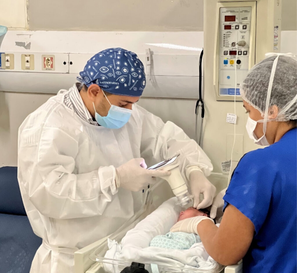
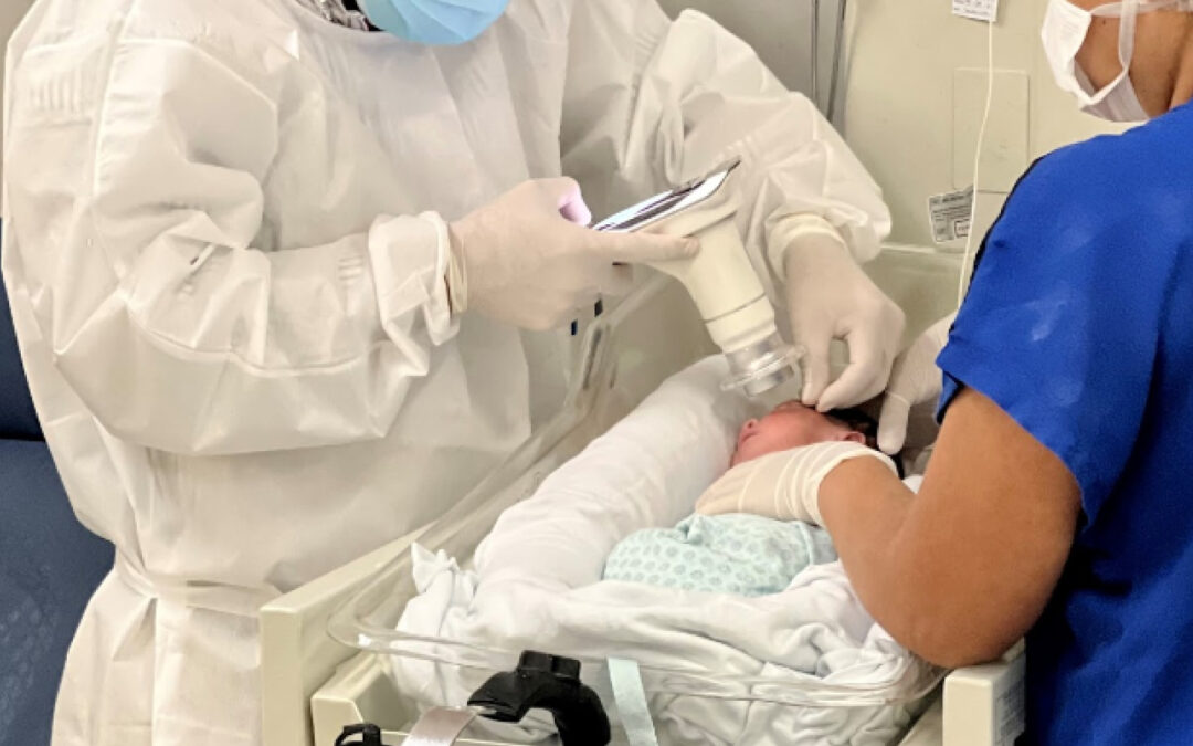
Image caption: Samuel Montenegro examining a newborn with the Eyer. Photo: personal archive.

ROP bears two dangerous characteristics: it’s silent, showing no visible symptoms, and it progresses rapidly. Therefore, adhering to international and national protocols for early diagnosis and treatment is crucial.

Montenegro explains that routine eye examinations for premature babies should be conducted four weeks after birth. “The child isn’t born with the disease, so it’s essential to assess during this period.”

The examination should be performed by an ophthalmologist experienced in evaluating preemies and knowledgeable about the disease to identify location and sequential retinal changes.
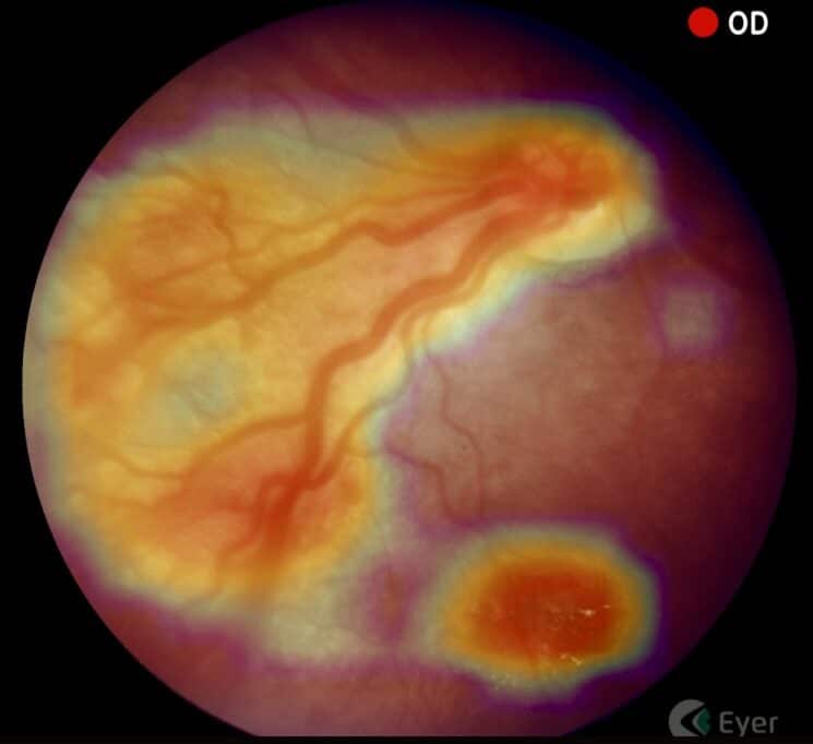
Image caption: Image taken with the Eyer. Photo: Samuel Montenegro.

Subsequent examination scheduling will be determined by findings from the initial examination.

After identifying ROP, Montenegro tracks and documents patients using a portable retinal camera, Eyer. This equipment, highly recommended for infant and child examinations due to its portability and high image quality, attaches to a smartphone and conducts retinal exams within minutes. It also makes images available on the online platform, EyerCloud, facilitating study and case progression monitoring for physicians.
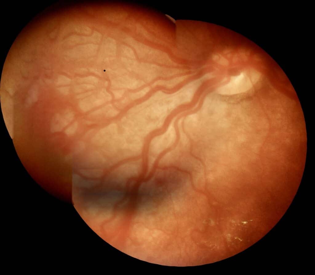
Image caption: Image taken with the Eyer. Photo: Samuel Montenegro.

“The device has been a game-changer as it greatly assists me in capturing the patient’s retina at that exact moment, in a practical, quick, and high-quality manner,” he states. Previously, the specialist used a retina mapping lens with smartphone assistance for videos. “Then I’d freeze the image, take a screenshot, and store it on the computer. It was quite labor-intensive,” he recalls.

Premature Retinopathy Treatment

Montenegro explains that treatment is most effective when ROP is identified early. “The secret to managing this disease lies in early diagnosis and immediate treatment when necessary.”

Currently, retinal ablation with a laser is the gold-standard treatment. Depending on the stage, there are alternatives, such as anti-VEGF injections and cryotherapy. “In this disease, we’re fighting blindness. Therefore, we apply laser therapy to prevent blindness in cases where it’s the best indication. However, this may permanently restrict the field of vision,” he points out.

Children with ROP receive follow-up care from a multidisciplinary team: pediatric ophthalmologist, retina specialist, occupational therapist, and physiotherapist. This follow-up extends beyond retinal ablation, aiming to achieve early visual stimulation.

“Newborns diagnosed with ROP are at a higher risk of developing ophthalmological issues in the future, such as strabismus, amblyopia, and refractive errors. Therefore, ophthalmological follow-up after discharge is recommended,” he emphasizes.
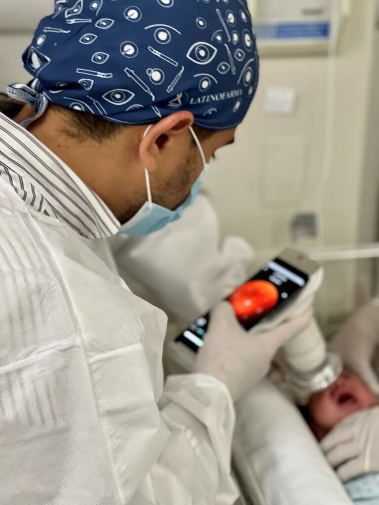
Image caption: Samuel Montenegro examining a newborn with the Eyer. Photo: personal archive.

ROP Brazil

Montenegro is part of a project called ROP Brazil, which aims to share knowledge and further study premature retinopathy.

Various surveys indicate that the proportion of blindness caused by ROP is greatly influenced by the level of neonatal care (availability of human resources, equipment, access, and quality of care), as well as the presence of effective screening and treatment programs. Consequently, there’s significant variability in disease occurrence between developed and developing countries.

“That’s why, understanding more about the disease is essential to decrease cases of preventable childhood blindness in the country,” he states.

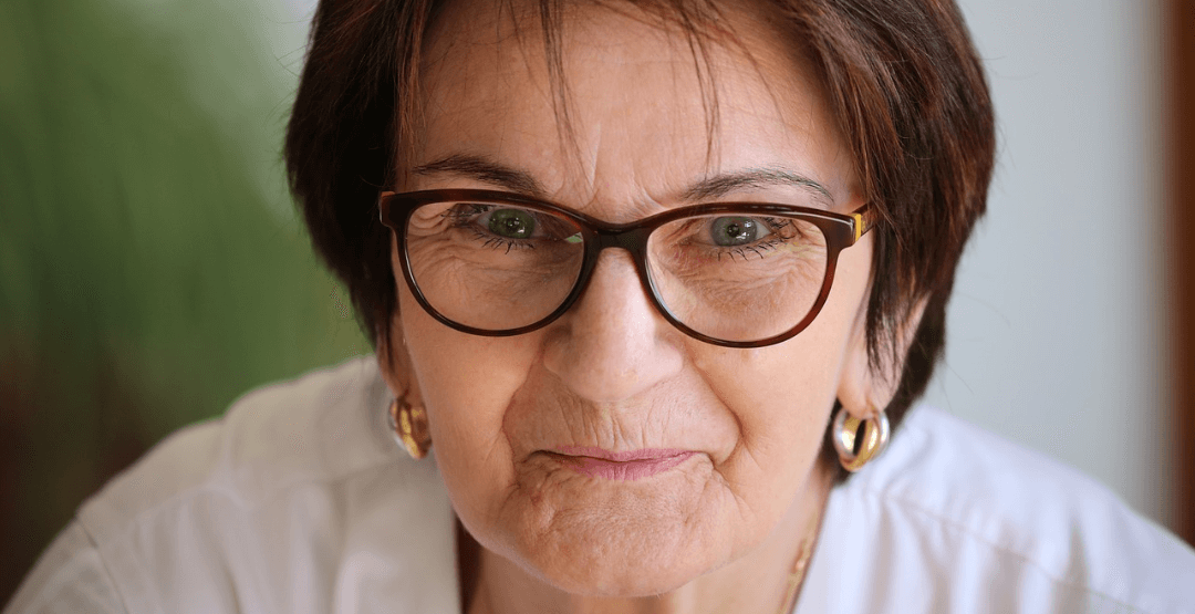
Glasses and coronavirus: study indicates a reduction of the risk of contamination by up to three times

Glasses and coronavirus: study indicates a reduction of the risk of contamination by up to three times

Since last year, scientists raised the hypothesis that glasses help to bar the contamination by the new coronavirus (SARS-CoV-2) through the eyes. This is because this region is also an open door for the virus, as well as a source of contagion.

Now, a recent study found out that the risk of infection by covid-19 in people who wear glasses may be twice to three times lower.  The research, conducted in India, was published in the website medRxiv.

Learn more about the preliminary work, how it was done, th results and next steps. It is worth noting the new investigations are necessary, but, as the disease is new, it is important to follow studies in the area.


Glasses and coronavirus: the research


The research evaluated 304 people, 223 men and 81 women, aging from 10 to 80, residents in Northern India, during two weeks.  Everyone presented symptoms of covid-19.

58 patients (19%) reported they frequently wore eyeglasses or sunglasses when exposed to sunlight.  Through a questionnaire, volunteers stated they touched the face 23 times and the eyes 3 times per hour, in average.


Glasses and coronavirus: results

óculos e coronavírus


According to the scientists, the results related to extended use of sunglasses were significant.  The current contamination rate of 1,35 of the population, in general, dropped to 0,48 among those who regularly wore glasses.  The calculated risk ratio, according to the study, was 0,36%.

That is, the chance of covid-19 infection dropped three times.

Researches believe that the low transmission ratio is due to two factors: glasses protect against contaminated droplets in the air and less frequent touches in the eye while wearing them.


Glasses and the coronavirus: Chinese resarch


A study carried out I n China and publish in the journal JAMA Ophthalmology in September 2020, pointed that wearing glasses for a long time reduces the chances of the new coronavirus infection by five times.

Scientists evaluated 276 positive covid-19 patients.  Factors are the same: less touches in the eyes and protection against airborne droplets.


Conclusion

In fact, scientists remark that the relation between wearing eyeglasses and the new coronavirus is still inconclusive.  This is because the study has not been revised by peers yet. Sampling was small and there was also difficulty to measure how long people wore glasses during the pandemic.

New investigations are necessary to evaluate the effects of glasses, such as protection against covid-19. However, as the disease is new, it is important to follow studies in the area.


Follow Phelcom’s blog and stay on top of main news in the area of eye care.


Subscribe

Icon Button Request A QuoteRequest a Quote

Fechar

Ico Back

Later

Request a quote

Fill out the form below and we will contact you shortly.

    Ico Form Success

    Request a Quote

    Our team will contact you shortly.

    Close

    …complete suas informações

      By clicking the Complete button I allow Phelcom to contact me regarding the above request and I declare that I have read and accept the terms of Privacy Policy and Terms of Use.
      Ico Back

      Depois

      Ico Form Success

      Obrigado por completar suas informações

      FECHAR

      Ico Back

      Depois

      Solicite e comece SEU TEST DRIVE

      Por favor, preencha o formulário abaixo que entraremos em contato.

        x

        Solicitar Orçamento

        Ao clicar em Solicitar, concordo com a Política de Privacidade e os Termos de Uso, permito que a Phelcom me contate e reconheço que o Eyer é um dispositivo médico (Anvisa classe II) exigindo a supervisão de um profissional com CRM para uso e interpretação das imagens.
        Ico Form Success

        Obrigado!

        Nosso time comercial logo entrará em contato para finalizar o processo.

        FECHAR

        Ico Back

        Later

        Request more information

        Please fill out the form below and we will contact you.

          x

          Request EyerMaps

          By clicking the Request button I allow Phelcom to contact me regarding the above request and I declare that I have read and accept the terms of Privacy Policy and Terms of Use.
          Ico Form Success

          Thank you!

          Our commercial team will contact you shortly to finalize the process.Our commercial team will contact you shortly to finalize the process.
          CLOSE