The retinoscope defines the prescription, but in modern eye care, correlating functional data with high-quality fundus photo-documentation is what truly elevates patient care.
The retinoscope remains one of the most iconic tools for objective refraction. For any eye care professional (ECP), mastering the play of light and shadow in retinoscopy is an art form that ensures refractive precision. It gives us the “how much”—the refractive power.
For decades, that information was enough. Today, however, a patient is more than just a “refractive error”; they are a clinical history in motion. Rapidly progressing astigmatism isn’t just a shift in power—and a standard retinoscopy is a fleeting assessment that leaves no permanent record. How, then, do we track and correlate a precise refraction with the structural health of the eye over time?
Correlating Function and Structure: The New Frontier
In a high-volume practice, efficiency demands that functional evaluation (refraction) and structural assessment (documentation) happen seamlessly. Specialists need to know: Does the patient’s complaint or the retinoscope’s finding match the actual ocular structure?
Retinoscopy may be flawless, but if there is a subtle macular change or suspicious optic nerve cupping, refraction alone doesn’t tell the whole story. Bridging this gap requires a tool that harmonizes functional refraction with structural records, integrating everything into a single, streamlined patient workflow.
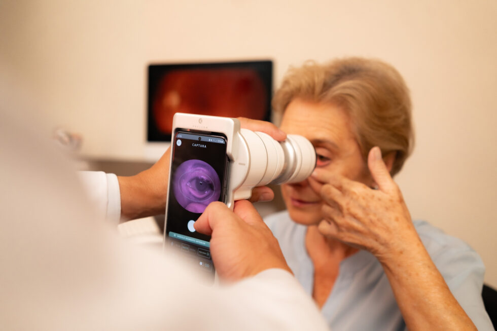
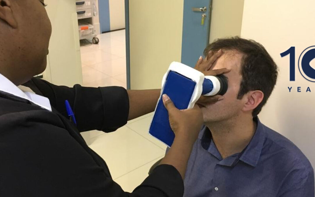
This is the core philosophy behind platforms like the Eyer2. As a portable fundus camera and imaging system, it delivers an immediate connection between refractive data and retinal health.
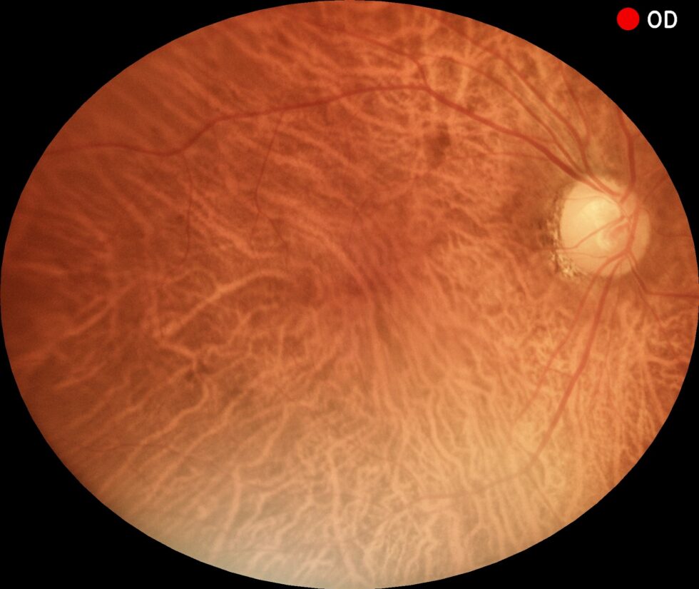
Color fundus photography captured by the Eyer2 portable fundus camera.
Imagine this workflow: The patient undergoes a refractive assessment via retinoscopy. Then, right there in the exam chair, the Eyer2 captures high-definition images of the fundus or ocular surface. What was once just a static data point on a chart is now paired with visual documentation of the retina, macula, and optic nerve.
Instead of juggling two isolated tests—retinoscopy (functional) and fundoscopy (structural)—the specialist now operates within a unified workflow. The precision of the retinoscope gains the context of high-definition documentation. The prescription found through retinoscopy is validated and enhanced by the image captured by the Eyer2.
Meet the Eyer2
The Eyer2 handheld fundus camera supports the diagnosis of over 50 conditions, including glaucoma, cataracts, diabetic retinopathy, age-related macular degeneration (AMD), retinoblastoma, hypertensive retinopathy, retinopathy of prematurity (ROP), and ocular toxoplasmosis.
The device also enables the detection of various ocular surface diseases and conditions, such as blepharitis, eyelash abnormalities, Meibomian Gland Dysfunction (MGD), hordeola, conjunctival and eyelid tumors, advanced cataracts, foreign bodies, burns, corneal lesions, and various types of keratitis (resulting from dry eye, contact lens wear, infections, or ulcers).
Meibography scan captured via the Eyer2 portable retinal camera, evaluating meibomian gland structure for evaporative dry eye.
Key Features & Benefits:
All-in-One Portable Platform: A handheld ocular imaging system capable of performing six different types of exams in a single, non-mydriatic device.
High-Quality Color Fundus Photography: Features a 55° field of view in a single image, making it easy to detect peripheral retinal lesions.
Instant Green-Channel Imaging: Automatically generated immediately following color capture.
Infrared Posterior Segment Imaging: Allows for deeper retinal evaluation with enhanced patient comfort; essential for diagnosing choroidal nevi and assessing evaporative dry eye.
Stereo Disc Photography: Provides 3D visualization of the optic nerve head and cupping.
Panoramic Fundus Imaging: Captures wide-field views up to 120°.
Advanced Analytics: Built-in editing tools and graphics for precise Cup-to-Disc ratio (CDR) analysis.
HD Ocular Surface Documentation: High-definition photo-documentation to track and monitor disease progression.
Cobalt Blue Light: Optimized for the evaluation and documentation of corneal lesions and staining patterns.
Unmatched Mobility: Perfect for multi-clinic practices, remote primary care, and examinations of bedridden or neonatal patients.
Cloud Ecosystem (EyerCloud): Seamless integration with an online platform for comprehensive exam management and secure data storage.
About Phelcom
Phelcom Technologies is an American-Brazilian company combining physics, electronics, and computing to make vision care simpler, more connected, and intelligent.
The company was founded in 2016 by three young researchers: José Augusto Stuchi (computer engineer), Flavio Pascoal Vieira (electronics engineer), and Diego Lencione (physicist). Inspired by Diego’s brother, who struggled with a severe vision condition from childhood, the trio set out to develop a portable retinal camera integrated with a smartphone.
In 2019, Phelcom launched its first product, the Eyer portable retinal camera. Five years later, the company introduced the Eyer2, a comprehensive photo-documentation platform capable of capturing high-quality images of both the posterior and ocular surface segments.
Today, with a 10-year track record, Phelcom’s technology has benefited over two million people across multiple countries, including the United States, Japan, Chile, Colombia, the United Arab Emirates, and Brazil, and has been deployed in more than 200 social outreach initiatives worldwide.
In a modern clinical setting, speed and mobility are vital, but seamless communication is everything. A medical device shouldn’t act like an isolated island. When diagnostic images are trapped on standalone equipment or require a complicated, manual export process, clinical workflows stall and patient care suffers.
The Eyer2 solves this bottleneck through native multi-protocol integration. While highly regarded for its portability and ability to capture stunning, 55° wide-field color retinal images without pupil dilation, the Eyer2’s true superpower is its ability to communicate directly with the eye care industry’s leading software ecosystems.
The EHR & PACS Ecosystem: True Compatibility
Operating on an intuitive, smartphone-integrated Android platform, the Eyer2 utilizes standard Wi-Fi to transmit files instantly. Rather than forcing your practice to adapt to proprietary software, the Eyer2 adapts to you, boasting native compatibility with the most trusted Electronic Health Records (EHR) and Picture Archiving and Communication Systems (PACS) in eye care today.
Supported Platforms At-A-Glance
Table references EHR and PACS systems/ platforms supported with Eyer as well as their purpose.
Flexible Formats and IT-Friendly Protocols
The Eyer2 bridges the gap between patient care and IT requirements by supporting multiple data communication protocols, including DICOM, DICOMWeb, SMB, and FTP. Depending on your department’s specific imaging needs, the Eyer2 allows you to save and download high-resolution exams in two primary formats:
DICOM Images: Ideal for full integration into PACS networks like Harmony or Forum. The Eyer2 embeds rich patient metadata directly into the image file, making tracking and compliance completely hands-free.
JPEG Images: Perfect for a quick snapshot, a patient education handout, a research slide, or a fast upload to a local, non-DICOM desktop directory.
Note for Network Administrators & IT Teams
The Eyer2 is engineered specifically to interact with advanced image management software and standard file-sharing architectures. Because our platform prioritizes rich, high-resolution visual data, the system handles data transport via robust DICOM/DICOMWeb protocols and secure network folder syncing (SMB/FTP). This ensures complex image files and metadata are attached accurately to the patient’s medical chart, bypassing the limitations of text-only messaging protocols.
The EyerCloud Platform
Engineered for high-volume clinical workflows, Phelcom Technologies’ EyerCloud platform combines unparalleled convenience, security, and stability by leveraging the trusted data infrastructure of Amazon Web Services (AWS). Every Eyer fundus camera purchase includes immediate access to the platform with an expanded free capacity of 10,000 images per clinic, allowing providers to securely access, sync, and share exams in real-time across any device.
To support growing practices while maintaining rigorous global standards, including full HIPAA and LGPD compliance, EyerCloud is introducing flexible premium subscription plans. These cost-effective paid tiers directly fuel enterprise-grade server performance, continuous workflow feature updates, and advanced encryption for a fraction of the cost of in-house IT, averaging less than one cent per image. Whether clinics choose to remain on the generous free tier or upgrade to a premium plan as their data needs expand, EyerCloud delivers a transparent, ready-to-use solution that ensures clinical workflows remain efficient, protected, and entirely uninterrupted.
The EyerCloud database
Ironclad Security: FDA Clearance and HIPAA Compliance
Introducing a connected device to a medical network requires absolute confidence in data security. The Eyer2 platform was built from the ground up to respect strict data privacy regulations.
FDA 510(k) Cleared: The Eyer2 is officially FDA cleared, solidifying its status as a trusted, professional-grade diagnostic tool.
HIPAA-Compliant Infrastructure: When syncing data to our cloud management system, EyerCloud, your information is securely backed up via Amazon Web Services (AWS)—the global benchmark for secure healthcare data infrastructure. End-to-end encryption keeps Protected Health Information (PHI) secure whether at rest or in transit.
Advanced Access Controls: Secure logins and customizable user access levels ensure that your clinic stays fully compliant with medical privacy standards.
Team Spotlight: Jeff from Technical Support
Jeff is our technical support analyst and brings an extensive background in healthcare IT and medical device network integration. He specializes in helping clinics onboard their new Eyer2 systems, configuring local DICOM networks, mapping FTP/SMB servers, and ensuring that your image pipeline flows smoothly into systems like ModMed, Forum, or Harmony.
“Joining Phelcom at a time when portable eye care is expanding so rapidly is incredibly exciting,” says Jeff. “Because the Eyer2 natively supports DICOM and standard file sharing, mapping it to systems like NextGen is incredibly clean. I’m here to handle the background technical setup so clinicians can focus purely on what they do best: patient care.”
Whether you need assistance establishing a server link or training your clinical staff on the EyerCloud interface, Jeff and our dedicated support team have you covered.
About Phelcom
Phelcom Technologies is an American-Brazilian company that employs Physics, Electronics, and Computing to make visual health simpler, more connected, and smart.
Founded in 2016 by three young researchers: José Augusto Stuchi, a computer engineer; Flavio Pascoal Vieira, an electronic engineer; and Diego Lencione, a physicist. Inspired by co-founder Diego’s brother, who had struggled with a severe vision condition since childhood, the three founders set out to develop a portable retinal camera with an integrated smartphone.
In 2019, Phelcom launched its first product, the Eyer, a portable retinal camera. Five years later, the company introduced the Eyer2, a photo documentation platform capable of capturing high-quality images of both the posterior and anterior segments.
Today, with 10 years of history, Phelcom’s technology has benefited over two million people across multiple countries, including the United States, Japan, Chile, Colombia, the United Arab Emirates and Brazil. It has also been used in over 200 social outreach initiatives.
Beyond its role in daily clinical workflows, the Eyer handheld fundus camera and EyerMaps Artificial Intelligence have become instrumental in advancing global ophthalmic research. Recently, two landmark studies were selected for presentation at the world’s most prestigious vision research conference: The Association for Research in Vision and Ophthalmology (ARVO). Notably, one of these studies also received the “Best Paper” award at the Association of University Professors of Ophthalmology (AUPO) annual meeting.
Eyer’s Artificial Intelligence in the Real World
The paper “Real-world performance of an offline, automatic algorithm for diabetic retinopathy detection embedded in a handheld smartphone-based retinal camera on two ethnically diverse populations,” led by Dr. Fernando Malerbi, evaluated Eyer’s offline AI for Diabetic Retinopathy (DR) screening in challenging, real-world environments. The study will be presented on May 3, 2026.
Overcoming Algorithmic Bias
For AI to be truly effective, it must perform reliably across all demographics, not just in a controlled lab setting. “In addition to the initial tests we perform to verify if the algorithm provides correct answers, it is crucial to reproduce them in populations with different characteristics—for example, in our study, with diverse ethnic backgrounds. This ensures representativeness in the database, optimizes the tool’s performance, and mitigates the risk of it succeeding in one scenario but failing in another,” explains Dr. Malerbi.
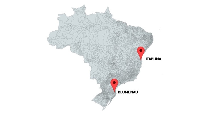
To prove this robustness, the study analyzed retinal images from 1,257 diabetic patients across two demographically distinct Brazilian regions:
Itabuna (Bahia): A population predominantly of African descent, characterized by a higher prevalence of DR and shorter diabetes duration.
Blumenau (Santa Catarina): A population predominantly of European descent.
Map highlighting Itabuna and Blumenau, the cities where patient samples were collected.
Professional Results in the Hands of Volunteers
A key differentiator of this study was that image acquisition was performed largely by non-medical volunteers with varying experience levels during high-volume screening events.
Even without specialized medical training, the results were impressive. “The relevance of this study also lies in its excellent accuracy metrics and image quality. In more than 90% of the cases, it was possible to acquire adequate, high-quality images,” notes Dr. Malerbi. He attributes this success to Eyer’s intuitive design, which simplifies framing, lighting, and focus.
The study confirmed that Eyer’s AI is accurate, consistent, and free of geographic or ethnic bias, demonstratimg that low-cost, user-friendly devices can play a massive role in preventing global blindness.
The Technical Challenge: AI at the Edge
For Paulo Prado, Phelcom’s AI and Mobile Software Coordinator, the project was a masterclass in engineering with a purpose. “Participating in this project was a very meaningful experience, connecting my background directly to people’s health. One of the most important aspects was developing an algorithm capable of running offline and embedded in the device without compromising accuracy, which posed a massive technical challenge,” Prado reports.
Prado reinforces that sample diversity was crucial to validate the engineering team’s work. “Validating the system across two such distinct populations demonstrates its robustness in real-world scenarios and helps ensure the technology is truly useful in clinical practice. For me, it was incredibly rewarding to contribute to a solution that aids in the early detection of diabetic retinopathy, expanding access to disease screening, especially in areas with less medical infrastructure.”
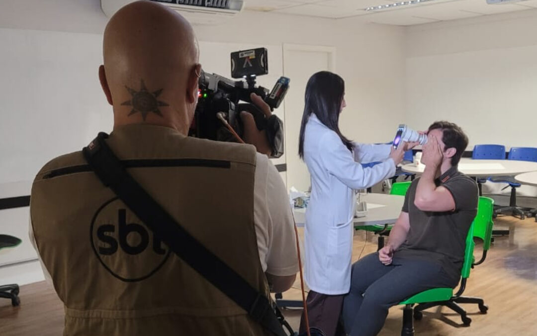
Paulo Prado taking a retinal image with Eyer during the Diabetic Retinopathy Screening Event in Itabuna, 2022.
Eyer at the Bedside: Top Honors at AUPO
A second study, “Handheld Non-Mydriatic Fundus Camera for Bedside Inpatient Ophthalmology and Neurology Consultations,” led by neuro-ophthalmology specialist Dr. Valerie Biousse and researchers at Emory University, highlighted Eyer’s utility in hospital settings.
In scenarios where transporting patients to traditional tabletop devices is impossible, Eyer’s portability allowed for rapid, accurate bedside diagnoses. The clinical impact was so profound that the study was named Best Paper at the AUPO conference.
Shaping the Future of Precision Ophthalmology
The 2026 ARVO Annual Meeting centers on the theme: “Achieving precision ophthalmology through innovative vision research,” a topic thoroughly aligned with the studies conducted using Eyer. The meeting will take place from May 3 to May 7, 2026.
“This event is considered the largest and most important scientific meeting for ophthalmology and visual science research in the world,” Dr. Malerbi summarizes. “It is where the main ideas are presented and validated. Solutions that will enter the market or become available as treatments in the future are presented at ARVO. It inherently carries this pioneering character.”
For Phelcom, having Eyer validated in these studies proves the company’s alignment with the future of global “precision ophthalmology.” As Dr. Malerbi concludes: “It is truly important to be present at this event, both from the perspective of a scientific author and that of a company with such robust research and development.”
Diego Lencione, Co-founder and CTO of Phelcom, sees these accolades as a sign of the company’s maturity. “It is incredible to witness the evolution of Phelcom’s products and our growing relevance on the international stage. In recent years, we have achieved FDA regulatory clearance for our products, and year after year, we see our presence expanding in the global market. Undoubtedly, part of this success stems from our efforts and investments in Research and Development, which culminate in this highly relevant work to be presented at ARVO 2026, combining our expertise in the design and manufacturing of ophthalmic devices and artificial intelligence solutions that truly add value for doctors, patients, and society as a whole.”
About Phelcom
Phelcom Technologies is an American-Brazilian company that employs Physics, Electronics, and Computing to make visual health simpler, more connected, and smart.
Founded in 2016 by three young researchers: José Augusto Stuchi, a computer engineer; Flavio Pascoal Vieira, an electronic engineer; and Diego Lencione, a physicist. Inspired by co-founder Diego’s brother, who had struggled with a severe vision condition since childhood, the three founders set out to develop a portable retinal camera with an integrated smartphone.
In 2019, Phelcom launched its first product, the Eyer, a portable retinal camera. Five years later, the company introduced the Eyer2, a photo documentation platform capable of capturing high-quality images of both the posterior and anterior segments.
Today, with 10 years of history, Phelcom’s technology has benefited over two million people across multiple countries, including the United States, Japan, Chile, Colombia, the United Arab Emirates and Brazil. It has also been used in over 200 social outreach initiatives.
In late 2017, industrial designer Peter Martins von Zweigbergk received a call that would spark a long-term partnership with the newly founded medical- tech startup Phelcom Technologies.
On the line was CEO and co-founder José Augusto Stuchi, inviting him to shape a vision that would eventually redefine portable retinal imaging. Von Zweigbergk, who already knew the founding trio– Stuchi, Diego Lencione, and Flávio Pascoal Vieira– from previous ventures, was immediately sold. “Two things drew me in: my passion for startups and the sheer potential of the product.” he recalls. “A portable fundus camera that captures high-quality images in minutes without pupil dilation is a game-changer.”
Peter Martins von Zweigbergk, Design and Brand Advisor
Defining the Identity
Before sketching a single line, Von Zweigbergk proposed a strategic workshop to align the prototype with a long-term brand vision. He challenged the team on positioning, strategy, and even the product’s original name. It was during these sessions that “Eyer” was born– transforming a promising piece of tech into a cohesive brand identity.
Tasked with leading the product design, Von Zweigbergk looked beyond aesthetics. He proposed an architecture inspired by smartphone ergonomics– a radical departure from traditional, bulky medical equipment.
“At first, clinicians found it unusual. A fundus camera held like a phone was unheard of,” says the designer. “We took the first concepts into hospitals to ensure it wasn’t just ‘cool’– it has to be stable, comfortable, and intuitive enough for a professional to operate with a single thumb.”
One of the greatest technical hurdles was power. By observing laptop peripherals, the team implemented USB-C power transfer, allowing the device to draw from the smartphone’s system and eliminating the need for a cumbersome secondary internal battery.
If the original Eyer was about establishing a new category, the Eyer2 was about refining it for a changing tech landscape As smartphone manufacturers moved cameras from the center to the corners, the Eyer2 had to abandon its signature symmetry.
“Medical equipment usually relies on symmetry for visual balance,” Von Zweigbergk explains. “For Eyer2, we transitioned to a more ‘technical’ and geometric aesthetic. The sharper lines helped accommodate the asymmetrical layout while signaling that this wasn’t just an update– it was a new generation.”
He also pushed for a “capture-first, data-later” user journey. He argued that in a fast-paced clinical environment, a doctor should be able to snap a diagnostic-quality photo instantly and worry about the paperwork once the patient is taken care of.
A Global Competitor
From day one, the goal was to compete with global giants. “If you aren’t a U.S- based company, you have to be twice as good to compete on the same stage,” Von Zweigbergk notes.
Today, the Eyer platform is a modular powerhouse. The Eyer2 features magnetic attachments, a 4,000 mAh battery capable of 60 exams per charge, and the ability to perform both anterior and posterior segment exams.
A Culture of Excellence
For Von Zweigbergk, the success of Eyer lies in Phelcom’s internal culture. He credits the founders’ willingness to listen, pivot, and treat design as a core business pillar rather than an afterthought.
“It’s a rare combination,” he concludes. “Partners who perfectly complement one another and are genuinely driven to build something with a lasting global impact.”
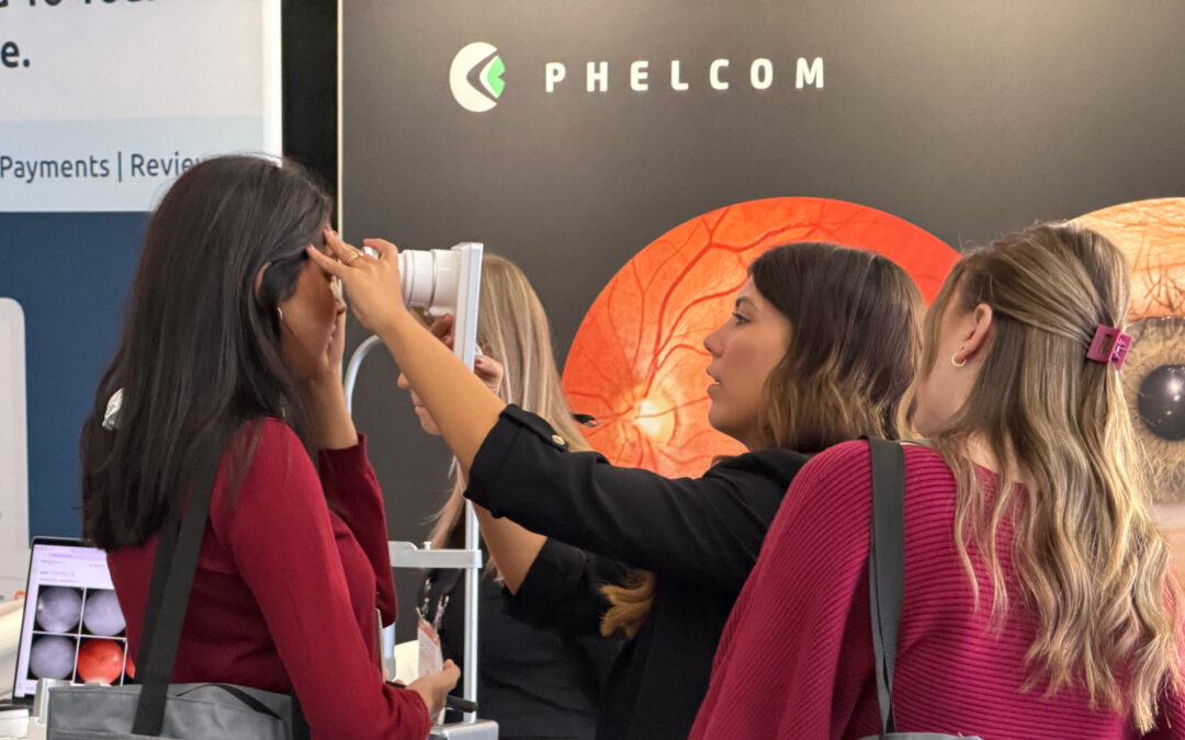
This year represents a major milestone in our journey to make eye care simpler, more connected and more intelligent for everyone, everywhere. Join us at any of our international stops to see our latest innovations in action.
Explore our full 2026 calendar below to find dates and locations – we look forward to seeing you at the Phelcom booth!
Ophthalmology and Health CONFERENCES Phelcom will attend worldwide iN 2026
We look forward to another amazing year of strengthening connections, sharing clinical insights, and showcasing how our technology transforms vision care and improves patient outcomes across the globe. We’ll see you on the road in 2026!
Ophthalmology and Health CONFERENCES Phelcom attended iN 2026
For Phelcom, 2025 was a pivotal year defined by global scientific validation and critical mission expansion. With the publication of a key scientific article about the Eyer portable fundus camera datasets and the expansion of our operations into several new countries, we reinforced our commitment to increasing access to vital eye care and contributing to the prevention of visual impairment and blindness globally.
Explore the key milestones leading up to Phelcom’s 10th anniversary!
Scientific Recognition: Publication in Nature Portfolio
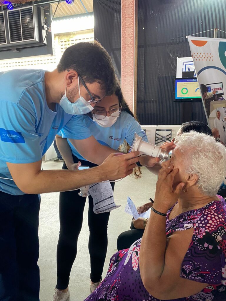
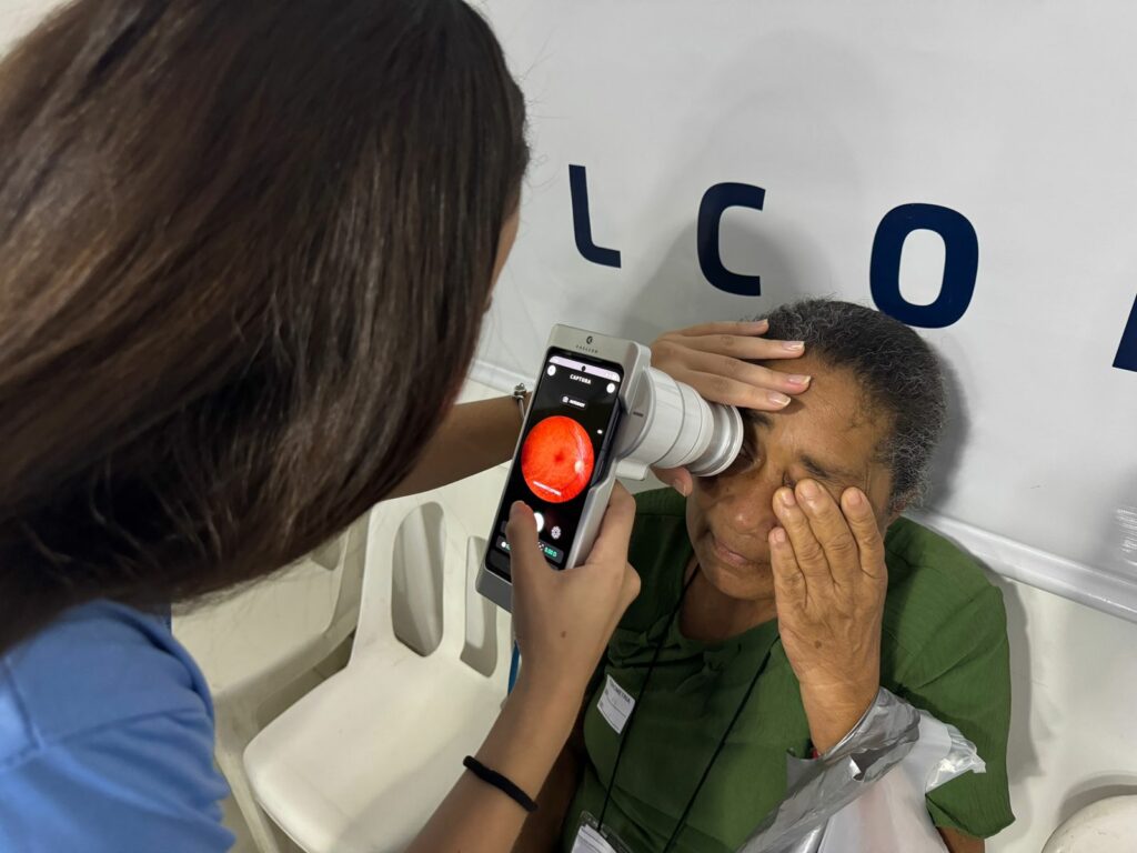
Capture being performed with Eyer during the Itabuna Diabetes Screening Campaign in 2022
2025 marked a major milestone for the Eyer technology, as its datasets were featured in one of the world’s most respected scientific journals.
Scientific Data, a journal from the Nature portfolio, published the article“A portable retina fundus photos dataset for clinical, demographic and diabetic retinopathy prediction,” introducingmBRSET: the first-ever publicly available dataset of diabetic retinopathy images captured using a handheld fundus camera in a real-world, high-burden setting. The images were collected during the 2022 Itabuna Diabetes Screening Campaign (Mutrião do Diabetes) in Bahia, Brazil, demonstrating the real-world utility and quality of the Eyer device.
Driving Impact: Screening Campaigns Across Brazil
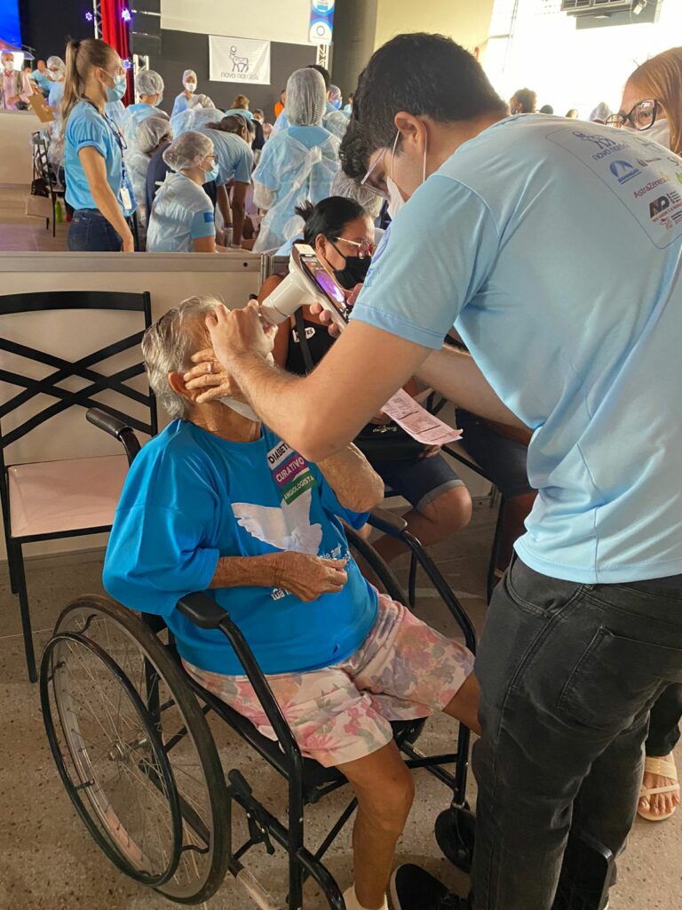
Capture being performed with Eyer2 during the Itabuna Diabetes Screening Campaign in 2025
Phelcom’s commitment to preventative eye care was demonstrated through our participation in major screening campaigns across Brazil, including initiatives in Itabuna (BA), Blumenau (SC), and São Paulo (SP). Our equipment played a pivotal role in ensuring efficient and effective care delivery.
The Diabetes Screening Campaign in Blumenau, registered over 1,300 scheduled visits. Using the Eyer, approximately 190 cases of diabetic retinopathy were identified at varying severity levels, with patients subsequently referred for treatment and further testing. Eyer’s Artificial Intelligence technology also allowed for the immediate discharge of approximately 290 patients (screening negatively for pathology), significantly optimizing clinical patient throughput.
In Itabuna, where more than 2,000 consultations were performed, we showcased innovation beyond the lens. The campaign utilized “Mutirômetro,” a dedicated app developed by Phelcom to completely digitize the patient journey and streamline the care processes from start to finish.
Phelcom’s technology also supported the 28th National Free Diabetes Campaign, a massive effort promoted by the National Association for Diabetes Care (ANAD) in partnership with FENAD. This event, held on November 1st, provided critical educational, physical activity promotion, and specialized care, further proving the Eyer’s utility in large-scale health initiatives.
Global Growth: Expansion into Latin America
Phelcom booth at Faco Extrema 2025, held in Buenos Aires, Argentina, from August 21 to 23.
2025 was the year Phelcom solidified its commitment to making high-quality eye care accessible across Latin America. Through powerful strategic partnerships, we successfully expanded the commercial availability of the Eyer portable fundus camera. Our technology is now officially accessible to eye care providers and patients in the key markets of Chile, Argentina, and Colombia.
The strong and positive receptivity from the Latin American market underscored the adaptability and essential reach of the solutions Phelcom develops. This expansion proves that innovative, portable technology like the Eyer is uniquely positioned to address high-burden health challenges across diverse global regions.
Investing in the Next Generation: The “Eyes to the Future” Contest
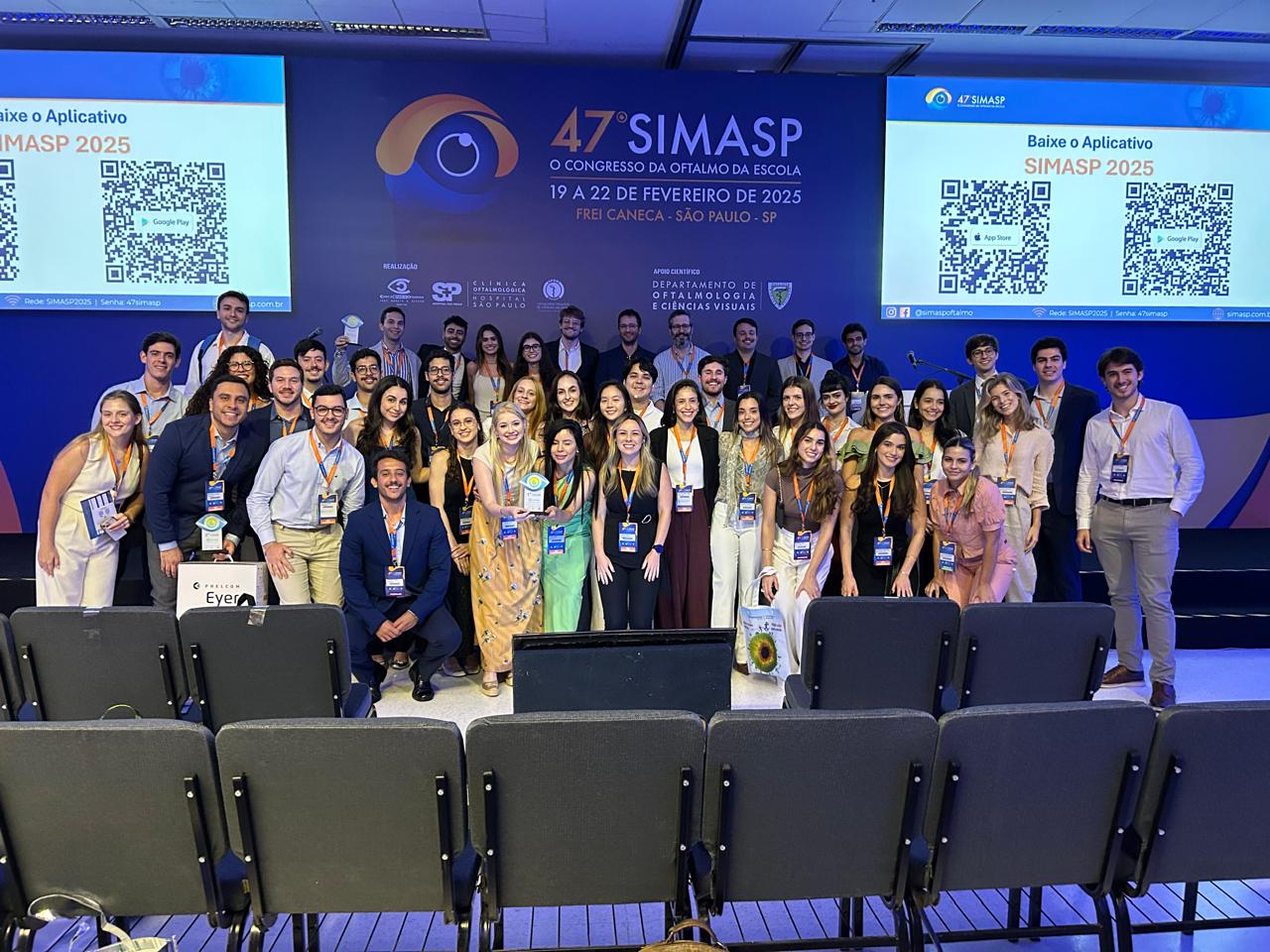
Participating leagues of the 2nd edition of the “Eyes to the Future” (De Olhos para o Futuro) contest during the award ceremony at SIMASP 2025.
Phelcom is dedicated not just to building great technology, but also to training the next generation of medical leaders. The second edition of our “Eyes to the Future” (De Olhos para o Futuro) contest, held in partnership with the Brazilian Association of Academic Ophthalmology Leagues (ABLAO), achieved significant social impact while also educating future doctors. The contest mobilized 205 medical students across seven different states, resulting in 2,486 vital eye screenings. More than 70 patients with severe conditions were diagnosed and referred for treatment with the support of the Eyer portable fundus camera and EyerMaps AI.
The winning projects powerfully demonstrated how portable technologies are key to expanding access to accurate diagnosis across diverse geographic and socioeconomic contexts in Brazil, proving that the future of eye care is both mobile and intelligent.
Strengthening Presence: Global Visibility and Market Reach
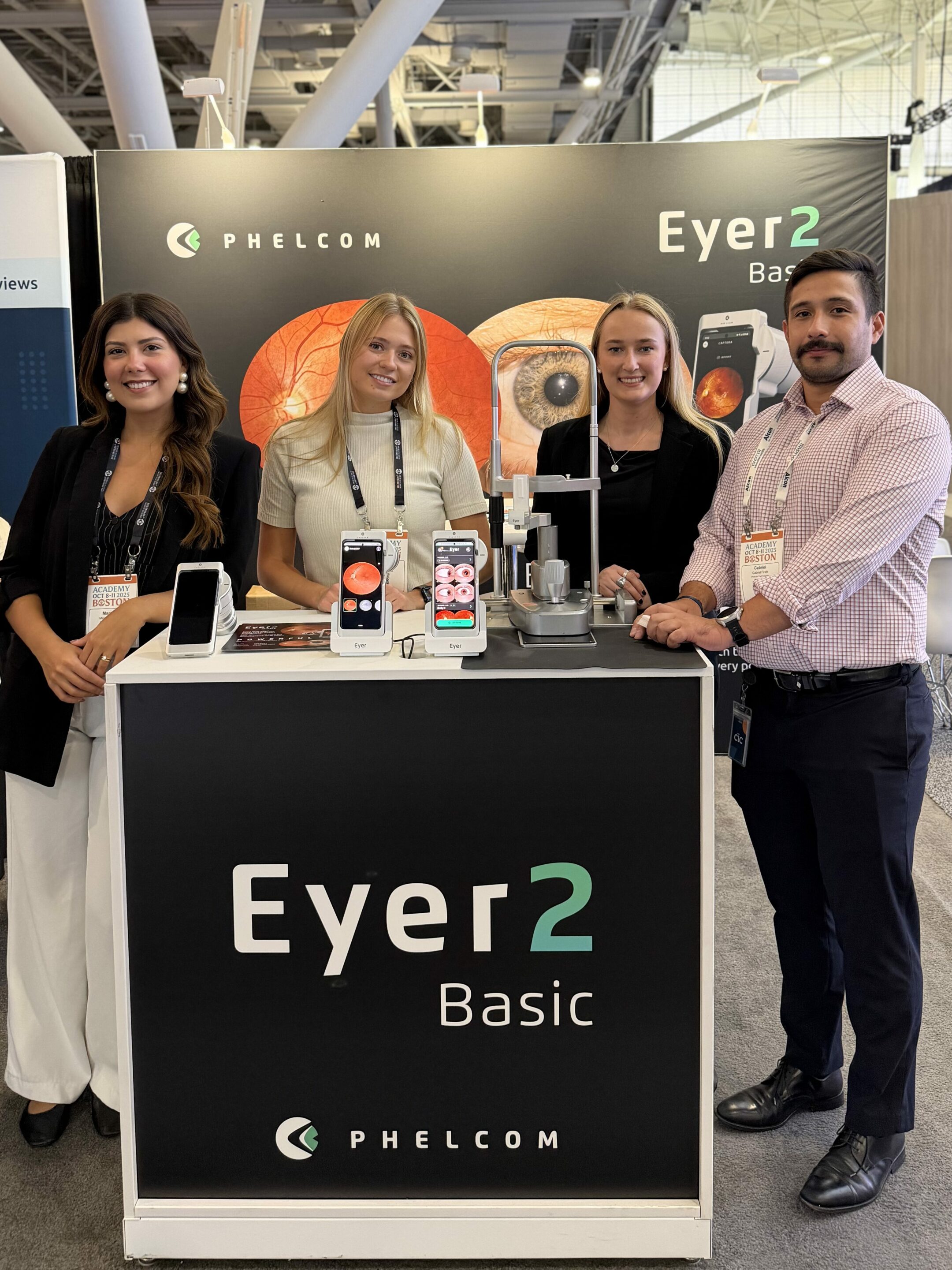
Phelcom booth at Academy 2025, held in Boston, United States, from October 8 to 11.
Phelcom’s achievements in 2025 were underscored by our intense market presence and global visibility. We intensified our participation in key ophthalmology events, featuring over 60 commercial exhibitions and institutional sponsorships at major conferences across Brazil, The United States, and Latin America. This visibility demonstrates our commitment to collaborating with and supporting the global eye care community.
Counting Down to a Decade of Innovation
The remarkable results achieved in 2025 serve as the perfect prelude to a significant milestone: in 2026, Phelcom will celebrate its 10th anniversary.
This coming year will be marked by numerous developments, innovations and celebrations. What began a decade ago as a project by three dedicated researchers has successfully transformed into a global benchmark for portability and intelligence applied to ophthalmology, with a strong, continued outlook for expansion and life-changing innovation.
About Phelcom
Phelcom Technologies is an American-Brazilian medtech company pioneering solutions in digital visual health. We leverage our deep expertise in Physics, Electronics, and Computing (Phelcom) to create technology that makes eye care simpler, smarter and more connected.
Phelcom was founded in 2016 by three dedicated researchers: Computer Engineer José Augusto Stuchi, Electronic Engineer Flavio Pascoal Vieria, and Physicist Diego Lencione. Inspired by the vision struggles of co-founder Diego Lencione’s brother, the founders set out to democratize eye diagnostics by developing a portable retinal camera with an integrated smartphone.
In 2019, Phelcom launched its first product, the Eyer, a portable retinal camera designed for mobility and ease of use. In 2024, the company introduced the Eyer2 a comprehensive photo documentation platform capable of capturing high-quality images of both the posterior and anterior segments of the eye.
To date, Phelcom’s innovative technology has benefitted over two million people across multiple countries, including the United States, Japan, Chile, Colombia, the United Arab Emirates, and Brazil. Our devices have also played a crucial role in over 100 social outreach initiatives, reinforcing our core mission to prevent visual impairment and blindness.