As the popular saying goes, the eyes are said to be the windows to the soul. When we apply this to the realm of healthcare, we might adapt it to say, “the eyes are the windows to the body.” This is because various diseases that affect our body manifest in the eyes.
Ophthalmologic exams can identify signs of abnormalities within our body, aiding in patient diagnosis. For instance, retinography and fundoscopy can detect infectious, chronic, vascular, hematologic, rheumatic, neurological disorders in addition to eye-related conditions.
In neurology, headaches, cerebral aneurysms, multiple sclerosis, and intracranial hypertension — the latter of which can be related to brain tumors can all impact the structure of the orbit and eyeball.
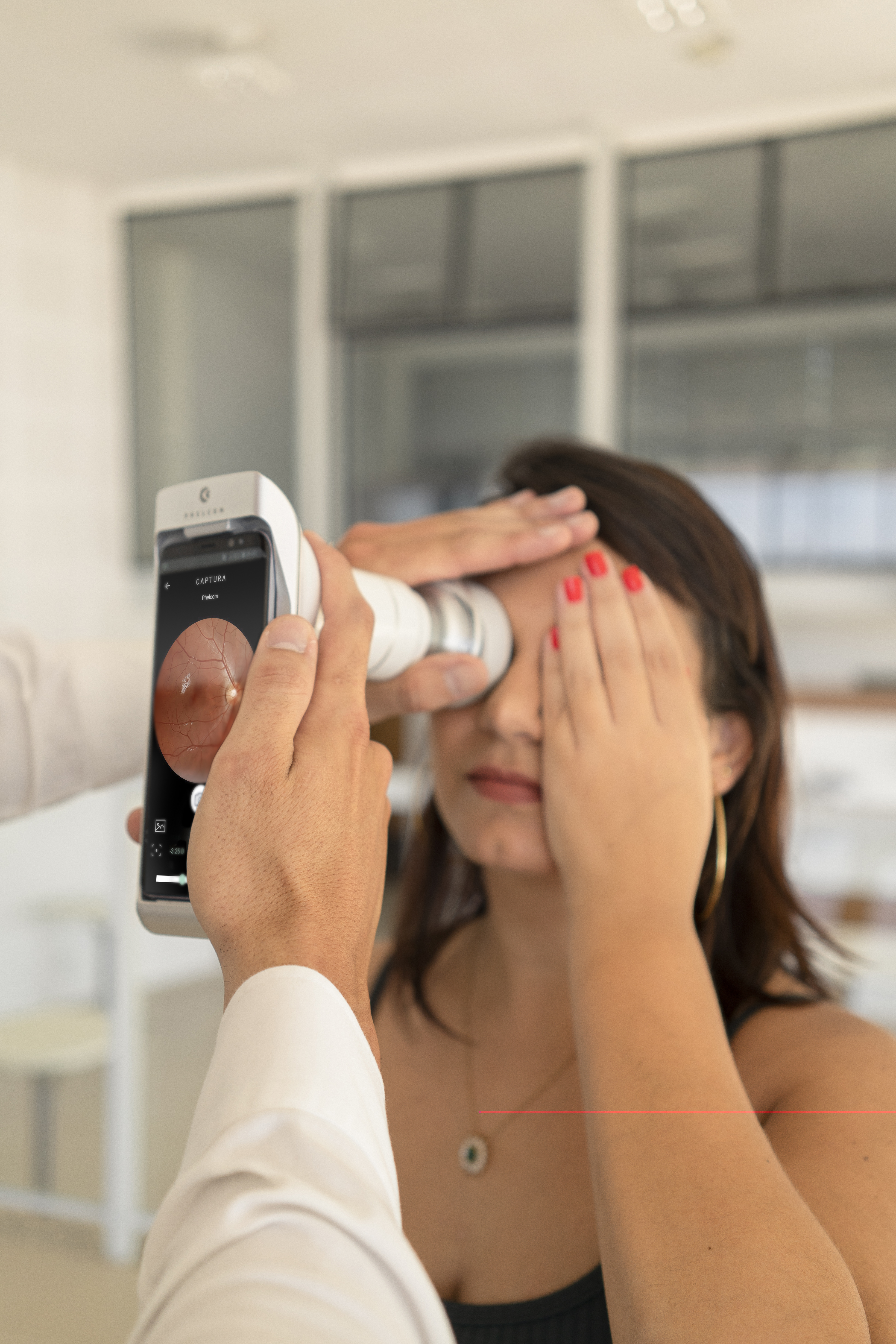
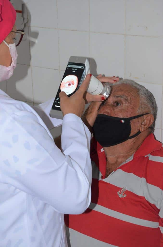
Since last year, neurologist Marcos Christiano Lange has been using Eyer, the portable retinal camera, to map a patient’s retina from the initial consultation. The device uses a built-in smartphone to conduct high-quality retinal exams within minutes, without the need for pupil dilation.
“Eyer is a significant help in screening. Besides being more convenient, when the attention map indicates potential abnormalities outside my field, I advise the patient to consult their ophthalmologist or refer the exam to a partnering ophthalmologist,” he explains.
Neurologist Marcos Christiano Lange
This was how one of Lange’s patients was diagnosed with Age-Related Macular Degeneration (AMD). “She reported worsening vision, attributing it to possible cataracts. Upon examination, I observed protein accumulation in the macula and referred her to an ophthalmologist, who diagnosed AMD and initiated treatment. If I had performed a traditional eye fundus examination as a neurologist, focusing solely on the optic nerve, I wouldn’t have detected these findings,” he recalls.
Heatmap
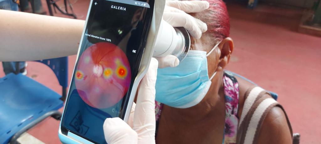
Recently, Phelcom introduced a new feature: EyerMaps, an innovative Artificial Intelligence (AI) system that accurately detects any suspected retinal abnormalities.
Within seconds of capturing a photo of the eye’s fundus, if a suspicion of an abnormality is detected, the AI generates a new image with an attention map (heatmap) highlighting potential retinal abnormalities.
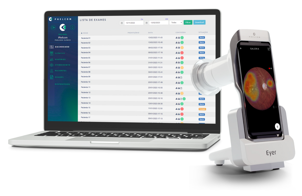
Synchronized with EyerCloud, Phelcom’s cloud-based system for patient data and exam management, the tool visually categorizes images and exams based on the probability of alteration, using color markers in the images and exams:
Green: Image or exam with low probability of abnormality (up to 30%);
Yellow: Image or exam with moderate probability of abnormality (31 to 70%);
Red: Image or exam with high probability of abnormality (71 to 100%).
The AI aids in diagnosing over 50 diseases, including diabetic retinopathy, hypertensive retinopathy, papilledema, and headaches. “I like to keep up with new technologies and am already familiar with Eyer. During a headache course I conducted, Phelcom provided the equipment for us to perform fundus assessments. After using it in practice, I decided to invest in acquiring the device,” he recalls.
Since then, the neurologist has conducted over 400 exams with Eyer, all stored in EyerCloud for reports and tracking. “As soon as I capture the image, I upload it to the platform and open it on the computer to show the patient and explain the details in case of suspected pathology,” he says.
Eyer for Neurologists
For Lange, investing in a portable retinal camera like Eyer is important for neurologists to have an expanded view of the retina. “Even though retinal diseases aren’t our specialty, we can still assist the patient. And that’s priceless,” he emphasizes.
Another advantage is following up with patients who exhibit papilledema and degenerative diseases, such as diabetic retinopathy and hypertensive retinopathy.
Eyer
Eyer is a portable retinal camera that performs high-quality retinal exams in a few minutes, without the need for pupil dilation.
The technology is currently available in Brazil, the United States, Japan, Chile, Colombia, and will become available soon in other countries.
Portability, connectivity, and integration of intelligent functions like EyerMaps, along with the more accessible price, contribute to increased access to retinal exams.
About Phelcom
Phelcom Technologies is a Brazilian medtech company based in São Carlos, São Paulo. The company’s story begins in 2016, when three young researchers – a physicist, an electronic engineer, and a computer engineer (PHysics, ELectronics, COMputing) – created a portable retinal camera integrated with a smartphone.
The idea for the first prototype arose from co-founder Diego Lencione’s interest in visual health, as his brother had a condition that severely compromised his retina and vision since childhood.
In 2019, Phelcom launched its first product on the Brazilian market: the portable retinal camera, Eyer. More than 2 million people in Brazil and around the world have been examined by it so far.
In four years, the company has participated in over 100 social initiatives and was recently ranked among Brazil’s top 10 most innovative companies by Forbes Brazil.
Premature Retinopathy (ROP) is an ocular condition that affects premature infants. According to retina disease and premature retinopathy specialist Samuel Montenegro, ROP is one of the primary causes of preventable childhood blindness. In Brazil, an estimated 13,500 cases occur annually, and among these cases, 1,000 infants may need treatment.
Therefore, identifying newborns in need of treatment early is crucial to reduce ROP-related blindness. Premature babies weighing up to 1.5 kilograms and/or born before 32 weeks’ gestation are a high risk group for ROP.
This period might extend to 35 weeks if the child experiences sepsis, intraventricular hemorrhage, respiratory distress syndrome, requires blood transfusions, or if the mother had a multiple pregnancy, even if the baby’s weight is above 1.5 kilograms.
This is because premature birth can disrupt the development of the baby’s retinal blood vessels. In these cases, vascularization might expand, twist, or even rupture. In advanced stages, this can lead to the formation of retinal scars or even retinal detachment, resulting in permanent vision loss.
The International Classification of ROP (ICROP) defines the disease by severity (stages 1-5), location (zones I-III), and extension in analog hours (1-12 h), with or without additional disease (arteriolar dilation and venous tortuosity), and the presence of which would indicate disease activity (4-5).
Check the table below:
Premature Retinopathy Classification
Stage 1
White line separating vascular from avascular retina
Stage 2
Elevated ridge
Stage 3
Fibrovascular proliferation from ridge
Stage 4
Proliferation leading to subtotal retinal detachment (4a, extrafoveal; 4b, total detachment, including fovea)
Stage 5
Total retinal detachment (open or closed funnel)
Threshold disease (defined by CRYO-ROP) (untreated cases show poor anatomical outcomes in 50% of cases)
Stage 3 retinopathy, zone I or II, with at least five contiguous hours or eight cumulative hours of extension, with an additional disease disease (arteriolar dilation and venodilation).
Type 1 pre-threshold disease (defined by ET-ROP)
Any ROP in zone I with an additional disease (aggressive posterior disease) Stage 3, zone I, without plus disease Stage 2 or 3 in zone II, with additional disease(s).
Type 2 pre-threshold disease (defined by ET-ROP)
Stage 1 or 2, zone I, without additional disease(s) Stage 3, zone 2, without additional disease(s).
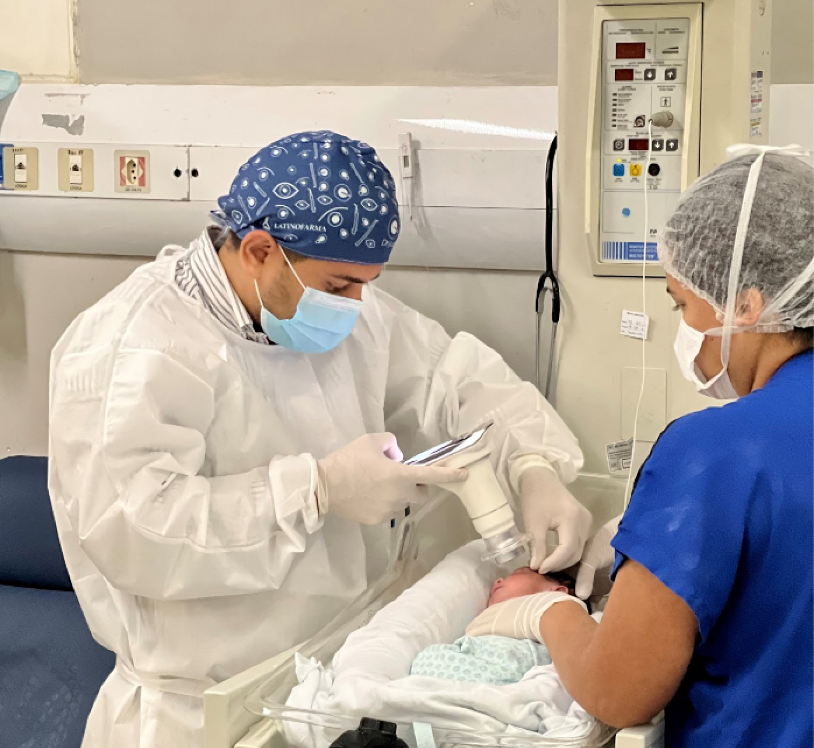
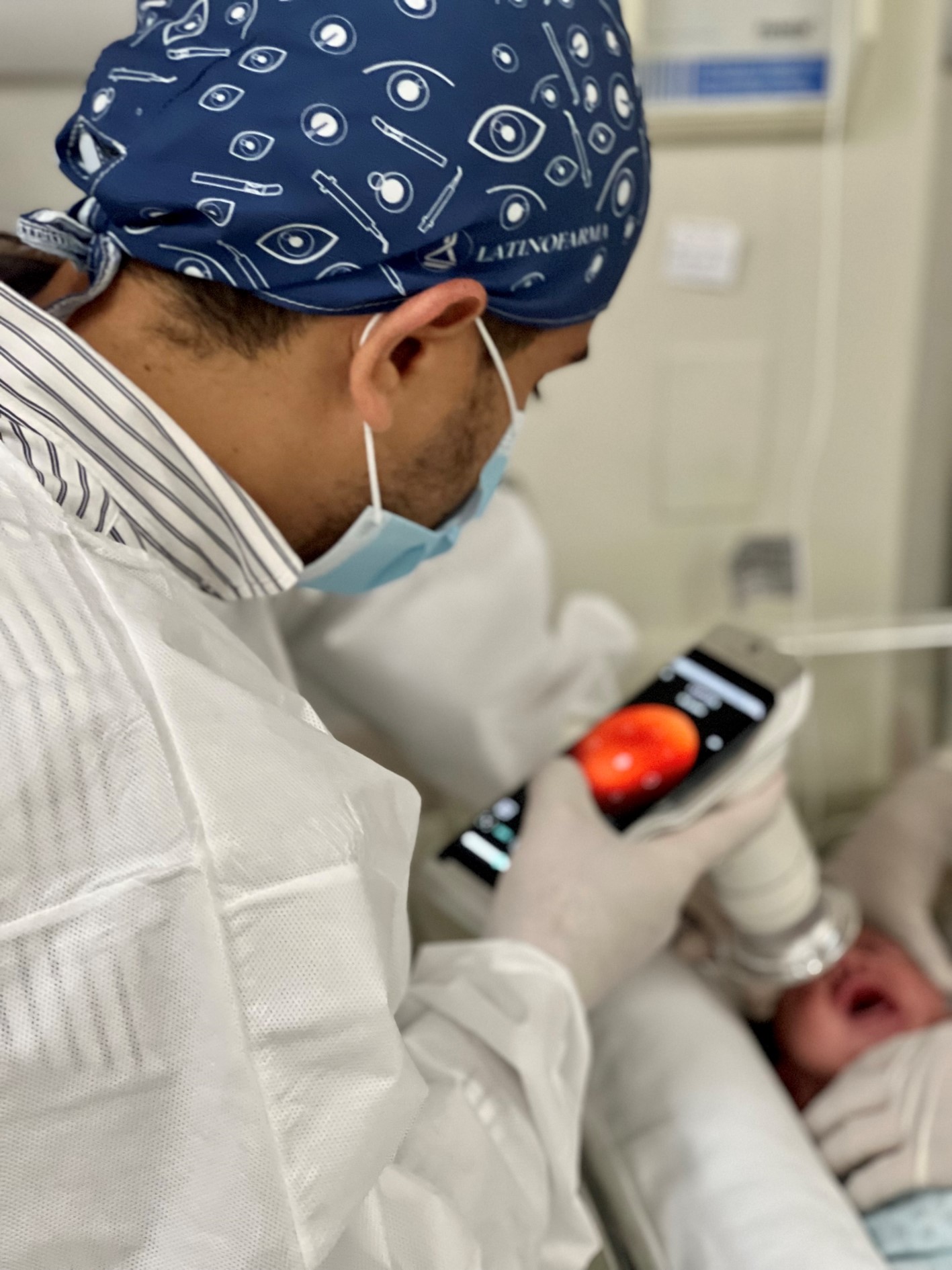
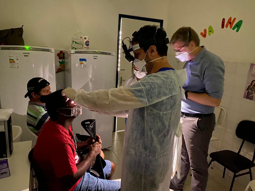
Image caption: Samuel Montenegro examining a newborn with the Eyer. Photo: personal archive.
ROP bears two dangerous characteristics: it’s silent, showing no visible symptoms, and it progresses rapidly. Therefore, adhering to international and national protocols for early diagnosis and treatment is crucial.
Montenegro explains that routine eye examinations for premature babies should be conducted four weeks after birth. “The child isn’t born with the disease, so it’s essential to assess during this period.”
The examination should be performed by an ophthalmologist experienced in evaluating preemies and knowledgeable about the disease to identify location and sequential retinal changes.
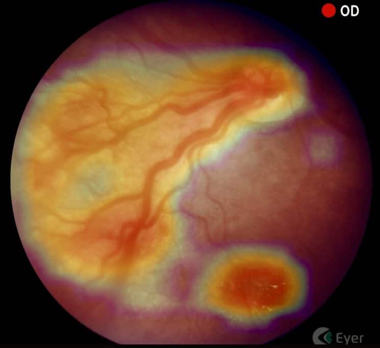
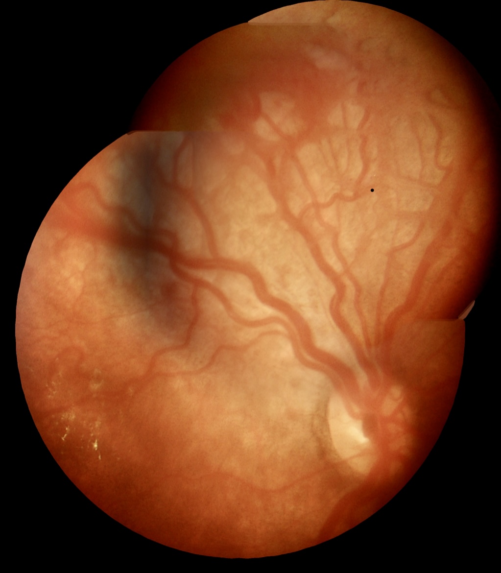
Image caption: Image taken with the Eyer. Photo: Samuel Montenegro.
Subsequent examination scheduling will be determined by findings from the initial examination.
After identifying ROP, Montenegro tracks and documents patients using a portable retinal camera, Eyer. This equipment, highly recommended for infant and child examinations due to its portability and high image quality, attaches to a smartphone and conducts retinal exams within minutes. It also makes images available on the online platform, EyerCloud, facilitating study and case progression monitoring for physicians.
Image caption: Image taken with the Eyer. Photo: Samuel Montenegro.
“The device has been a game-changer as it greatly assists me in capturing the patient’s retina at that exact moment, in a practical, quick, and high-quality manner,” he states. Previously, the specialist used a retina mapping lens with smartphone assistance for videos. “Then I’d freeze the image, take a screenshot, and store it on the computer. It was quite labor-intensive,” he recalls.
Premature Retinopathy Treatment
Montenegro explains that treatment is most effective when ROP is identified early. “The secret to managing this disease lies in early diagnosis and immediate treatment when necessary.”
Currently, retinal ablation with a laser is the gold-standard treatment. Depending on the stage, there are alternatives, such as anti-VEGF injections and cryotherapy. “In this disease, we’re fighting blindness. Therefore, we apply laser therapy to prevent blindness in cases where it’s the best indication. However, this may permanently restrict the field of vision,” he points out.
Children with ROP receive follow-up care from a multidisciplinary team: pediatric ophthalmologist, retina specialist, occupational therapist, and physiotherapist. This follow-up extends beyond retinal ablation, aiming to achieve early visual stimulation.
“Newborns diagnosed with ROP are at a higher risk of developing ophthalmological issues in the future, such as strabismus, amblyopia, and refractive errors. Therefore, ophthalmological follow-up after discharge is recommended,” he emphasizes.
Image caption: Samuel Montenegro examining a newborn with the Eyer. Photo: personal archive.
ROP Brazil
Montenegro is part of a project called ROP Brazil, which aims to share knowledge and further study premature retinopathy.
Various surveys indicate that the proportion of blindness caused by ROP is greatly influenced by the level of neonatal care (availability of human resources, equipment, access, and quality of care), as well as the presence of effective screening and treatment programs. Consequently, there’s significant variability in disease occurrence between developed and developing countries.
“That’s why, understanding more about the disease is essential to decrease cases of preventable childhood blindness in the country,” he states.
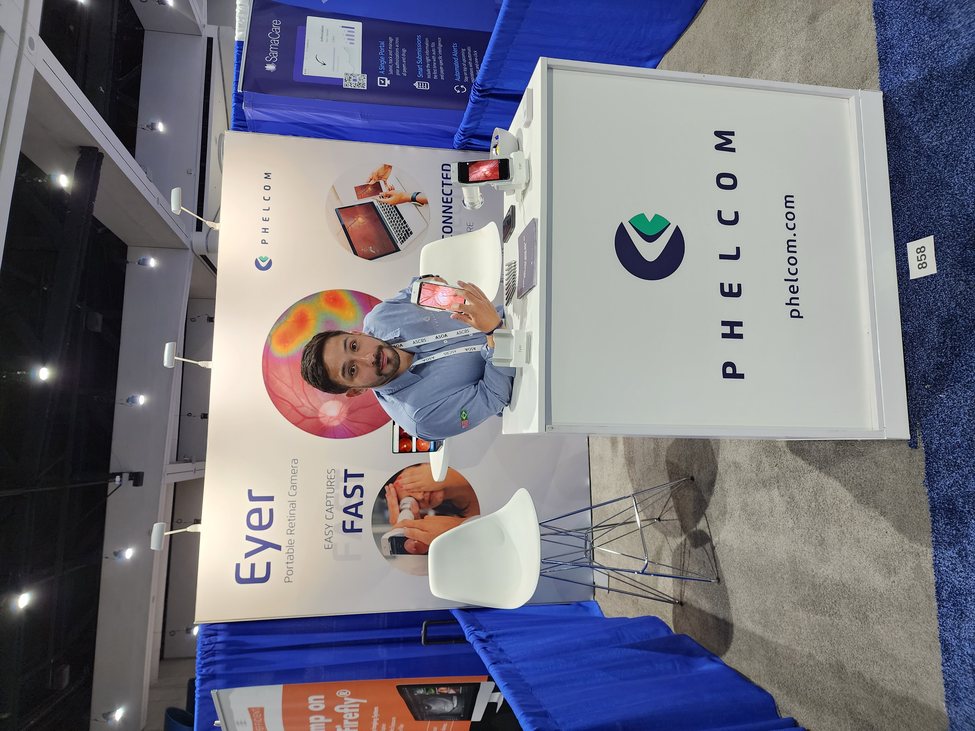
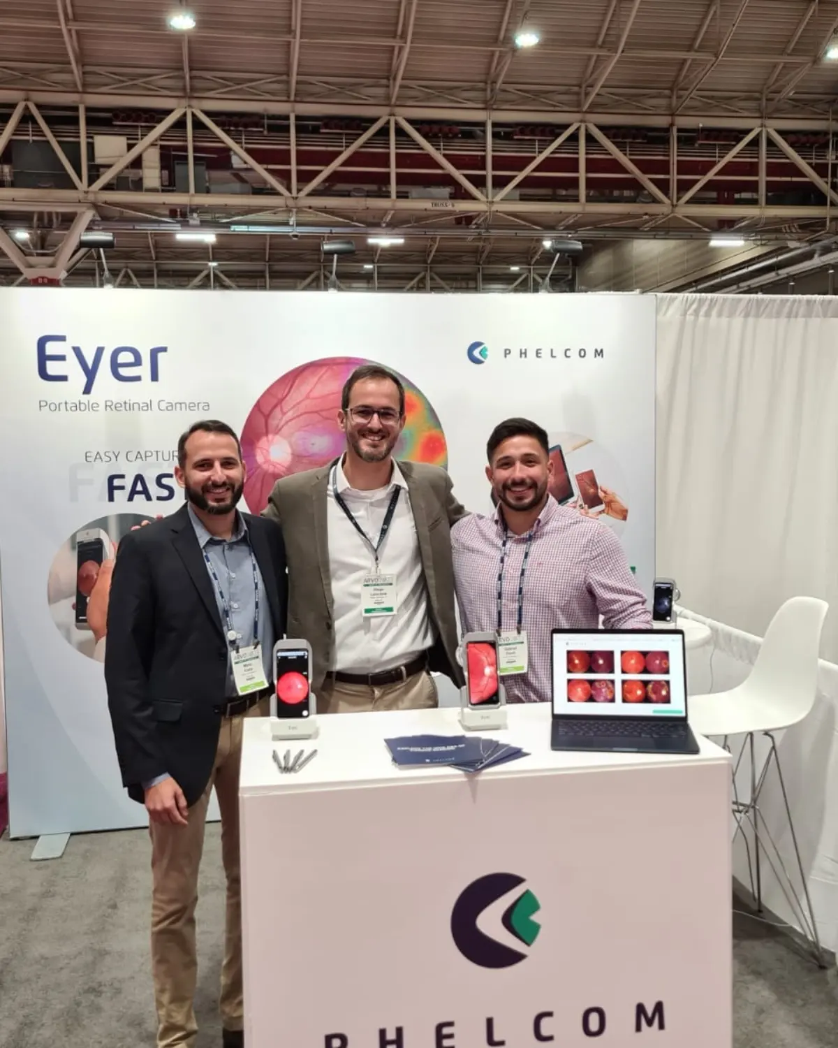
The company solidified its presence in the country by participating in significant events such as Vision Expo East, held from March 16 to 19 in New York, ARVO, from April 23 to 27 in New Orleans, and ASCRS, from May 5 to 8 in San Diego.
“These events bring forth many innovations, products, services, research, and advancements that not only keep us updated but also allow us to introduce Eyer to doctors, healthcare professionals, scientists, researchers, pharmaceutical companies, distributors, and potential partners in a relevant and swift manner,” emphasizes Phelcom’s International Manager, Mário Costa.
Phelcom showcased the portable retinal camera Eyer at the ARVO congress in 2023.
FDA Approves Eyer as 510(k)/FDA Cleared
Recently, the FDA (Food and Drug Administration) granted approval for Phelcom’s portable retinal camera, Eyer, as 510(k). Previously considered an exempt device, the technology was limited to patient photo documentation.
“Now, as we are FDA Cleared, Eyer can be used for diagnostic and telemedicine purposes. This new classification, the highest in our category, opens up more opportunities for our American clients to use our equipment both inside and outside the office,” Costa highlights.
Upcoming International Events
Phelcom will take part in several international trade shows and congresses in the second half of the year. Check out the schedule:
Phelcom Eyer is a portable retinal camera that attaches to a smartphone and conducts high-quality retinal exams within minutes without the need for pupil dilation.
This technology aids in diagnosing over 50 diseases, including glaucoma, cataracts, diabetic retinopathy, AMD, retinoblastoma, hypertensive retinopathy, and ocular toxoplasmosis. It is available in Brazil, the United States, Japan, Chile, Colombia, and will soon become available in other countries.
Portability, connectivity, and integration with intelligent functions like EyerMaps, combined with the technology’s accessible price point, contribute to increased access to retinal exams.
About Phelcom
Phelcom Technologies is a Brazilian medtech company headquartered in São Carlos, São Paulo. The company’s story began in 2016 when three young researchers – a physicist, an electronic engineer, and a computer engineer (PHysics, ELectronics, COMputing) – created a portable retinal camera integrated with a smartphone.
The idea for the first prototype arose from the interest of partner Diego Lencione in visual health, driven by his brother’s condition, which severely affected his retina and vision since childhood.
In 2019, Phelcom introduced its first product to the Brazilian market: the portable retinal camera Eyer. More than 2 million people in Brazil and around the world have been examined using Eyer so far.
In four years, the company has participated in more than 100 social initiatives and was recently named among the 10 most innovative companies in Brazil by Forbes.
Recently, Phelcom visited the MENA region to train Allm MEA on using and presenting the Eyer retinal camera. This non-mydriatic device attaches to a smartphone and performs high-quality retinal examinations in just a few minutes.
The training took place at the Allm MEA office in Dubai, United Arab Emirates. “We conducted onboarding with our distributors, who are already highly qualified to demonstrate the advantages of Eyer to customers in the region,” emphasizes Phelcom’s International Business Manager, Mário Costa.
In addition, the Phelcom team visited significant hospitals in the United Arab Emirates, such as the Al Maerid Health Center MOH in Ras Al Khaimah and the Al Qassimi Hospital in Sharjah. At the latter, Eyer was introduced to the entire ophthalmology team.
Phelcom provided training on the use and commercial presentation of the portable retinal camera, Eyer, to their local partner and distributor, Allm MEA.
Pilot Project in Egypt
Phelcom has also conducted training on the use of Eyer at Ain Shams University Virtual Hospital (AVH) in Cairo, Egypt. “We are participating in a pilot project focused on teleophthalmology at AVH, which is responsible for one of the largest telemedicine programs in the country,” Costa explains.
The hospital develops technology-enabled healthcare systems in Egypt, the Middle East, and Africa. The project involves international collaborators and beneficiaries specializing in telemedicine, e-health research, artificial intelligence, virtual reality, and innovative healthcare models.
Phelcom’s team during training on the use of Eyer at Ain Shams University Virtual Hospital.
Eyer is currently present in 6 countries.
Eyer was approved in the United Arab Emirates in the second quarter of this year. As a result, Phelcom now operates in six countries: Brazil, the United States, Japan, Chile, Colombia, and the United Arab Emirates.
Furthermore, the company is in the regulatory process for marketing Eyer in other locations such as Mexico, Egypt, and Saudi Arabia.
For Phelcom CEO, José Augusto Stuchi, being a Brazilian company operating in important global markets is a source of pride and great responsibility. “Internationalization undoubtedly expands our potential reach. However, it also increases our commitment to providing excellent quality products that truly benefit professionals,” he affirms.
Eyer
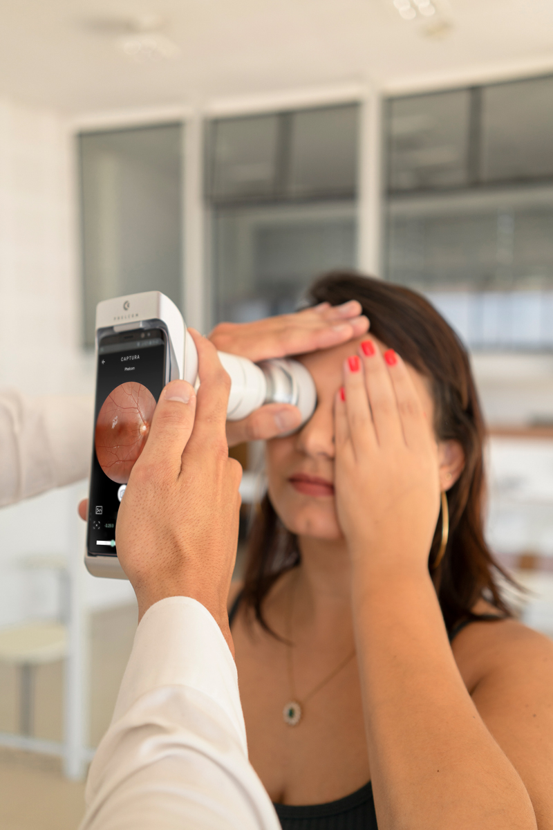
Retina examination conducted with Eyer.
Eyer is a portable retinal camera that attaches to a smartphone and performs high-quality retinal examinations in minutes, without the need for pupil dilation.
The technology aids in the diagnosis of over 50 diseases, including glaucoma, cataracts, diabetic retinopathy, AMD, retinoblastoma, hypertensive retinopathy, and ocular toxoplasmosis. To date, more than 10 million examinations have been conducted in Brazil, the United States, Chile, and Colombia.
The portability and affordable price of the technology democratizes access to retinal examinations, costing approximately ten times less than a conventional tabletop retinal camera that still requires integration with a computer.
About Phelcom
Phelcom Technologies is a Brazilian medtech company based in São Carlos, São Paulo. The company’s story began in 2016 when three young researchers – a physicist, an electronic engineer, and a computer engineer (PHysics, ELectronics, COMputing) – created a portable retinal camera integrated with a smartphone.
The idea for the first prototype emerged from the interest of co-founder Diego Lencione in visual health, as his brother has a condition that severely affects the retina and vision that he’s had since childhood.
In 2019, Phelcom launched its first product, the portable retinal camera Eyer, to the Brazilian market. Today, the technology has reached over two million people throughout Brazil and other countries it is present in..
In four years, the company has participated in over 100 social initiatives and was recently named one of the top 10 most innovative companies in Brazil by Forbes.
Brazilian General Data Protection Law (LGPD), in effect since May, 2021, regulates how to process personal data of citizens in Brazil. On the whole, it aims to keep data safety and eliminate sharing unauthorized by owner.
As telemedicine spreads in the country, medical offices, clinics and other healthcare institutions are legally obliged to follow LGPD rules. Otherwise, infringement fines may reach high values.
Therefore, it is imperative for data exchange, online consultations and exam delivery for reporting, among other medical practices, to occur in reliable and safe environments. Furthermore, the law considers data on patient healthcare as sensitive.
“The great change with LGDP relates to the concept of property of medical data. These data no longer belong to healthcare institutions. Now they exclusively belong to the patient. However, institutions are responsible for data security”. – explains Phelcom’s Data Engineering manager, Fernando Yamanaka.
For this reason, data security is a priority in Phelcom. All images made with Eyer handheld fundus camera and stored in EyerCloud, online platform to manage exams, are protected by Amazon Web Services (AWS). This computing services has the most credible database and cloud systems in the market.
“We store retina images from millions of patients. This part of the eye is known as an ‘eye fingerprint’, since it is unique for each person. That’s why we use the best information security practices to protect data from patients of our customers”, he says.
HIPAA
More than LGPD, Phelcom follows the rules of Health Insurance Portability and Accountability Act (HIPAA), a law on portability and accountability of health insurance in the United States.
Eyer is also sold in the USA. Recently, the FDA (Food and Drug Administration) approved the handheld fundus camera in the category 510(k), the highest for retinographs. Part of the regulatory process involved cyber security analyses of Phelcom’s device.
HIPAA Compliance establishes rules to protect electronic records and medical data of patients. With regards to the LGPD, it has similar goals: protect patient privacy, oblige entities to make medical records available when prompted and assure that patients are notified in case of leakage of sensitive data.
EyerCloud
Eyercloud is an online platform integrated to Eyer that allows storing and managing patients’ exams. All data the equipment captures can automatically synch to the system, so that they are uploaded with full security.
More than assuring data backup in a secure server, the user has all data organized in a friendly, functional and intuitive interface.
For instance, the tool enables evaluation of images by EyerMaps, an innovative system of Artificial Intelligence (AI) that detects any suspicious retinal abnormalities with high accuracy.
In a few seconds after capturing the fundus photo, if a suspicious alteration is detected, the AI generates a new image with a heat map, highlighting the potential abnormalities in the retina. Synchronized with EyerCloud, it classifies the captured images and exams in a simple and visual way, according to the probability of alteration, using colored markers:
Green: image or exam with low probability of alteration (up to 30%);
Yellow: image or exam with medium probability of alteration (31 to 70%);
Red: image or exam with high probability of alteration (71 to 100%).
Eyercloud helps doctors to make reports quickly, prioritize urgent cases and share all data, either printed or by e-mail, with the patients, by generating a protocol number and password.
“We invested in a complex infrastructure to secure the sensitive data of our patients. This is one of the main advantages of EyerCloud. At present, our platform stores about two million exams”, highlights Yamanaka
The manager explains that keeping sensitive data of patients in computers or storages of easy access and in unprotected systems may be risky. “Cloud systems with high security, as EyerCloud, are an essential investment to protect doctors, medical offices, clinics and hospitals from leakage of sensitive data and, consequently, from probable penalties from LGPD”.
Eyer Handheld Fundus Camera
Eyer is a portable fundus camera that works coupled to a smartphone and makes high-quality retinal exams in a few minutes, without need of pupil dilation.
The technology helps to diagnose more than 50 diseases, such as glaucoma, cataract, diabetic retinopathy, DMRI, retinoblastoma, hypertensive retinopathy and ocular toxoplasmosis. It is available in Brazil, United States, Japan, Chile, Colombia and, soon, in other countries.
Portability, connectivity and integration to intelligent functions as EyerMaps, as well as the lower cost of the technology, contribute to increase access to retina exams.
About Phelcom
Phelcom Technologies is a Brazilian medtech headquartered in São Carlos, upstate São Paulo. The company history started in 2016, when three Young researchers – a physicist, an electric engineer and a computing engineer (Physics, ELetronics, COMputing) – created a handheld fundus camera integrated to a smartphone.
The project of the first prototype was born from the interest of the partner Diego Lencione on visual health. His brother has a condition that severely compromised the retina and vision since childhood.
In 2019, Phelcom lauched its first product in Brazilian market: Eyer handheld fundus camera. Nowadays, the technology has already reached more than two million people all over Brazil and the countries it is present.
In four years, the company already took part of more than 100 social actions and, recently, has been ellected one of the 10 most innovative companies in Brazil by Forbes.
In partnership with the NGO Retina Global, doctors plan to perform digital retinal exams and retinal mapping in more than 15 thousand diabetic patients in the countryside of Sergipe.
Last year, the NGO Retina Global was interested in developing a social project to diagnose and treat diabetic retinopathy in Brazil. The North-American institution acts to create sustainable solutions in controlling retinal diseases in poor areas all over the world.
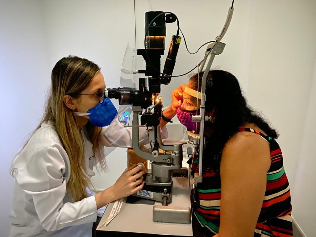
Thus, the partnership with the ophthalmologists Fernando Malerbi and Gustavo Melo gave birth to “Iluminar”, a project to track and treat diabetic retinopathy in 13 municipalities in the backlands of Sergipe. The group chose the region for its history of poorness related to drought and lack of ophthalmological assistance in public health.
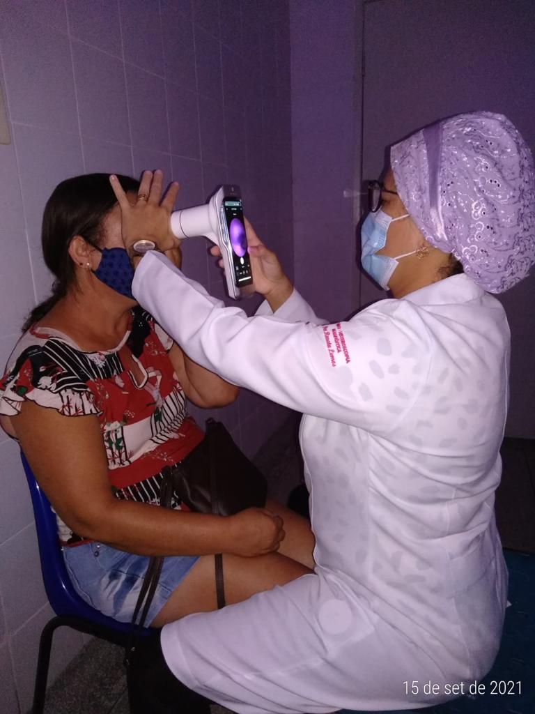
Up to now, more than 700 people have undergone retinal exams in the cities of Itabi, Graccho Cardoso and Canindé de São Francisco. “Approximately 150 patients have been submitted to a new retinal mapping exam with the team of retinologists on site. From those, 50 were sent to laser photocoagulation treatment”, highlights one of the project leaders, ophthalmologist Gustavo Melo.
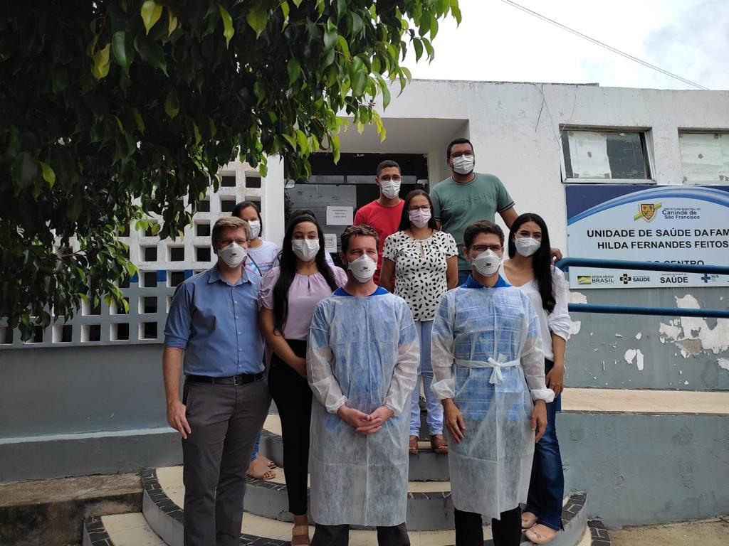
The joint efforts count on two healthcare technicians, around ten workers from Basic Healthcare Units (UBS) from each municipality and four volunteer ophthalmologists, part of them sending remote reports.
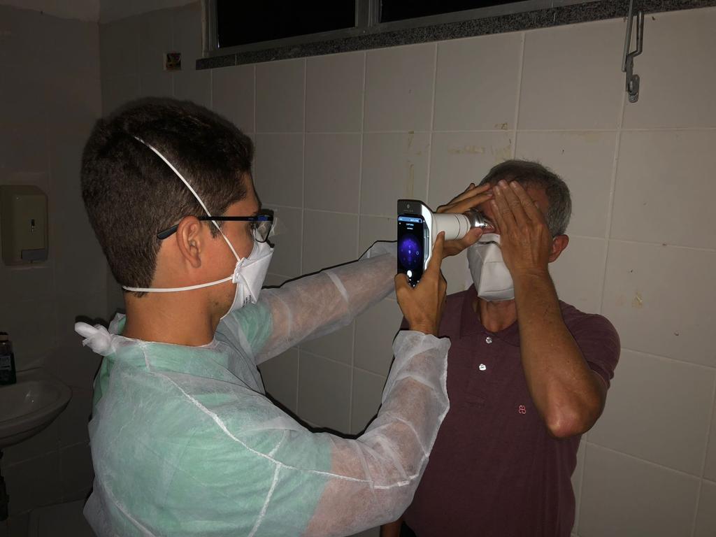
Retinal images are made with Phelcom Eyer smartdevice. Coupled to a smartphone, the equipment carries out high quality exams, in a few minutes without need of pupil dilation. Integrated to a cloud system, it makes data available automatically in EyerCloud online platform. This way, a doctor can generate a report from anywhere in the world.
“Eyer allows optimizing time and costs by tracking the diabetic population in the countryside so that they do not need to move to the cities that have equipment and specialists. Non-midriasis is another advantage, since 90% of the cases do not need to dilate the pupil”, explains Melo.
The smart device also offers the joint efforts of an artificial intelligence that identifies diabetic retinopathy and other eye diseases with more than 95% sensitivity in just three seconds. After that, the exam is sent to the ophthalmologist for checking and reporting. The AI is at the final test stage.
Next Stage
This month, the Project will be at Poço Redondo. It aims to provide care for 15 thousand diabetic patients in 13 cities in Sergipe, in a period of two years.
“The great differential is optimization of time, essential for early diagnosis, so that chances of diabetes-related blindness lower considerably. Raising awareness of both the population and public managers about the efficiency and lower cost of this way of tracking diabetic retinopathy may stimulate the creation of health policies to treat this disease and others that affect the retina”, analyzes Melo.


