Fundus imaging plays a crucial role in diagnosing and monitoring various eye diseases, including diabetic retinopathy. However, in many low- and middle-income countries, access to conventional retinal imaging equipment remains limited due to high costs and infrastructure requirements. Additionally, publicly available image datasets for doctors, researchers, and clinicians remain scarce, particularly for images captured with portable fundus cameras.
In this context, portable fundus cameras like the Eyer have emerged as a more accessible and cost-effective solution for eye screening and disease management. Mounted onto a smartphone, the Eyer captures high-quality retinal images in minutes—without requiring pupil dilation.
These devices can be used in a variety of settings beyond hospitals, such as community health screenings and remote telemedicine consultations. Additionally, they record detailed metadata typically unavailable in other datasets, including patient age, sex, diabetes duration, treatments, and comorbidities.
Recently, Scientific Data, a journal from the Nature portfolio, published the article “A portable retina fundus photos dataset for clinical, demographic, and diabetic retinopathy prediction,” introducing mBRSET: the first publicly available diabetic retinopathy dataset captured using handheld fundus cameras in real-world, high-burden environments. Among the authors of the study are ophthalmologist Fernando Korn Malerbi and Phelcom’s CEO, José Augusto Stuchi.
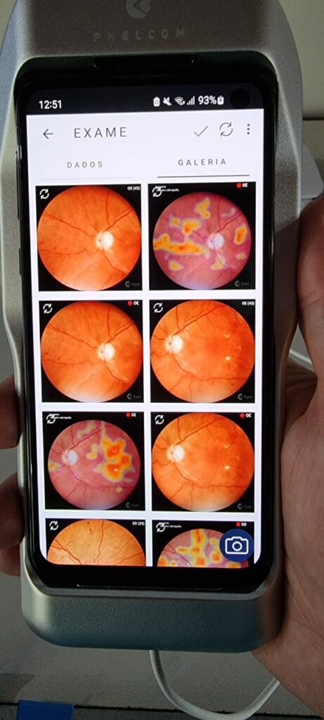
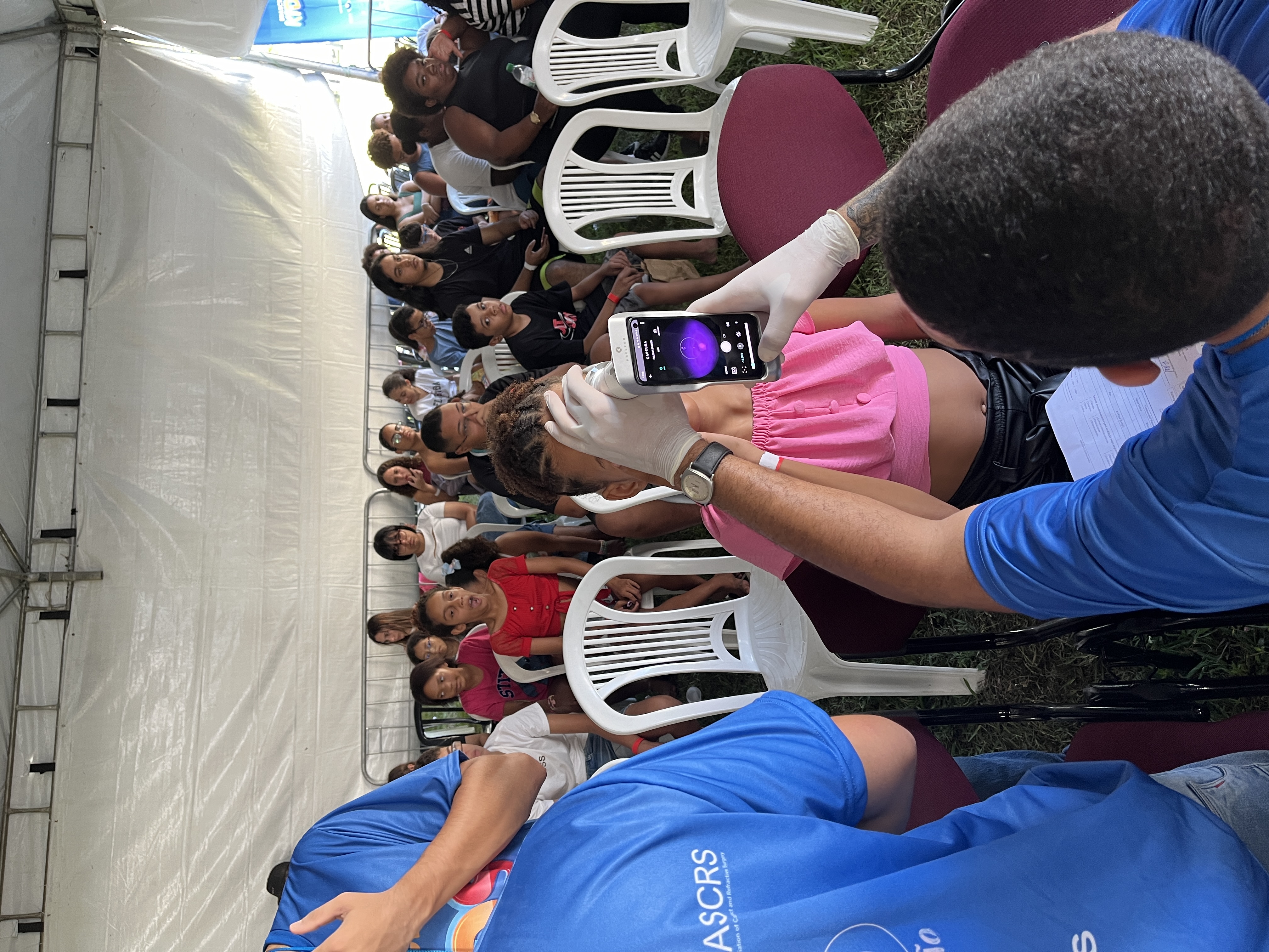
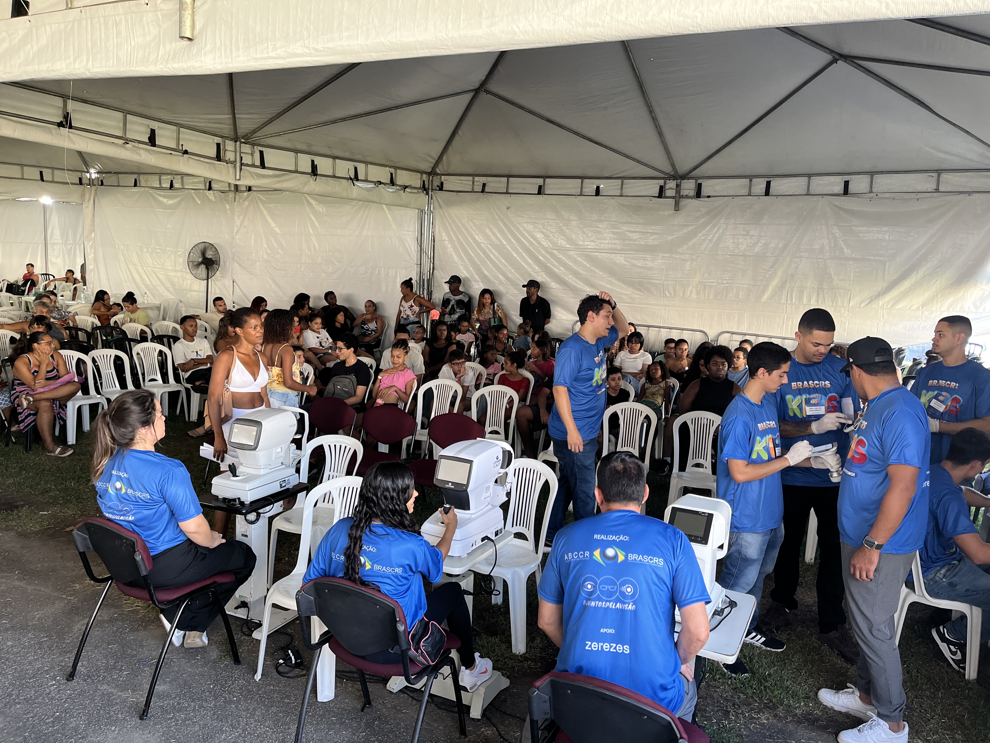
Fundus images captured with Eyer during the Itabuna Diabetes Campaign, held in 2022, highlighting the attention map generated by EyerMaps, indicating possible anomalies.
mBRSET: A Groundbreaking Dataset
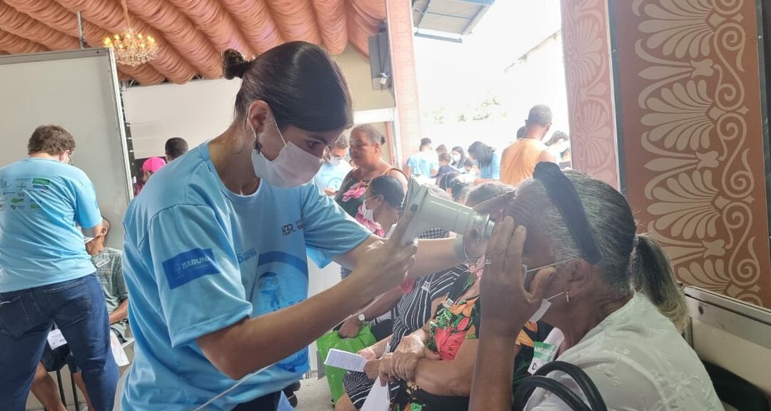
mBRSET consists of 5,164 from 1,291 patients of diverse backgrounds, all captured with the Eyer device during the 2022 Itabuna Diabetes Campaign in Bahia. Recognized as one of the world’s largest diabetes prevention and treatment initiatives, this campaign provides hundreds of patients with vital screenings—such as fundus examinations—and referrals for specialized treatment.
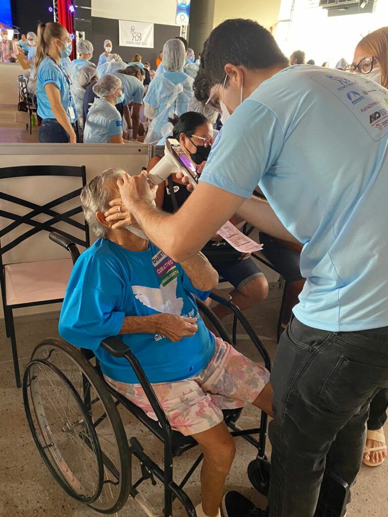
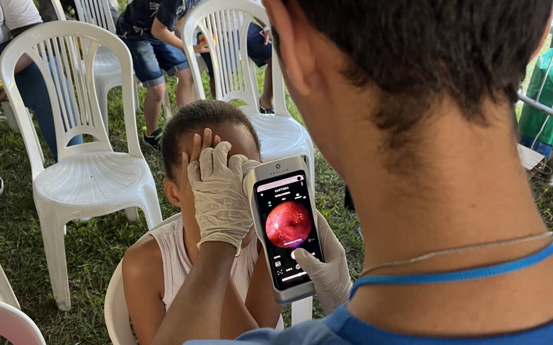
Exam performed with Eyer during the Itabuna Diabetes Campaign, held in 2022.
To validate the utility of mBRSET, state-of-the-art deep learning models were trained for benchmarking, demonstrating high accuracy in diagnosing diabetic retinopathy and macular edema, as well as predicting demographic data.
An analysis of 4,885 assessed images revealed that 3,759 images (76.79%) showed no signs of diabetic retinopathy (DR), 272 images (5.56%) indicated mild non-proliferative DR, 570 images (11.64%) exhibited moderate non-proliferative DR, 82 images (1.67%) showed severe non-proliferative DR, 427 images (8.69%) displayed signs of macular edema.
A Milestone for Ocular Health and Scientific Research
The significance of the mBRSET dataset can be outlined in 5 key aspects:
Representing Brazil’s Diverse Population
mBRSET helps reduce the underrepresentation of low- and middle-income country (LMIC) populations in ophthalmological datasets by including individuals from various ethnic and socioeconomic backgrounds in Brazil.
The First Public Dataset with Portable Camera Images
This is the first publicly available dataset featuring images captured with portable fundus cameras, reflecting the increasing adoption of this technology in resource-limited settings.
Data Collection in Real-World, High-Demand Environments
Images were captured in high-volume clinical settings, ensuring the dataset accurately represents real-world challenges in eye disease screening and management.
Inclusion of Detailed Demographic Data
mBRSET goes beyond retinal images, incorporating information such as gender, education level, and health insurance status. This enables researchers to evaluate AI algorithm performance across different subpopulations.
A Foundation for AI Development in Ophthalmology
This dataset serves as a critical resource for training and validating AI algorithms, fostering advancements in automated screening, diagnosis, and monitoring of diabetic retinopathy and other eye conditions.
Phelcom CEO José Augusto Stuchi emphasizes the dataset’s impact on the scientific and medical communities:
“The creation of this mBRSET marks a significant milestone in ocular health, particularly for regions with limited resources. By providing high-quality images captured with portable devices, we expand research opportunities and accelerate the development of AI-driven solutions that can revolutionize the diagnosis and treatment of eye diseases.”
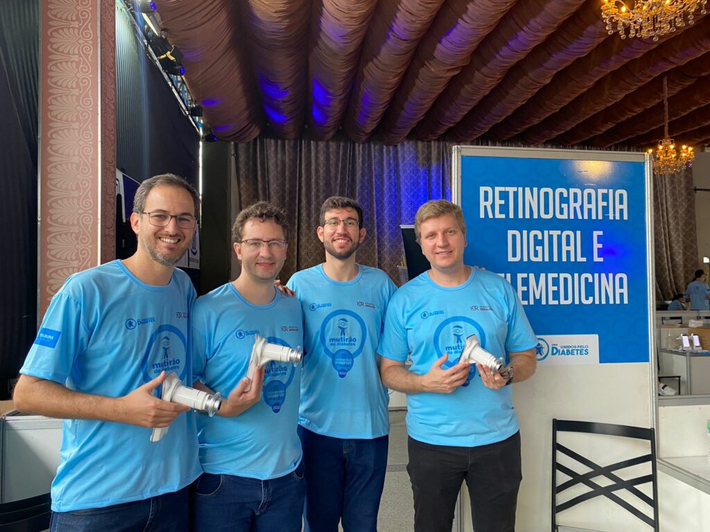
Diego Lencione, co-founder and CTO of Phelcom, Flavio Pascoal Vieira, co-founder and COO of Phelcom, Paulo Prado, coordinator of Mobile Software and AI at Phelcom, and José Augusto Stuchi, co-founder and CEO of Phelcom, during the Itabuna Diabetes Campaign, held in 2022.
Eyer
The Eyer is a portable fundus camera that attaches to a smartphone, enabling high-quality retina exams in just minutes—without the need for pupil dilation.
The technology supports the diagnosis of more than 50 diseases, including: glaucoma, cataracts, diabectic retinopathy, retinoblastoma, hypertensive retinopathy, retinopathy of prematurity, ocular toxoplasmosis.
Recently, Phelcom launched Eyer2, an enhanced version of the device featuring new built-in tools for expanded diagnostic capabilities. In addition to posterior eye imaging, Eyer2 enables the detection of anterior segment conditions such as: blepharitis and other eyelash abnormalities, meibomian gland dysfunction, styes, conjunctival and eyelid tumors, advanced cataracts, foreign bodies and burns, corneal injuries, keratitis caused by dry eye, contact lenses, infections and ulcers.
About Phelcom
Phelcom Technologies is a Brazilian medtech company based in São Carlos, São Paulo. Founded in 2016 by three young researches—a physicist, an electronics engineer, and a computer engineer—the company developed a portable retinal camera integrated with a smart phone.
The first prototype was inspired by co-founder Diego Lencione’s personal experience, as his brother struggled with a severe vision condition from childhood.
In 2019, Phelcom launched its first product, the Eyer portable retinal camera, in Brazil. Five years later, the company introduced the Eyer2, a platform capable of capturing high-quality images of both the posterior and anterior segments.
To date, Phelcom’s technology has benefited over two million people across Brazil and multiple countries, including the United States, Japan, Chile, Colombia and the United Arab Emirates. It has also been used in over 100 social outreach initiatives.
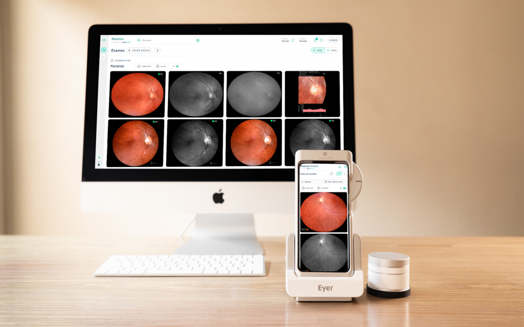
In November, Phelcom launched an upgraded version of Eyercloud, the online platform for managing ophthalmic examinations conducted using Eyer and Eyer2 portable fundus cameras. With significant advancements in data security, accessibility and design, the new version enhances the user experience, streamlines processes, and accelerates diagnoses, effectively meeting the needs of doctors and healthcare professionals.
Key updates in the new Eyercloud include the ability to view exams in grid or list format, simplified editing fields, and a refreshed design with more visible buttons and intuitive navigation. Additionally, users can now register patients and associate exams directly through the platform.
“We applied the new design language already present in Eyer2, unifying the Eyercloud interface with that of the device. Our aim is to offer an equally efficient and agile user experience on both platforms”, explains Phelcom’s marketing consultant and product designer, Edgard Regolão Junior.
Junior also emphasizes one of the key enhancements: the immediate display of retinal images on the homepage. “When the user accesses the platform, they can see the latest images and exams taken. This facilitates the quick analysis of the verification and classification already done by EyerMaps. This artificial intelligence detects possible abnormalities in seconds and displays a heatmap on the images. This feature speeds up the identification of alterations and allows professionals to make faster and more accurate diagnoses,” he adds.
Accessibility and security
Enhanced navigation is one of the most significant changes in EyerCloud. The platform is now simpler and faster to access, requiring fewer clicks and eliminating unnecessary steps. Additionally, data entry has been streamlined, allowing users to add, edit, and save information more intuitively and efficiently, making the tool more user-friendly for everyday use.
“The system has also been updated to enhance security, ensuring compliance with information and data protection regulations.,” says Phelcom’s Data Engineering Manager, Fernando Mineo Raze Yamanaka.
All the information stored on EyerCloud is protected by Amazon Web Services (AWS), a world reference in cloud solutions. The platform also strictly follows HIPAA guidelines (Health Insurance Portability and Accountability Act), guaranteeing maximum data protection and reliability.
Continuous improvement and customer support
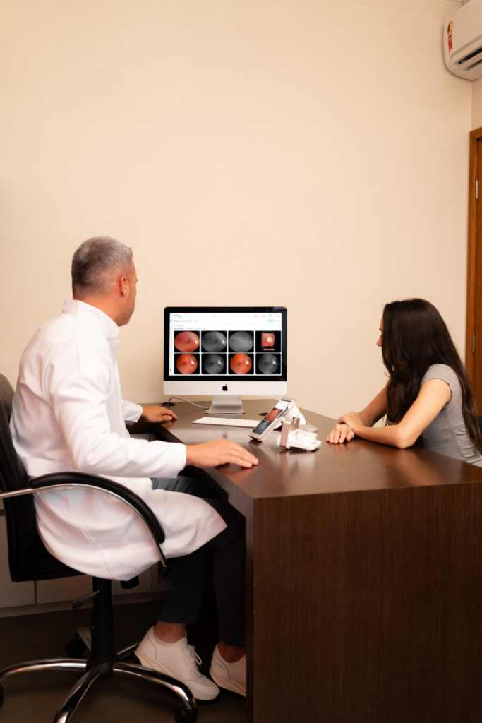
EyerCloud facilitates communication with patients by displaying and sharing exams in a clear and accessible way.
Customers who have questions or suggestions can count on Phelcom’s support for quick and efficient assistance.
Yamanaka explains that the company keeps an organized list of user feedback and requests, prioritizing the most requested improvements to optimize the platform’s ongoing development. “We will continue to invest in improvements following this feedback. EyerCloud will become increasingly dynamic, with new features, filters and search methods to make it even easier for professionals to work with it”.
EyerCloud: safe and efficient management of ophthalmic exams
EyerCloud is an online platform designed to streamline the management of ophthalmic exams, delivering enhanced security, efficiency and convenience. Featuring an intuitive and user-friendly interface, the system enables seamless storage and sharing of exams and reports conducted with the Eyer and Eyer2 portable fundus cameras. Automatic synchronization ensures data is securely uploaded to the cloud, offering both reliability and accessibility.
This integration streamlines the workflow for healthcare professionals by ensuring exams are always accessible, well-organized, and securely protected. It also facilitates swift communication and efficient monitoring of results.
Discover the advantages of EyerCloud:
Connectivity
The platform is easily accessible via an Eyer or Eyer2 device, smartphone, tablet or computer. Even without an internet connection, exams are automatically saved and seamlessly uploaded to the cloud once internet connection is restored. Additionally, EyerCloud is fully compatible with leading electronic medical record systems, enabling smooth integration into the workflow of clinics and hospitals.
Artificial Inteligence
When integrated with EyerMaps, EyerCloud displays exams using a color-coded system to identify potential alterations, simplifying the prioritization of urgent cases:
Green: low probability of alteration (up to 30%).
Yellow: medium probability of alteration (31% to 70%).
Red: high probability of alteration (71% to 100%).
Transparency
Facilitate communication with patients by displaying and sharing exams in a clear and accessible way. EyerCloud enhances understanding of diagnoses and treatment progress, fostering greater patient trust and engagement.
Mobility
With EyerCloud, healthcare professionals can carry out examinations in the field while ophthalmologists report remotely, from anywhere in the world. This flexibility extends the reach of ophthalmic services and speeds up care.
One of the main improvements in the new EyerCloud is the immediate display of retinal images on the home page.
Security
The data stored in EyerCloud is protected by Amazon Web Services (AWS) infrastructure, guaranteeing maximum security and compliance with HIPAA (Health Insurance Portability and Accountability Act). In addition, the platform allows you to manage different levels of access, ensuring that each professional only sees the information they need.
About Phelcom
Phelcom Technologies is a Brazilian medtech company based in São Carlos, the interior of São Paulo. The company’s story began in 2016, when three young researchers – a physicist, an electronics engineer and a computer engineer (Physics, Electronics, Computing) – created a fundus camera integrated with a smartphone.
Phelcom’s first prototype was inspired by the personal experience of one of the partners, Diego Lencione, whose brother faced a serious condition that severely compromised his vision since childhood.
In 2019, Phelcom launched its first product to the Brazilian market: the Eyer portable fundus camera. Five years later, it launched the Eyer2, a platform that makes it possible to document the posterior and anterior segments with high quality images.
Today, Phelcom’s technology has benefited more than two million people worldwide, and has also been used in more than 100 social projects.
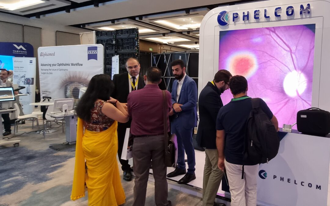
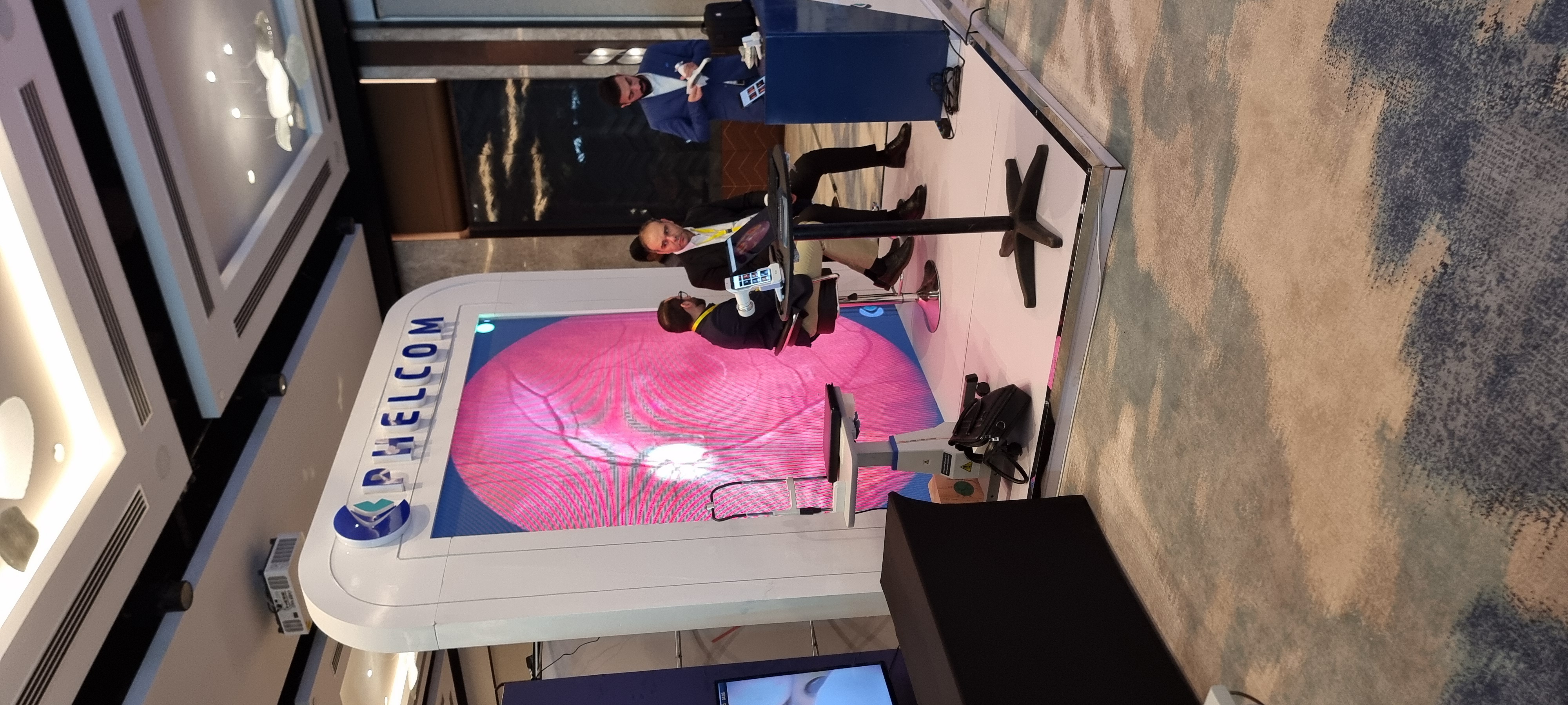
Phelcom Technologies recently participated in the 6th Middle East Ophthalmology Meeting (MEOM), held from October 3rd to 5th in Dubai, United Arab Emirates. Recognized as one of the most significant events in the ophthalmology field within the Middle East, MEOM offered a strategic opportunity for Phelcom to solidify its international expansion and bolster its position as a leading competitor in the global ophthalmic device market.
Partnering with its local distributor, Allm MEA, Phelcom aims to strengthen relationships with regional clients and explore new business opportunities.
“The response from local professionals has been overwhelmingly positive. Many emphasized the significant impact of the Eyer portable retinal camera in clinical practice, screening exams and for patients with special needs, those who are bedridden or have physical disabilities. Even for those who don’t use Eyer as their primary equipment, its complementary role in various clinical settings was evident,” says Mário Costa, Phelcom’s International Business Manager.
The Eyer, a portable, non-mydriatic fundus camera that attaches to a smartphone that performs high-quality retinal examinations in just a few minutes. In addition, Phelcom offers the EyerMaps AI platform, capable of detecting potential retinal abnormalities within seconds.
As an international hub, Dubai attracts professionals not only from the UAE but from across Asia, Southeast Asia, and Europe. “This allowed us to extend Phelcom’s reach into new markets and unlock future opportunities, including research collaborations and social impact initiatives,” Costa adds.
Phelcom’s booth at MEOM 2024.
Innovation and Development in the Middle East
Dubai is renowned for its dedication to innovation, future-focused thinking, and technological advancement, particularly in healthcare. “From the moment we arrived, we could see the emphasis on cutting-edge technologies and innovative care models. This is a region that highly values tech-driven development, which has fueled its growth. For us, being in an environment that fosters such progress is incredibly enriching,” Costa remarks.
The local culture, with its focus on precision and excellence, aligns perfectly with Phelcom’s values of ensuring top quality across all stages of product development and service delivery. “Our participation in MEOM 2024 not only strengthened Phelcom’s presence in the Middle East but also provided new insights to continue innovating and expanding our global reach,” Costa says.
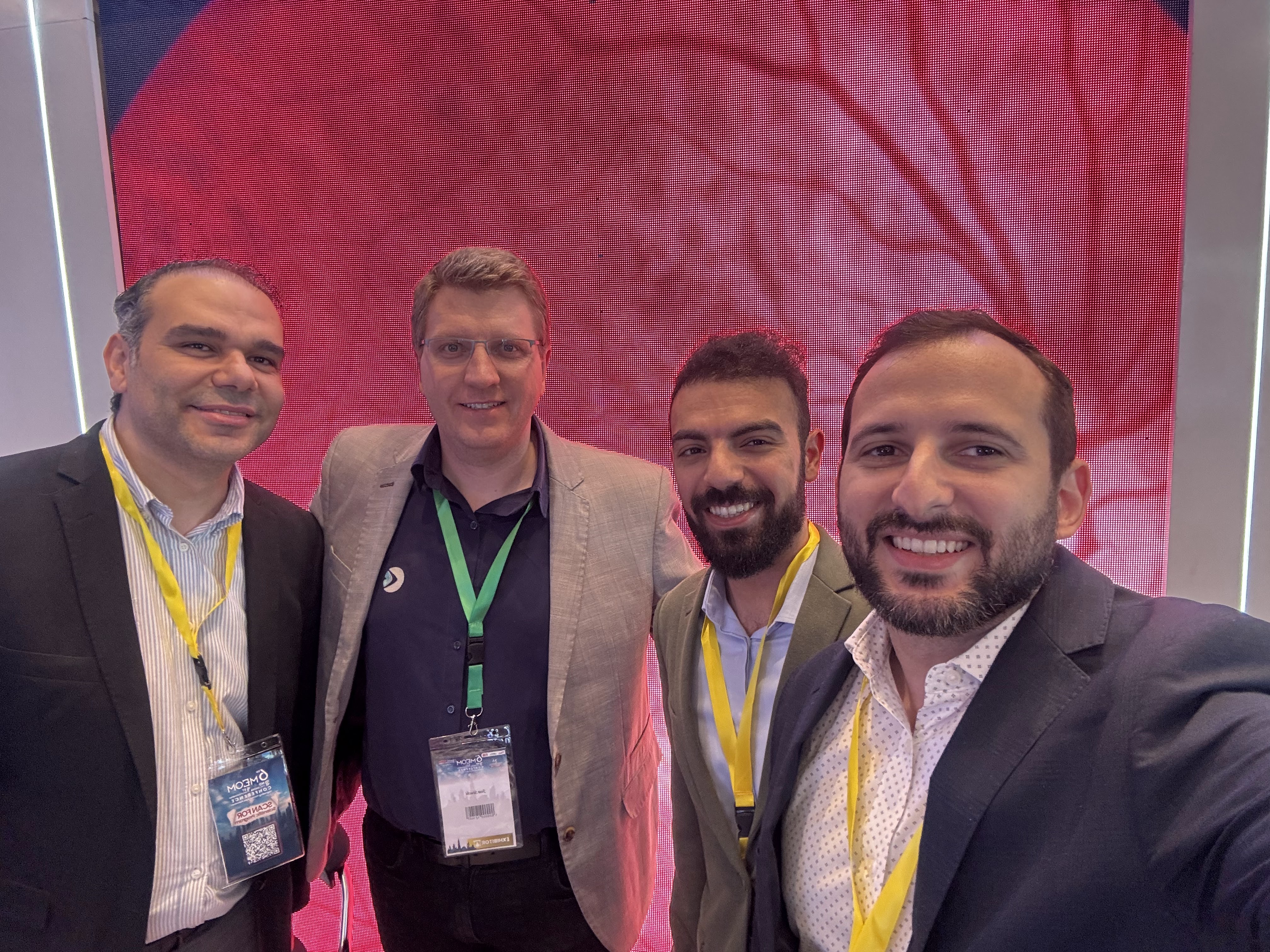
Representatives of Phelcom and Allm MEA at MEOM 2024.
Eyer expands presence across six coutries
The Eyer was approved for sale in the UAE in the second quarter of last year. Today, Phelcom is active in six countries: Brazil, the United States, Japan, Chile, Colombia, and the United Arab Emirates.
Additionally, Phelcom is in the regulatory approval phase to expand operations into new markets, including Argentina, Mexico, Vietnam, and Saudi Arabia.
For Phelcom’s CEO, José Augusto Stuchi, being a Brazilian company operating in key global markets is both a source of pride and a significant responsibility. “Our global expansion enhances our capabilities, but it also reaffirms our commitment to delivering high-quality products that make a meaningful difference in the daily practice of ophthalmology professionals,” he explains.
About Phelcom
Phelcom Technologies is a Brazilian medtech company based in São Carlos, São Paulo. The company was founded in 2016 by three young researchers—a physicist, an electronics engineer, and a computer engineer (physics, electronics, computing) —who developed a portable retinal camera integrated into a smartphone.
The first Phelcom prototype was inspired by the personal experience of co-founder Diego Lencione, whose brother struggled with a severe vision condition since childhood. In 2019, Phelcom launched its first product, the Eyer portable retinal camera, in the Brazilian market. Five years later, the company introduced the Eyer2, a comprehensive eye platform capable of capturing high-quality images of both the posterior and anterior eye segments.
To date, Phelcom’s technology has benefited over two million people across Brazil and several other countries, and it has been utilized in over 100 social outreach initiatives.
Results of Renovatio, a non-profit organization that brings visual health and prescription glasses to those who need them the most, saw more than 410,000 eye treatments in 25 Brazilian states and three countries. They also donated more than 180,000 glasses.
According to the Brazilian Institute of Geography and Statistics (IBGE), one in six Brazilians has a vision problem. With an adapted 100m² bus, transformed into two modern mobile eye clinics, Renovatio is able to serve medical deserts, such as remote towns, indigenous villages and riverside communities.
“We believe that the most difficult places to reach are the ones that need us the most. Today, 71% of Brazilian municipalities don’t even have an ophthalmologist,” says ophthalmologist and Renovatio’s medical director, Bruna Gil Ferreira.
Since 2022, Renovatio has been using the Eyer portable fundus camera to screen patients. They currently have almost 50 units. The main factors for choosing Eyer were its portability, the fact that it is a Brazilian product and the EyerCloud cloud platform, which allows exams to be stored and shared remotely and in real time.
“The possibility of sending the images for analysis and getting immediate medical consultations, even in remote areas, contributes to a more agile and effective assessment of patients. Sometimes I’m in São Paulo and I can access, in real time, the examination of a patient being treated in the Xingu Indigenous Park,” Ferreira points out.
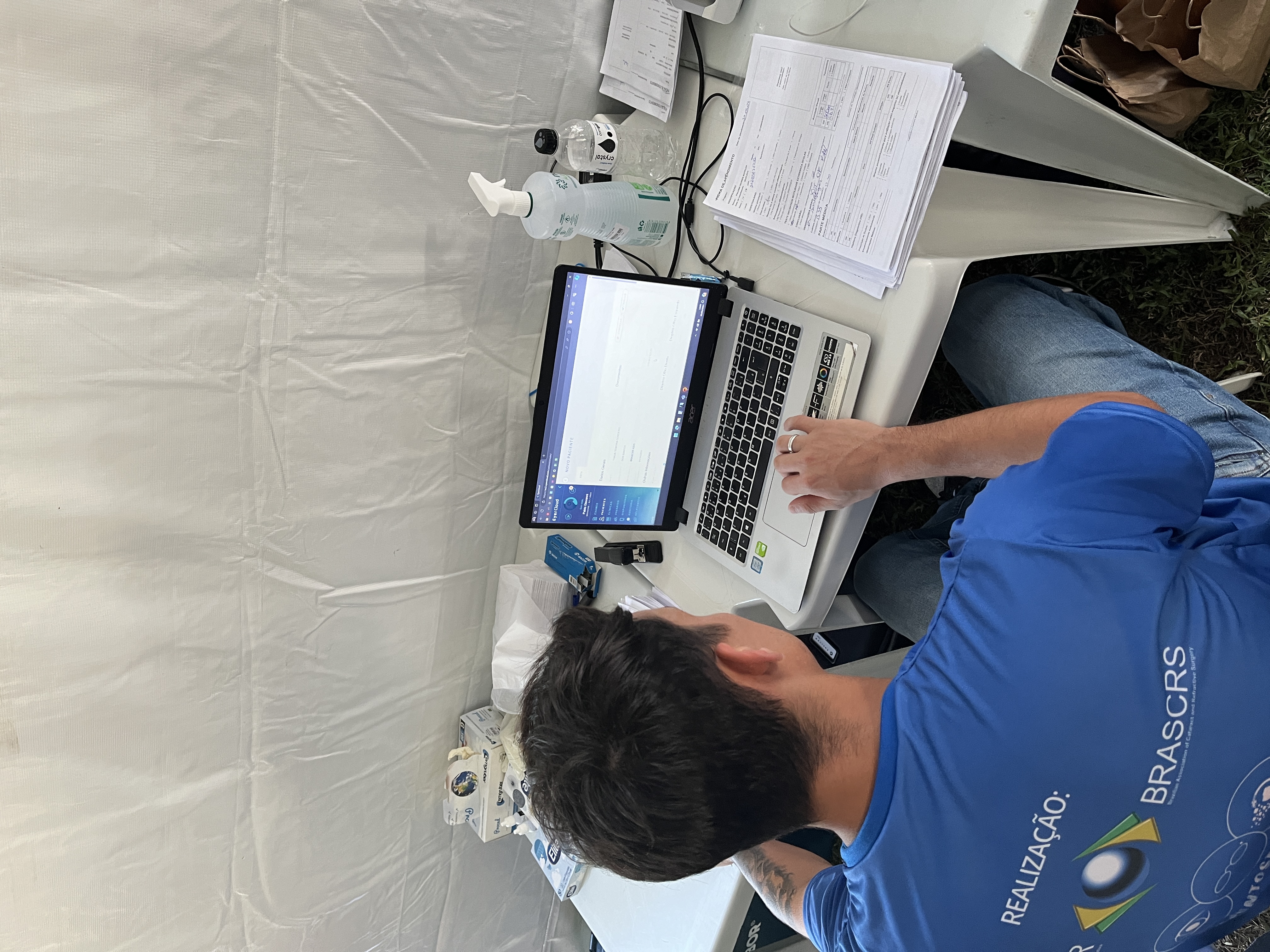
Renovatio invested in Eyer because of the EyerCloud cloud platform, among other factors.
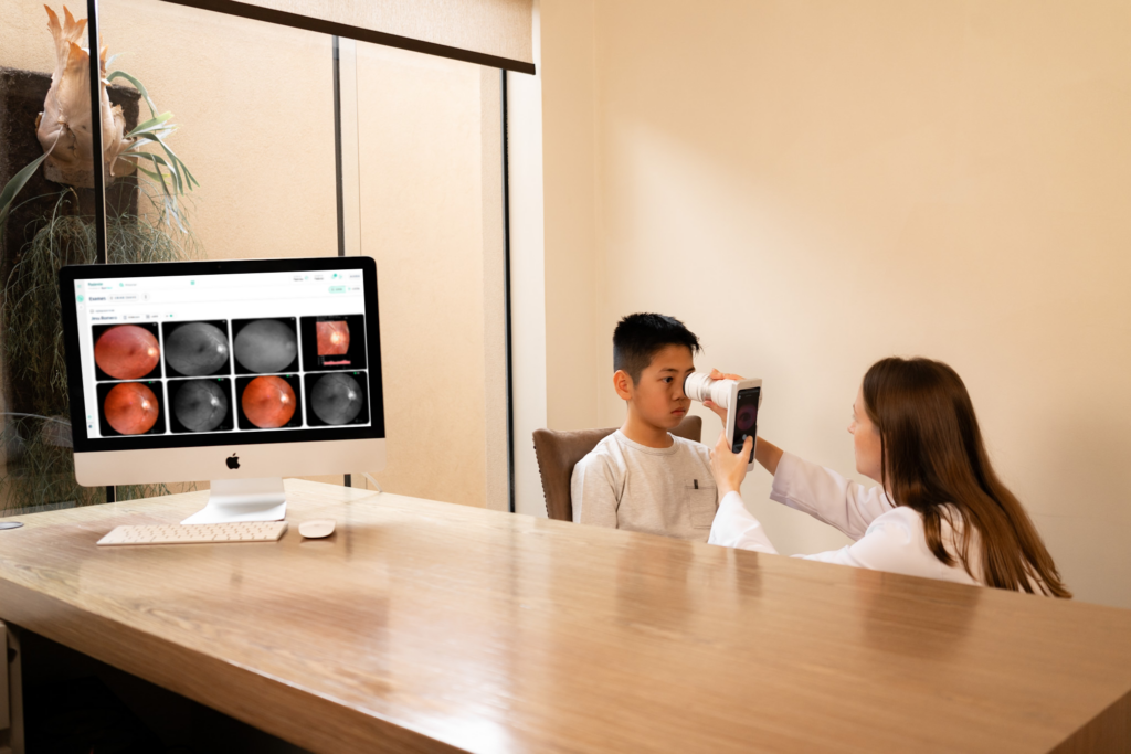
During the exams, the team evaluates the visual acuity, tonometry, keratometry and uses Eyer for fundus examinations. When Eyermaps artificial intelligence system detects possible retinal alterations, the patient is referred for a medical consultation with a specialist.
If an alteration is suspected, the AI generates a new image with a heatmap highlighting potential retinal abnormalities. The system can help detect diseases like glaucoma, Age-related Macular Degeneration (AMD), diabetic retinopathy, hypertensive retinopathy, vascular tortuosity, occlusions, papilledema, retinitis pigmentosa,nevus, and many more.
Ferreira points out that most social work in the ophthalmology area follows protocols based only on visual acuity and, as a result, other diseases are not diagnosed. “Including Eyer in our protocol has contributed greatly to identifying silent diseases that can go unnoticed in screenings focused only on vision.”
A child being examined with Eyer during a social action by the NGO Renovatio.
Chorioretinitis caused by toxoplasmosis
By storing all the fundus images in EyerCloud, Renovatio carried out a comparative study of the frequency of chorioretinitis scars caused by toxoplasmosis in different regions of Brazil.
In total, 9,648 fundus images were evaluated:
Distrito Federal (DF) – 2.496;
Tocantins (TO) – 2.426;
São Paulo (SP) – 1.862;
Minas Gerais (MG) – 1.602;
Rio Grande do Norte (RN) – 742;
Rio de Janeiro (RJ) – 502.
The majority of exams (89.04%) showed no alterations. Of the 10.95% of images with alterations, 7.09% contained suspicions and confirmations of chorioretinitis scars:
Tocantins (TO) – 62,7%;
Minas Gerais (MG) – 14,7%;
Distrito Federal (DF) – 10,07%;
São Paulo (SP) – 8%;
Rio de Janeiro (RJ) – 2,7%;
Rio Grande do Norte (RN) – 1,3%.
Among the results obtained in all states, there was a higher prevalence in women (74.7%) and a higher number of cases in the left eye (37.3%), followed by both eyes (33.3%) and then the right eye (29.3%).
It is also worth noting that the state of Tocantins had the highest incidence of the disease in the left eye (26.7%), in both eyes (17.3%) and in the right eye (18.7% of diagnosed cases).
The use of Eyer integrated with EyerCloud allowed Renovatio to carry out this important mapping, which can help with future management of toxoplasmosis, a pathology that can lead to loss of vision.
NGO Renovatio has already traveled to 25 Brazilian states providing free ophthalmic consultations and donating prescription glasses to those who need them most.
Eyer
Eyer is a portable fundus camera that works with an integrated smartphone to perform high-quality retinal examinations in just a few minutes without the need for pupil dilation.
Currently, more than 15 million tests have been carried out in Brazil, the United States, Chile, Colombia and Japan. The technology is also present in the United Arab Emirates and is in the regulatory process of being marketed in Mexico, Egypt and Saudi Arabia.
Phelcom recently launched Eyer2, a true visual examination platform that allows you to capture the posterior and anterior segments with high quality images.
With new embedded tools and improved functions, the new equipment makes it possible to capture various conditions of the anterior segment of the eye, such as blepharitis,eyelash alterations, meibomian gland dysfunction, styes, conjunctival tumors, eyelid tumors, advanced cataracts, foreign bodies, burns, corneal lesions and keratitis in general, caused by dry eye, contact lenses, infections and ulcers.
Portability, connectivity and integration with intelligent functions such as EyerMaps, together with the technology’s affordability, are contributing to increased access to retinal examinations.
About Phelcom
Phelcom Technologies is a Brazilian medtech company based in São Carlos, in the interior area of São Paulo. The company’s story began in 2016, when three young researchers – a physicist, an electronics engineer and a computer engineer (physics, electronics, computing) – created a portable fundus camera integrated with a smartphone.
The first prototype project was born out of partner Diego Lencione’s interest in visual health, as his brother has had a condition that has severely compromised his retina and vision since childhood.
In 2019, Phelcom launched its first product in the Brazilian market: the Eyer portable fundus camera. Today, the technology has reached more than two million people throughout Brazil and in the countries where it is present and has been used in more than 100 community screenings.
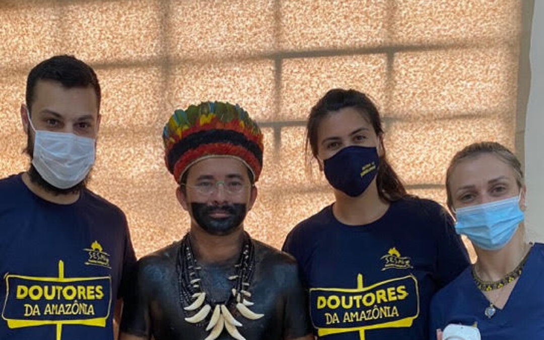
In October of last year, the NGO Médicos da Amazônia undertook a mission with a special objective: to provide complete, free eye care to approximately 500 indigenous people from the Marmelos Village, in Amazonas.
The NGO offers health acess to Brazilian indigenous communities through specialized care, modern and advanced techniques and equipment, and highly trained professional, always respecting the ancestry of their cultures and values. Since its foundation, in 2015, it has carried out more than 64 thousand medical and dental consultations and procedures.
To document the patients’ fundus and anterior segment, the voluteer medical team used the Phelcom Eyer smartdevice. The device works in conjuction with a smartphone and performs high-quality retinal examinations in a few minutes without the need for pupil dilation. As it is integrated with the clooud, it automatically males the data available on the Eyercloud online platform for analysis by a specialist anywhere. In other words, it enables remote diagnosis.
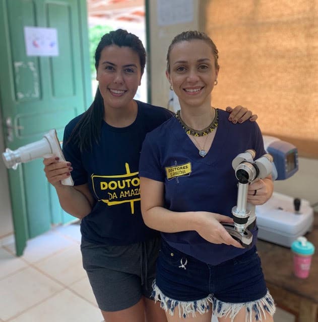
“As well as being portable and not needing internet at the time of the examination, Eyer optimizes care and does all the documentation for patients, wich allows them to be followed up correctly. This is essential for any action in remote areas”, says ophthalmologist Jade Fernandes de Melo, one of the project’s volunteers.
The main retinal diseases diagnosed by the NGO were diabetic retinophaty, glaucoma, Age-related macular disease (AMD) and asteroid hyalosis, among others. The doctors also detected cataracts, refractive alterations and pterygium. The patients were referred to indigenous Health for treatment.
Melo evaluates the device as easy to use, self-taught, with excellent image quality and essential in primary care. “The Eyer can make a difference to many people’s lives by bringing ophthalmic access to remote communities with a lack of health infrastructure”, Melo believes.
Doctor Iddi Ndyabawe, ophthalmologist and a passionate ROP specialist in Uganda, East Africa, was responsible for the very first ROP study in the country, published by BMC Ophthalmology in 2023. He’s currently involved in Adult Retina services as well, working in a screening project for diagnosing CMV Retinitis in HIV patients with meningitis admitted at Kiruddu National Referral Hospital and Mulago National Referral Hospital. He also offers Diabetic Retinopathy screening services to private patients at Kisubi Hospital.
Dr Iddi in the Aravind Eye Hospital Library, Coimbatore India
Dr. Iddi got to know about the Eyer Retinal Camera in February 2023, when Nurse Nancy Maria Douat Dietrich from Santa Catarina, Brazil came along with the high quality images device and joined the ROP screening activities at Kawempe National Referral Hospital.
“When Nancy saw my passion for ROP in Uganda, she communicated with Mr. Jose Augusto, CEO of Phelcom, who then sent me an Eyer Fundus camera by June 2023 as a support for my humble efforts in ROP “, recalls Dr. Iddi.
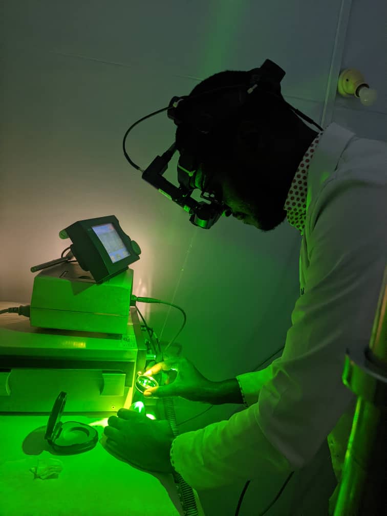
Dr Iddi training in laser therapy for ROP on an eye model
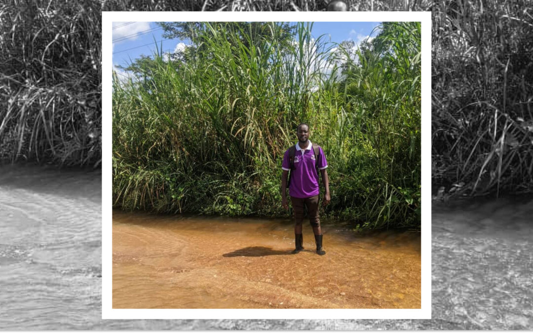
Dr. Iddi crossing the flooded roads to reach the village NICU to conduct ROP screening
According to Dr. Iddi, the main advantages of the Eyer Fundus Camera for his medical practice are:
Eyer takes clear resolution images for best diagnosis of posterior disease;
It is perfect at CDR assessment;
Eyer can easily pick out the hyperemic disc in optic neuritis that could easily be missed;
EyerMaps heat map is perfect in pointing where the clinician should pay more attention.
Dr. Iddi mentions as well that the portability and non-mydriatic use of Eyer Fundus Camera, which allows him to perform examinations in different scenarios. He recalls one particular Sunday when a relative came in complaining of reduced vision. He was able to discover a diagnosis of toxoplasmosis right there in his own home.
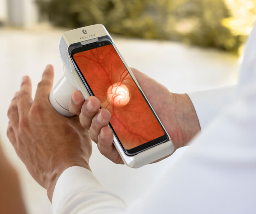
About the Eyer
Eyer Portable Fundus Camera
The Eyer is a portable fundus camera that works in conjunction with a smartphone and performs high-quality retinal examinations in a few minutes without the need for pupil dilation.
The technology supports the diagnosis of more than 50 diseases, including glaucoma, cataracts, diabetic retinopathy, AMD, retinoblastoma, hypertensive retinopathy and ocular toxoplasmosis. Currently, more than 10 million tests have been carried out in Brazil, the United States, Chile and Colombia.
The technology’s portability and affordability democratize access to retinal examinations. It costs approximately six times less than a conventional tabletop fundus camera, which still needs to be integrated with a computer.
About Phelcom
Phelcom Technologies is a Brazilian medtech company based in São Carlos, in the interior area of São Paulo. The company’s story began in 2016, when three young researchers – a physicist, an electronics engineer and a computer engineer (physics, electronics, computing) – created a portable fundus camera integrated with a smartphone.
The idea for the first prototype was realized by Diego Lencione’s interest in visual health, as his brother has had a condition that has severely compromised his retina and vision since childhood.
In 2019, Phelcom launched its first product on the Brazilian market: the Eyer portable fundus camera. Today, the technology has reached more than two million people across Brazil and worldwide.
In four years, the company has participated in more than 100 social actions and was recently named one of the 10 most innovative companies in Brazil by Forbes.