

The Lavinsky Clinic in Porto Alegre (RS) uses the Eyer portable fundus camera to take images of the posterior segment. The equipment is used more frequently in one of the units where optical coherence tomography (OCT) is not an option.
“For example, in cases of glaucoma, the OCT exam provides fundamental structural information for diagnosis and progression, but only the photo shows findings such as optic disc hemorrhage. By combining the two tests, we can better diagnose and monitor the progress of the disease. In this way, retinal imaging is essential for glaucoma analysis”, says glaucoma specialist, researcher from Wills Eye, and coordinator of the Diagnostic Unit at the Lavinsky Clinic, Fábio Lavinsky.
Fábio Lavinsky is a glaucoma specialist, researcher from Wills Eye, and coordinator of the Diagnostic Unit at the Lavinsky Clinic.
The images taken with the Eyer can be accessed on the EyerCloud online platform, allowing for complete documentation, patient follow-up and remote reports.
The fundus images help to identify other retinal problems as well. “That’s why Eyer can be differential in the diagnosis of glaucoma, as it takes an exceptional picture”, he points out.
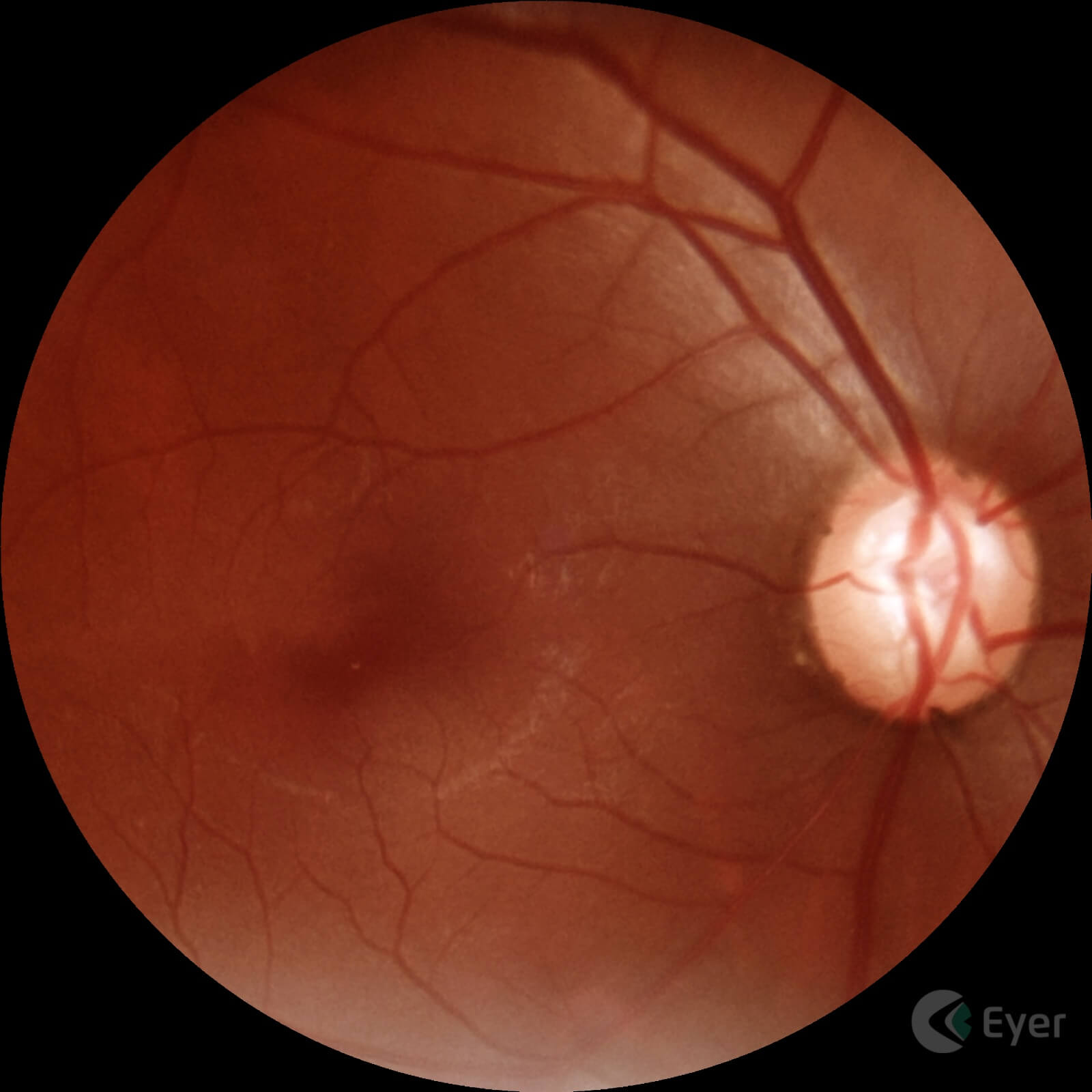
Retinal image of a patient diagnosed with glaucoma taken with Eyer.
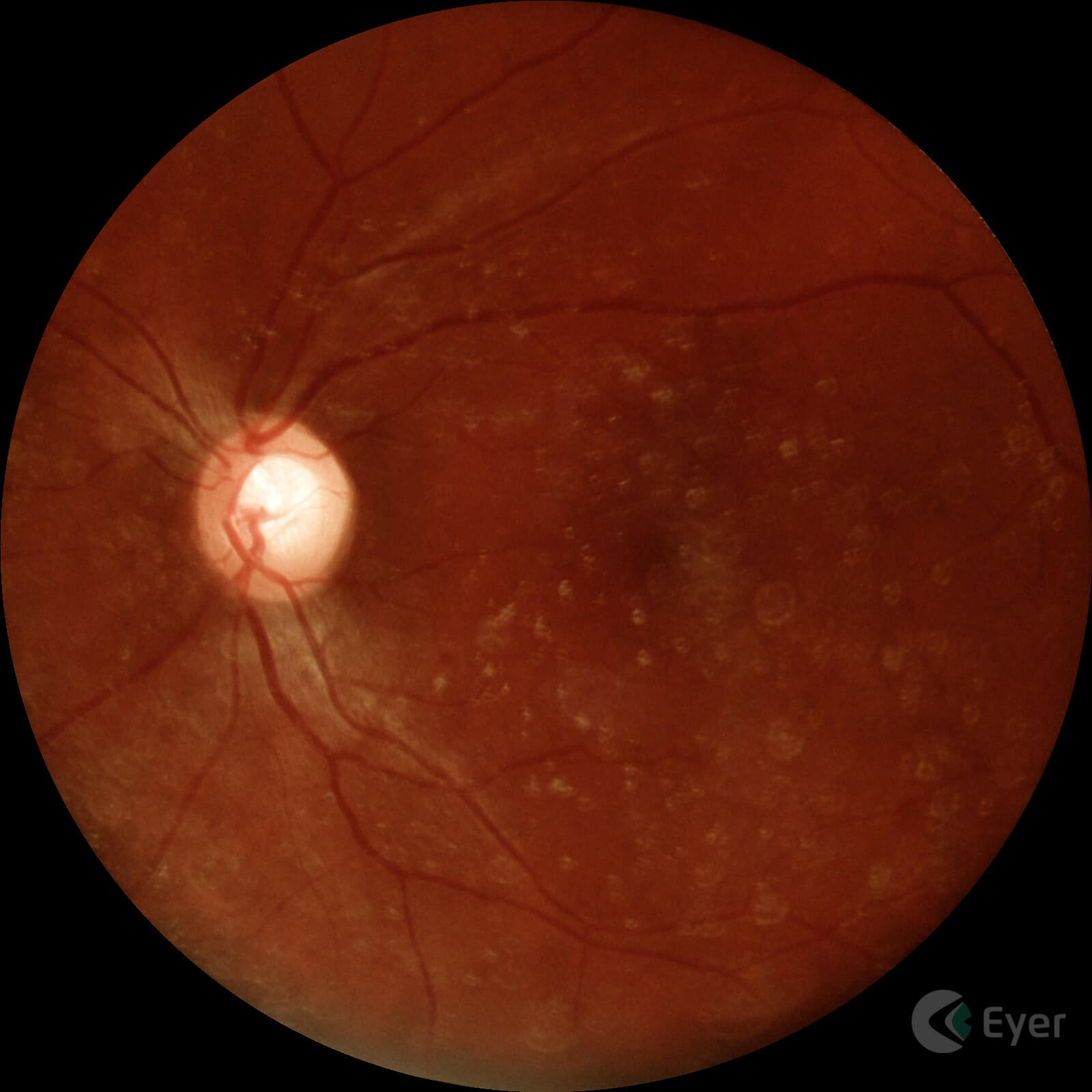
Retinal image of a patient diagnosed with glaucoma taken with Eyer.
Lavinsky also uses the EyerMaps artificial intelligence (AI) system, embedded in Eyer, which detects potential retinal abnormalities in real time. If a suspected abnormality is identified, the AI generates a new image with a heatmap highlighting potential abnormalities in the retina. “For clinics that don’t have OCT, this can be a guideline for ordering the test.”
The ophthalmologist points out that AI does not yet accurately identify changes in the optic nerve such as cupping, however, Eyer offers tools for diagnosing the disease.
Stereophoto and CDR – Cup to Disk Ratio
The stereophoto is a video created by Eyer based on the superimposition of retinal images with the nerve and macula in the center of the image. This way, doctors can have a clearer view of the region of interest for diagnosing glaucoma.
Stereophoto taken with Eyer.
The Cup to Disk Ratio (CDR) tool shows the percentage ratio between the optic nerve and the excavation, a measurement which, together with other clinical and diagnostic findings, can indicate the presence or risk of the disease.
With just a few clicks and by selecting the circles that outline the nerve and the excavation on the device’s screen, Eyer not only calculates the CDR, but also the neural RIMA measurements and the ISNT graph, metrics that support the diagnostic interpretation of the exam quickly and more accurately.
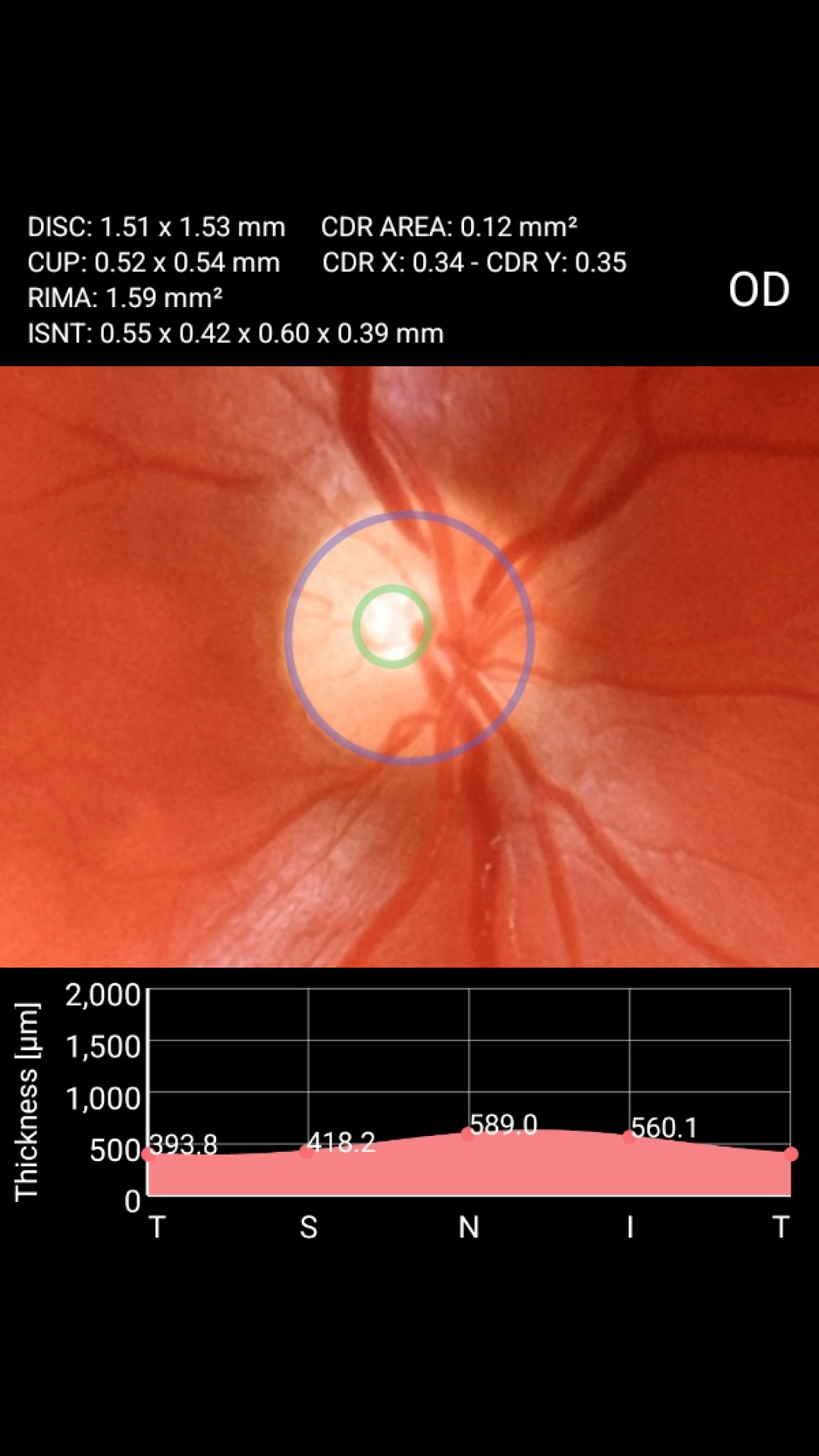
CDR – Cup to Disk Ratio tool built into Eyer.
Education
Lavinsky is currently in the United States working as a researcher in imaging and ophthalmology at Wills Eye Hospital in Philadelphia, Pennsylvania. But he is also a member of the medical faculty at the University of Vale do Rio dos Sinos (Unisinos), in São Leopoldo (RS).
As well as bringing better clinical results for patients, he believes that Eyer can help teach future doctors. “The Eyer should be like a stethoscope for medicine. Courses should have devices in internal medicine teams and in other related specialties to take pictures of patients with systemic pathologies with ocular repercussions, in the classroom, as well as promoting discussions with students about findings”, he reflects.
Lavinsky explains that the direct ophthalmoscope has a very small field of vision. Although it has some advantages, such as three-dimensionality and showing the optic nerve clearly, the Eyer comes out on top because it has excellent optics and high-quality images that can be viewed digitally by several students. “More specifically, the Eyer should be part of the semiology course. It would be fantastic, something almost revolutionary in education.”
For the doctor, the technology should not only be used in the field of ophthalmology, but also in those where diseases have ocular manifestations, such as endocrinology, cardiology and neurology. “Using EyerCloud, ophthalmologists could provide diagnostic support to colleagues in related specialties. This would greatly speed up the diagnosis and management of important ocular complications, having a positive impact on patients’ clinical outcomes”, he says.
Fábio Lavinsky has no commercial relationship with or receives compensation from Phelcom. He was interviewed for this article because of his expertise and experience in the medical field, with the aim of sharing relevant information about glaucoma and ophthalmic technologies.


