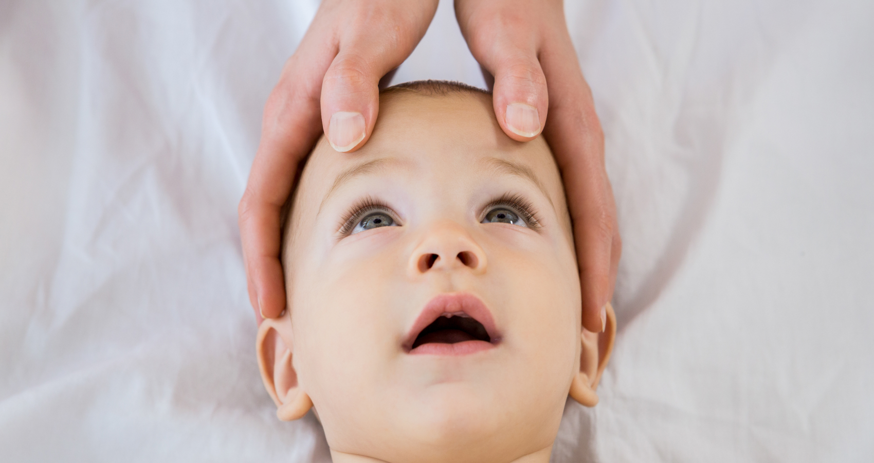
Undoubtedly, one of the obstacles in pediatric ophthalmology is being able to carry out examinations on little ones. After all, it can be difficult to keep a child still for examinations.
This difficulty is routine for ophthalmologist, Dr. Patrícia de Freitas Dotto, MD, PhD at the Dr. Jeser Amarante Faria Children’s Hospital, at the São José Municipal Hospital (HMSJ) and in her office, all in Joinville (SC), and at the Lavinsky clinic in Porto Alegre (RS).
“The main challenge is the non-mydriatic fundoscopic evaluation of children between ten months and three years old without sedation or restraint. That’s why it’s essential to create a playful and calm environment that brings peace of mind to the whole family. In this context, photographic documentation, or even fundoscopic imaging, in real time allows parents to be involved in the care, which improves the doctor-patient relationship and, consequently, has a positive impact on the child’s follow-up and/or treatment.”
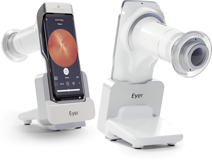
Dotto says that she uses the Eyer portable fundus camera to speed up examinations on pediatric patients. The equipment, which is very suitable for examining infants and children due to its portability and high image quality, works in conjunction with a smartphone and performs retinal examinations in a few minutes. These photographs are then available on the EyerCloud online platform, making it easier to study and monitor the progression of cases.
“The field of vision is excellent. Under medicated mydriasis, it is also an excellent complementary propaedeutic tool for assessing the mid-periphery of the retina, particularly small hyperpigmented lesions of the choroid (nevus) and retina (melanocytomas), using it as an infrared scan,” she explains.
In her office, with help of the Eyer Slit Lamp Adapter, the ophthalmologist attaches the device to her slit lamp allowing exams to be performed more easily. “Although I really enjoy using it this way, it’s magical to use it in ‘mobile’ form in ICUs, operating rooms, home care and within the office itself, such as evaluations of children on their mother’s lap or in the waiting room,” she points out.
The doctor also uses the Eyer in the emergency room, in adult and child care, for documenting infectious diseases and Retinopathy of Prematurity (ROP) in neonatal ICUs severely debilitated adults in ICUs, as well as inter-consultations for neurology, neurosurgery, nephrology, cardiology, and in medical expertise for the National Civil Aviation Agency (ANAC) and the Santa Catarina Court of Justice (TJSC). “I literally carry Eyer in my bag. It makes my job a lot easier,” she says.

The ophthalmologist, Dr. Patrícia de Freitas Dotto, MD, PhD treats infants and children at the Dr. Jeser Amarante Faria Children’s Hospital, the São José Municipal Hospital (HMSJ) and at her office, all in Joinville (SC).
Shaken Baby Syndrome
With daily use of the Eyer in various locations, Dotto has witnessed several remarkable cases, such as the care of a child suffering from Shaken Baby Syndrome. The syndrome occurs when the baby is shaken intensely, causing permanent brain damage or even death.
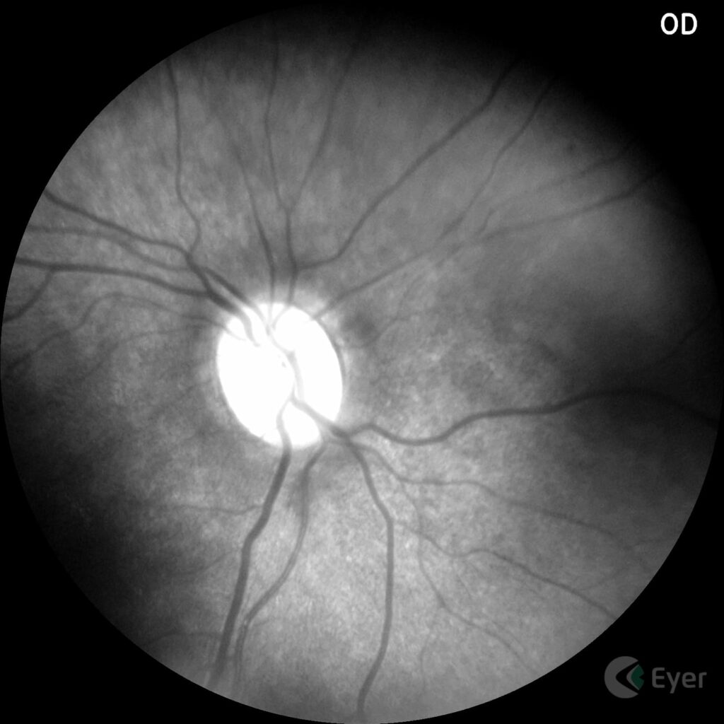
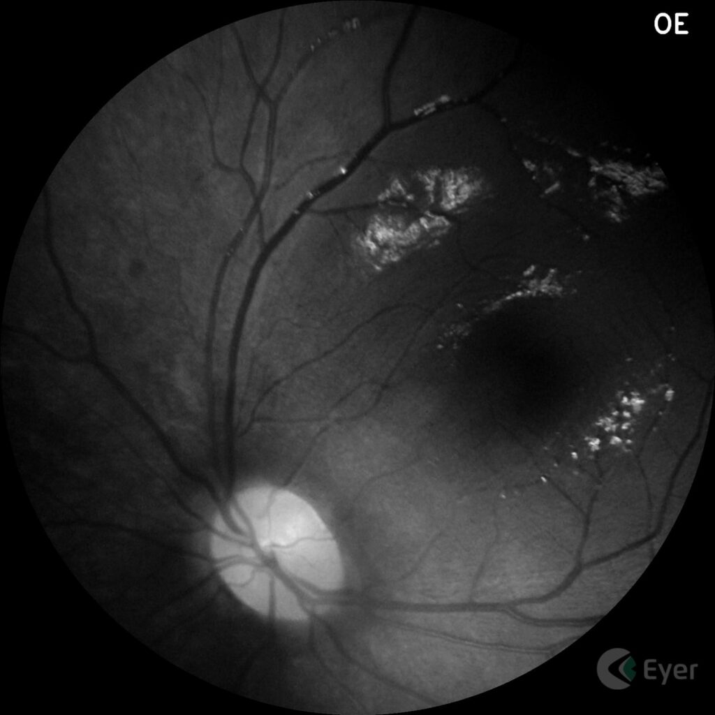
Here’s the doctors report on the case of shaken baby syndrome:
“The child had been followed up for a few months in hospital for convulsions and severe malnutrition. The suspicion of Shaken Baby was raised when neuroimaging alterations (hematoma) were observed during the complementary investigation of status epilepticus.
During the ophthalmological assessment, when I examined her visual acuity (grating), I found that the vision in one eye was outside the normal range for her age, despite the fact that she had no changes in ocular motility.
When I laid her on the gurney for the retinal mapping, she started crying desperately. It was a very difficult examination and I was struck by the fact that the child eventually let go, almost gave up, and then reacted again, without showing any change in her level of consciousness.
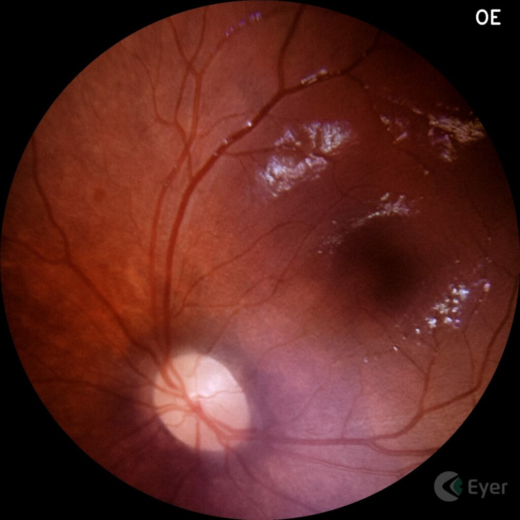
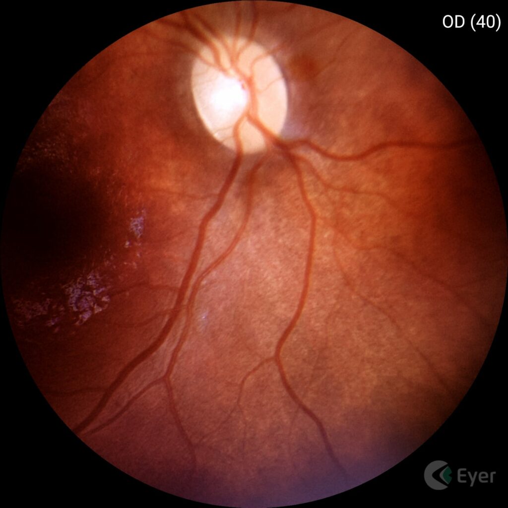
It was at one of these moments that I managed to photograph the fundus of the eye and identify retinal lesions, indicating serious damage to the central nervous system. In one particular case, the hemorrhage was very small, probably because it was being reabsorbed, and I could only be sure of the diagnosis because of the examination. It was a miracle for me and for her.
I was devastated that day, but very grateful to HaShem and Phelcom [the company that invented the Eyer]. The case was really investigated from a social point of view, the assaults were confirmed and the appropriate measures were taken.
We saved a life. And in my religion, Judaism, saving a life means saving the whole world.”
Next, see the images of this case taken by Dotto with Eyer:
More cases
Dotto performs retinography on all patients, as she considers it important the normal state of the retina. “We often look for illnesses and forget how important it is to make sure the fundus of the eye is normal, because problems happen throughout life and it’s important to make sure of the moment when the state of eye health is lost,” she says.
She highlights the case of a 22-year-old woman who was admitted to the ER with compromised visual acuity, severe hypertensive retinopathy and serous detachment of the macula. The team suspected Autoimmune Nephropathy (IgA). “Unbelievably, four or five days after pressure control (in the ICU), she progressed to 20/20 visual acuity and total resolution of the serous detachment.”
Another situation involved a patient in the ER with anterior uveitis and an apparent normal USG. After remission, when mapping the retina, the doctor noticed a peripheral blackened lesion. “Using the Eyer, I was able to document it. We redid the USG thinking it was a melanoma. And it was indeed a melanoma at the level of the ciliary body,” she says.
In children, the ophthalmologist recalls the care of two patients with slight vitreous hemorrhage: “After improving the transparency of the media, it was possible to record the presence of a slight vascular malformation at the level of the optic nerve,” she recalls.
Among the most commonly diagnosed diseases in babies are ROP, congenital infections, optic nerve hypoplasia and retinal and optic nerve colobomas. “I also use the Eyer a lot to quantify the size of the optic nerve and the excavation. This helps with the differential diagnosis of suspicious glaucomas, particularly in short-sighted children,” she concludes.
Below are some images taken by the ophthalmologist with the Eyer:
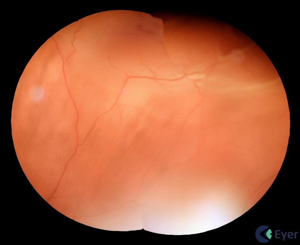
Patient with retinal displacement.
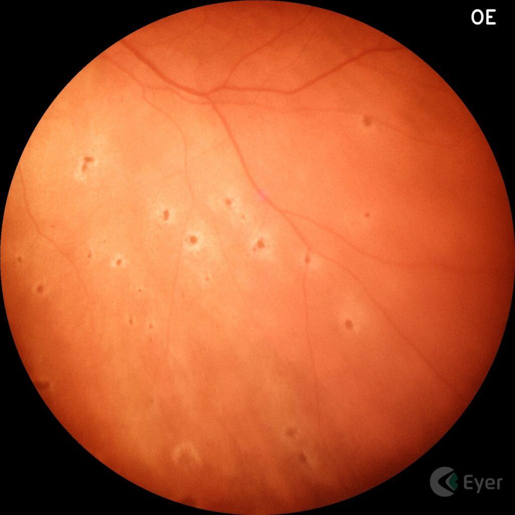
Patient with microembolization in the choroid due to covid.
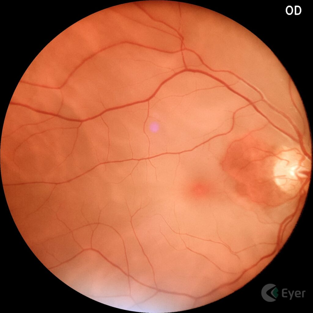
Patient with arterial occlusion.
The Eyer
The Eyer is a portable fundus camera that works in conjunction with a smartphone and performs high-quality retinal examinations in a few minutes without the need for pupil dilation.
The technology supports the diagnosis of more than 50 diseases, including glaucoma, cataracts, diabetic retinopathy, AMD, ROP, retinoblastoma, hypertensive retinopathy and ocular toxoplasmosis. Currently, more than 10 million tests have been carried out in Brazil, the United States, Chile, Colombia and Japan.
It was recently approved in the United Arab Emirates and is in the regulatory process of being marketed in Mexico, Egypt and Saudi Arabia.
Portability, connectivity and integration with intelligent functions such as EyerMaps, together with the technology’s affordability, contribute to increasing access to retinal examinations.

