Minutes. This is the average time that the Brazilian client, who value immediate feedback, wait for Phelcom’s customer support department to solve technical issues, clarify doubts, request support materials or schedule training.
In other countries, even if the customer usually prefers a different approach, requesting support via email, agility is still Phelcom’s trademark. “Normally, the foreign clients don’t call us as soon as the problem appears, but when they have time between their appointments.” explains Phelcom’s customer support analyst, Leonardo Chagas Pizzo.
To guarantee this agility, Phelcom chose to have its own local team in Brazil. “Unlike other services, our clients don’t wait in digital lines. We’re here to solve their problems quickly and try to reduce the bureaucracy”, says Phelcom’s technical support, Hygor Nunes.
As a result, the team has developed a close relationship with many clients. “Today, we are able to identify each person’s profile, being able to serve them in a personalized way, according to their necessities”, explains Phelcom’s customer support coordinator, Rodrigo Cabrera.
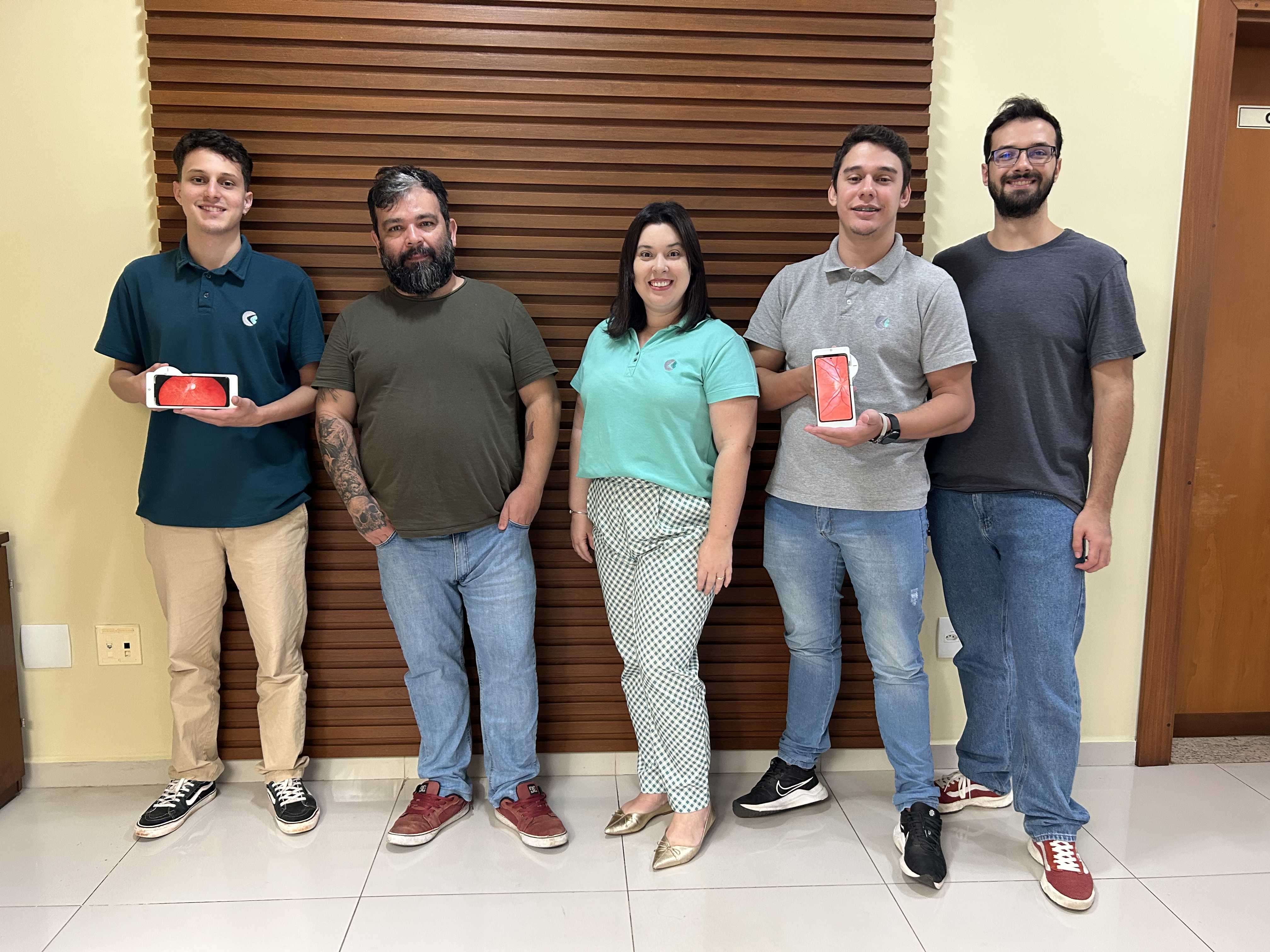
Phelcom’s customer support team in Brazil
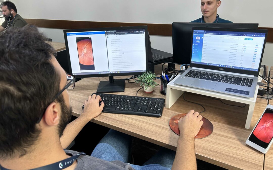
Trainings
After purchasing a Phelcom Technologies product, the support customer team gets in touch to schedule online training on how to use the equipment and its on board technologies, such as the online exam management platform, EyerCloud and the artificial intelligence system EyerMaps, that accurately detects various changes in the retina.
“Eyer is a multifunctional fundus camera that offers a variety of features and it’s important to train our clients to make the most of all the device’s functionalities.”, says Phelcom’s customer support intern, Pablo Sathler.
However, training is optional. “We make our schedule available so that the client can choose the best day and time for them. Besides that, we also ask for at least two people to be present so that they can have the experience of making images with each other, aligning possible doubts about use.”, emphasizes Phelcom’s customer support analyst, Tatiane Fragalli.
Cabrera explains that, in addition to the initial training, clients can take as many courses as they wish during the one-year Eyer warranty period. “Often, clients want to ask questions about how to edit images or configure circuits, for example. So we prepare customized training courses according to their needs.”
Phelcom also continuously makes educational and support materials available to answer the most frequently asked questions, such as folders, videos in the form of information pills and infographics with detailed guidelines and practical tips to maximize the use of Eyer. To get access, just contact the Customer Support department via email support@phelcom.com.
For technical support, Phelcom uses a communication system for opening tickets directly on this link: phelcom.com/servicecenter, which guarantees more agility and transparency.
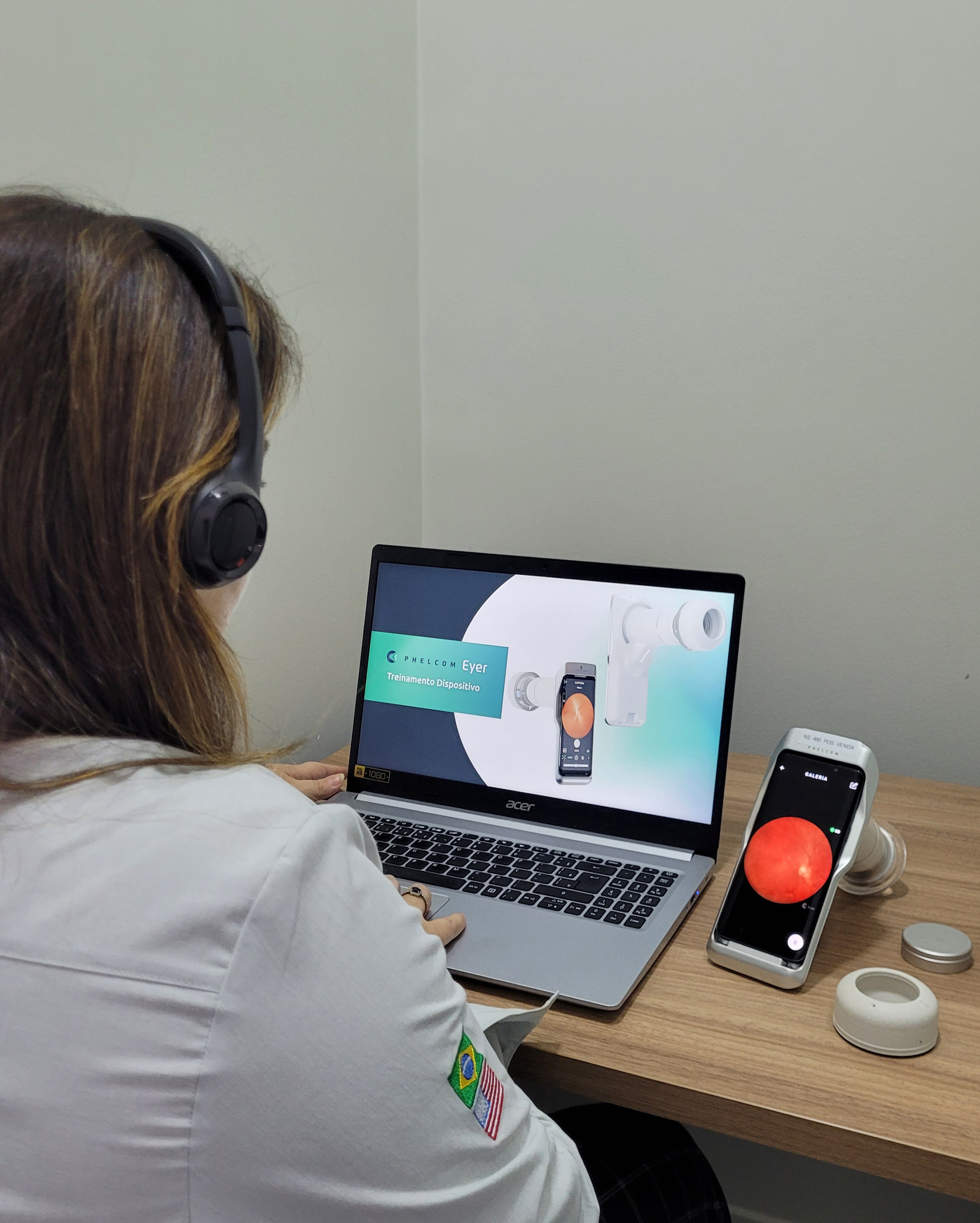
Tatiane Fragalli provides online training on how to handle the Eyer family of products.
Eyer
Eyer supports the diagnosis of more than 50 diseases, including glaucoma, cataracts, diabetic retinopathy, AMD, ROP, retinoblastoma, hypertensive retinopathy and ocular toxoplasmosis. Currently, more than 10 million tests have been carried out in Brazil, the United States, Chile, Colombia and Japan.
It was recently approved in the United Arab Emirates and is in the regulatory process of being marketed in Mexico, Egypt and Saudi Arabia.
Portability, connectivity and integration with intelligent functions such as EyerMaps, together with the technology’s affordability, are contributing to increased access to retinal examinations.
Phelcom recently launched Eyer2, a portable fundus and eye camera that offers high-quality images of the posterior and anterior segments.
About Phelcom
Phelcom Technologies is a Brazilian medtech company based in São Carlos, in the interior area of São Paulo. The company’s story began in 2016, when three young researchers – a physicist, an electronics engineer and a computer engineer (physics, electronics, computing) – created a portable fundus camera integrated with a smartphone.
The first prototype project was born out of partner Diego Lencione’s interest in visual health, as his brother has had a condition that has severely compromised his retina and vision since childhood.
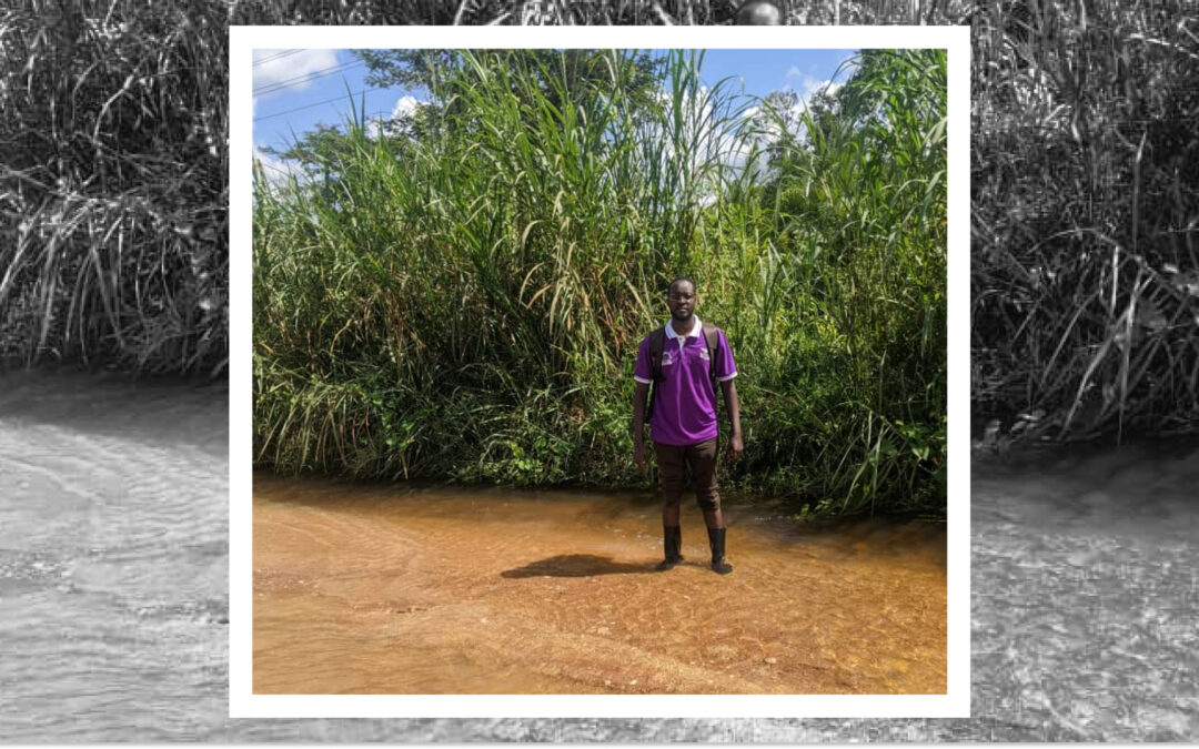
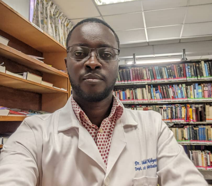
Doctor Iddi Ndyabawe, ophthalmologist and a passionate ROP specialist in Uganda, East Africa, was responsible for the very first ROP study in the country, published by BMC Ophthalmology in 2023. He’s currently involved in Adult Retina services as well, working in a screening project for diagnosing CMV Retinitis in HIV patients with meningitis admitted at Kiruddu National Referral Hospital and Mulago National Referral Hospital. He also offers Diabetic Retinopathy screening services to private patients at Kisubi Hospital.
Dr Iddi in the Aravind Eye Hospital Library, Coimbatore India
Dr. Iddi got to know about the Eyer Retinal Camera in February 2023, when Nurse Nancy Maria Douat Dietrich from Santa Catarina, Brazil came along with the high quality images device and joined the ROP screening activities at Kawempe National Referral Hospital.
“When Nancy saw my passion for ROP in Uganda, she communicated with Mr. Jose Augusto, CEO of Phelcom, who then sent me an Eyer Fundus camera by June 2023 as a support for my humble efforts in ROP “, recalls Dr. Iddi.
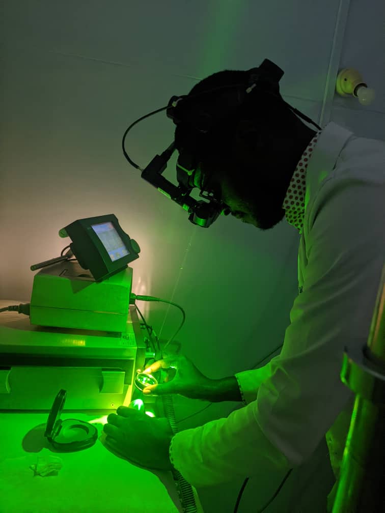
Dr Iddi training in laser therapy for ROP on an eye model
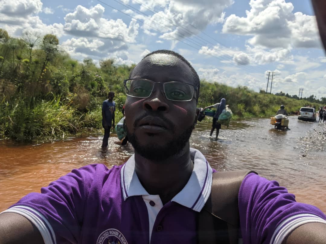
Dr. Iddi crossing the flooded roads to reach the village NICU to conduct ROP screening
According to Dr. Iddi, the main advantages of the Eyer Fundus Camera for his medical practice are:
Eyer takes clear resolution images for best diagnosis of posterior disease;
It is perfect at CDR assessment;
Eyer can easily pick out the hyperemic disc in optic neuritis that could easily be missed;
EyerMaps heat map is perfect in pointing where the clinician should pay more attention.
Dr. Iddi mentions as well that the portability and non-mydriatic use of Eyer Fundus Camera, which allows him to perform examinations in different scenarios. He recalls one particular Sunday when a relative came in complaining of reduced vision. He was able to discover a diagnosis of toxoplasmosis right there in his own home.
About the Eyer
Eyer Portable Fundus Camera
The Eyer is a portable fundus camera that works in conjunction with a smartphone and performs high-quality retinal examinations in a few minutes without the need for pupil dilation.
The technology supports the diagnosis of more than 50 diseases, including glaucoma, cataracts, diabetic retinopathy, AMD, retinoblastoma, hypertensive retinopathy and ocular toxoplasmosis. Currently, more than 10 million tests have been carried out in Brazil, the United States, Chile and Colombia.
The technology’s portability and affordability democratize access to retinal examinations. It costs approximately six times less than a conventional tabletop fundus camera, which still needs to be integrated with a computer.
About Phelcom
Phelcom Technologies is a Brazilian medtech company based in São Carlos, in the interior area of São Paulo. The company’s story began in 2016, when three young researchers – a physicist, an electronics engineer and a computer engineer (physics, electronics, computing) – created a portable fundus camera integrated with a smartphone.
The idea for the first prototype was realized by Diego Lencione’s interest in visual health, as his brother has had a condition that has severely compromised his retina and vision since childhood.
In 2019, Phelcom launched its first product on the Brazilian market: the Eyer portable fundus camera. Today, the technology has reached more than two million people across Brazil and worldwide.
In four years, the company has participated in more than 100 social actions and was recently named one of the 10 most innovative companies in Brazil by Forbes.
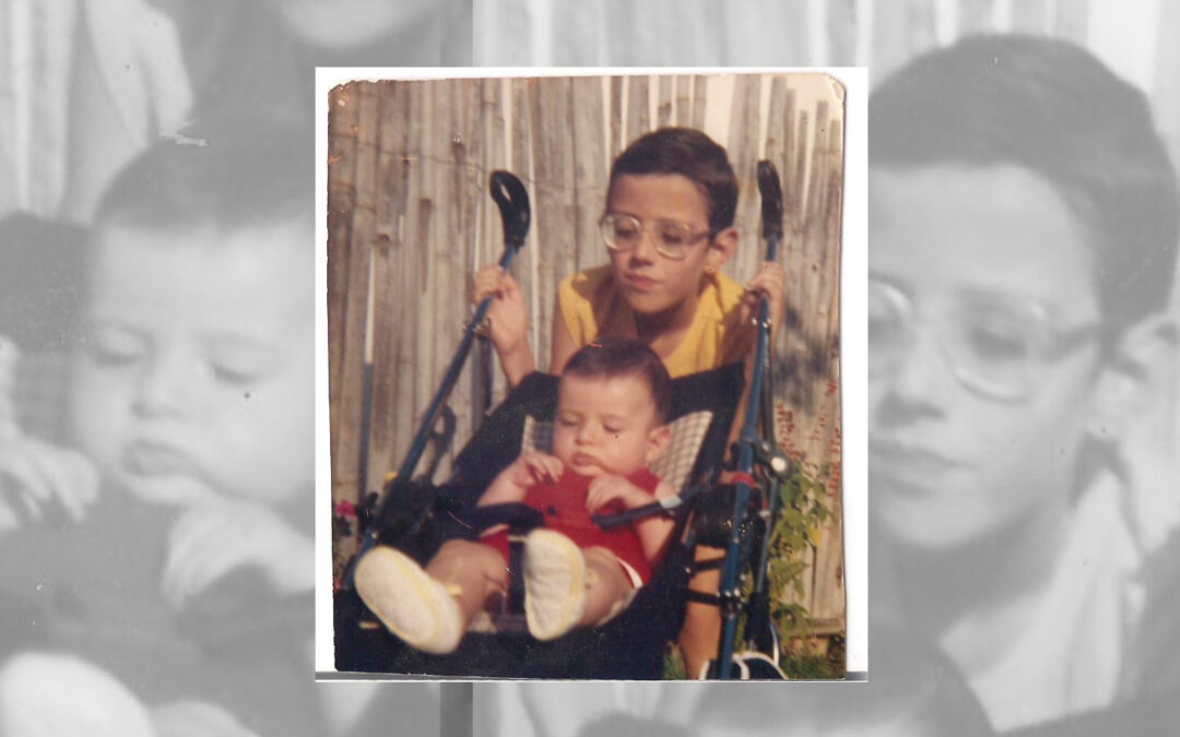
As a child, Phelcom’s Co-Founder and CTO Diego Lencione dreamed of becoming an ophthalmologist. His goal was to help people who suffered from retinal and vision impairment like his older brother, Welber Lencione.
Welber was diagnosed with high myopia when he was seven months old. During his childhood, it reached 17 degrees. The main difficulty with the disease is being able to see distant objects clearly. However, for those with high degrees of myopia, the symptoms are even more serious and have a significant impact on quality of life.
At school, seeing the small print on the blackboard was a real challenge for Welber. “For me to be able to see well, I have to get very close to my cell phone, computer or television. Without my glasses, I can’t even leave the house”, he explains.
High myopia can cause a series of eye complications and significant damage to vision. At the age of 11, Welber suffered retinal detachment in both eyes. At the time, he was admitted to the hospital for two months for treatment.
At 18 years old, he had another episode of retinal detachment, also in both eyes. However, laser surgery was available and he returned home the same day. Since then, he has had no further complications and his myopia has dropped to 11 degrees. “All this has matured me a lot. In the end, time goes by and things settle down”, he reflects.
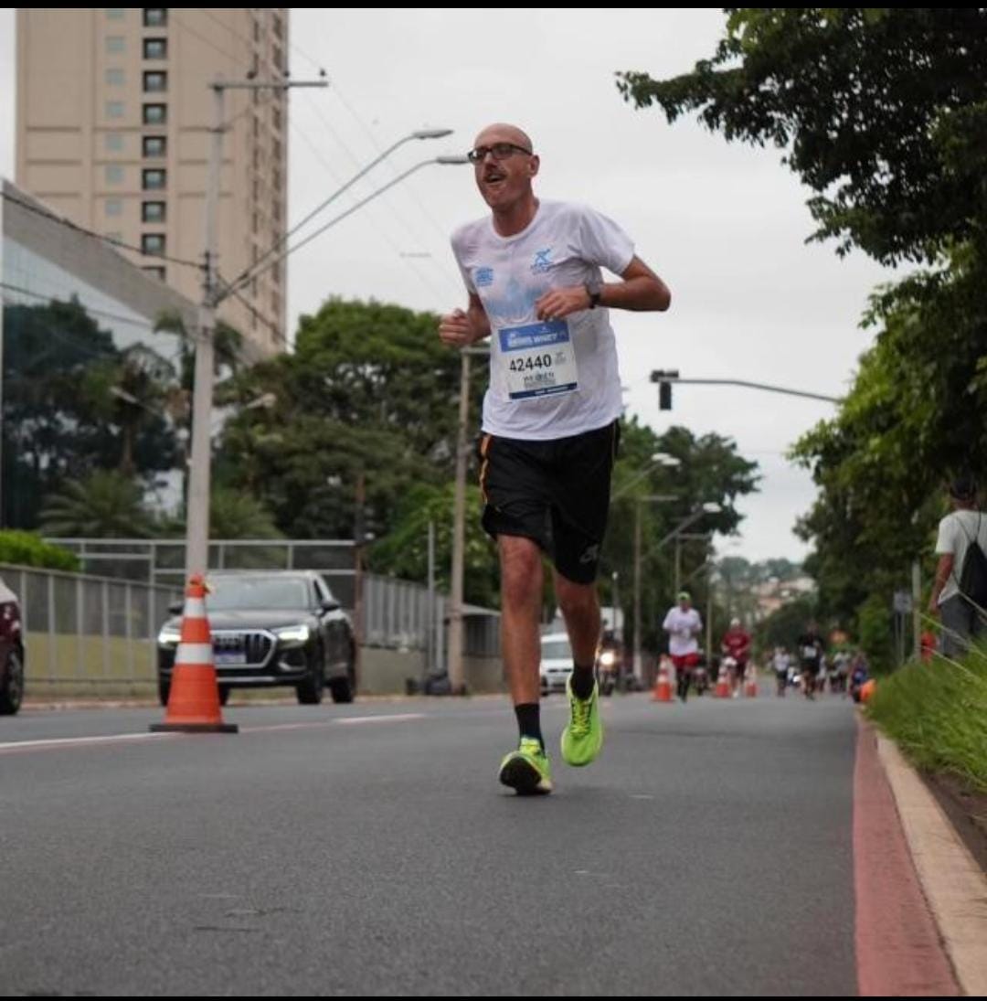
Welber currently has a productive life full of achievements. He works as a security guard at the University of São Paulo (USP) for 18 years, has a 12 year old son named Andrew, is a regular reader and recently became a marathon runner.
Welber Lencione during a competition
Running
Welber says he has always loved sports. Seven years ago, he started running. “I don’t have any difficulties related to my eyesight. I feel 100% myself when I run”.
The athlete has been taking part in competitions in and around São Carlos (SP) for three years. To prepare, he trains in his own neighborhood, day in and day out, in the company of some colleagues. In January, he achieved something he never had before: he completed a marathon in Ribeirão Preto (SP).
Welber finished the last 100 meters of the Ribeirão Preto Marathon alongside his son, Andrew.
Welber reveals that he wants to do it again, but in the famous “São Silvestre International Race”, which takes place every year on December 31st in the city of São Paulo. The obstacle? “It’s a very bad date,” he jokes.
Inspiration
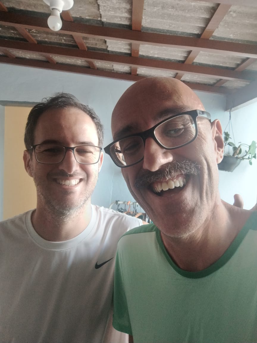
During Welber’s first surgery, Diego was four years old. He had to spend days away from his beloved brother and his mother, who stayed with her firstborn for two months in the hospital.
Throughout his childhood and adolescence, Diego followed his brother’s journey: “When I grew up, life took me into other areas and I ended up studying Physics. At university, I specialized in optics and, before I knew it, I was already working in research and development of ophthalmic equipment,” Diego recalls.
Phelcom’s Co-Founder and CTO, Diego Lencione, with his brother, Welber Lencione.
This gave rise to Phelcom and the idea of creating Eyer -a portable fundus camera that aims to help combat severe visual impairment and blindness. The first tests performed with the first prototypes were on Welber’s eyes. “It was an unforgettable moment. We created something that allowed us to clearly see the changes in his retina and that it would soon be useful for many other people as well. Many times throughout Phelcom’s history, I’ve found myself returning to this time, especially in the company’s most difficult moments”, he reveals.
His brother continues to be a source of inspiration for Diego. Recently, the youngest brother performed a new retinal examination on Welber with the help of EyerMaps, an Artificial Intelligence (AI) system that runs on board Eyer and detects any suspected retinal abnormalities with high accuracy.
The AI pointed to the presence of something new in the back of Welber’s eye. “My brother even suspected a new retinal detachment. But with further tests, the ophthalmologist concluded that it was a kind of membrane that had grown there and that it didn’t affect his vision”, he says, relieved.
About Phelcom
Phelcom Technologies is a Brazilian medtech company based in São Carlos, in the interior area of São Paulo. The company’s story began in 2016, when three young researchers – a physicist, an electronics engineer and a computer engineer (physics, electronics, computing) – created a portable fundus camera integrated with a smartphone.
The Lavinsky Clinic in Porto Alegre (RS) uses the Eyer portable fundus camera to take images of the posterior segment. The equipment is used more frequently in one of the units where optical coherence tomography (OCT) is not an option.
“For example, in cases of glaucoma, the OCT exam provides fundamental structural information for diagnosis and progression, but only the photo shows findings such as optic disc hemorrhage. By combining the two tests, we can better diagnose and monitor the progress of the disease. In this way, retinal imaging is essential for glaucoma analysis”, says glaucoma specialist, researcher from Wills Eye, and coordinator of the Diagnostic Unit at the Lavinsky Clinic, Fábio Lavinsky.
Fábio Lavinsky is a glaucoma specialist, researcher from Wills Eye, and coordinator of the Diagnostic Unit at the Lavinsky Clinic.
The images taken with the Eyer can be accessed on the EyerCloud online platform, allowing for complete documentation, patient follow-up and remote reports.
The fundus images help to identify other retinal problems as well. “That’s why Eyer can be differential in the diagnosis of glaucoma, as it takes an exceptional picture”, he points out.
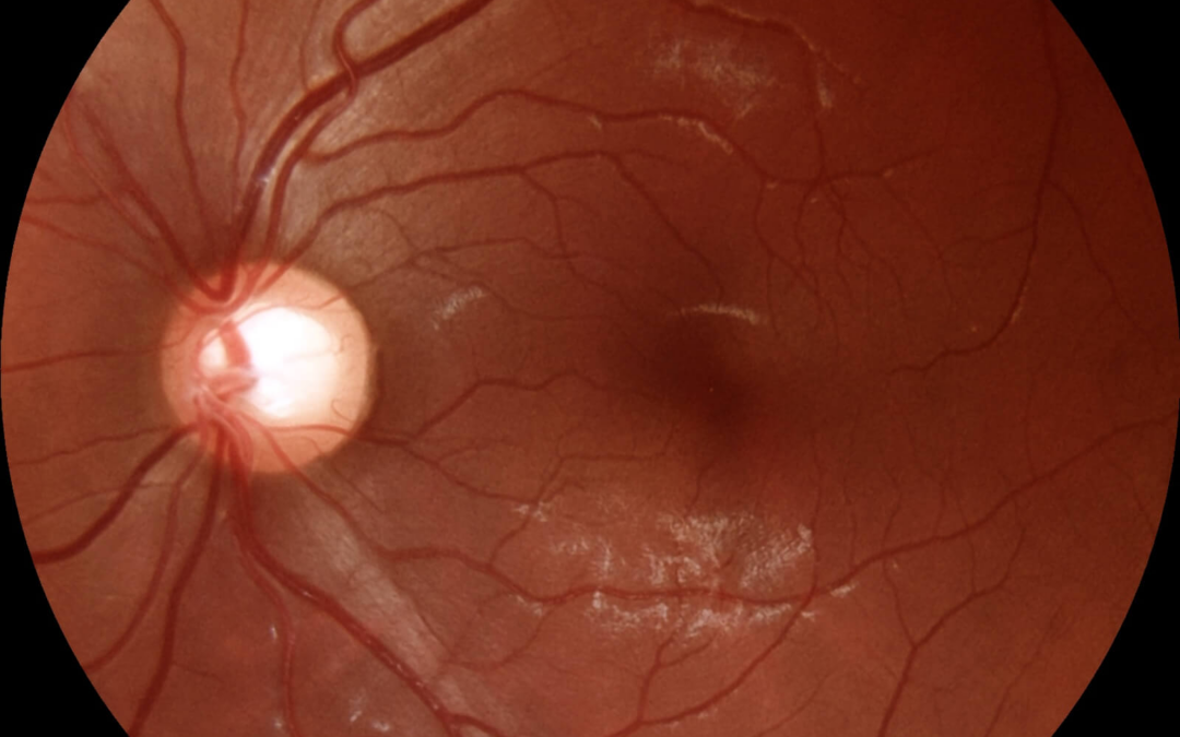
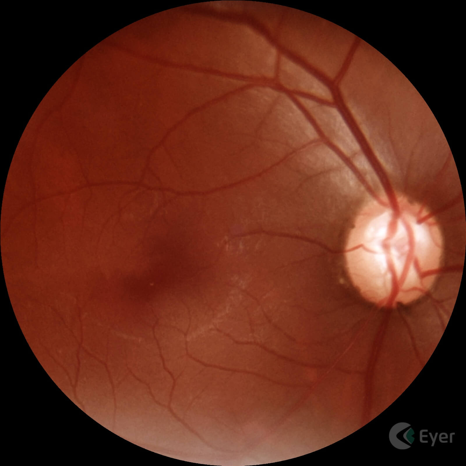
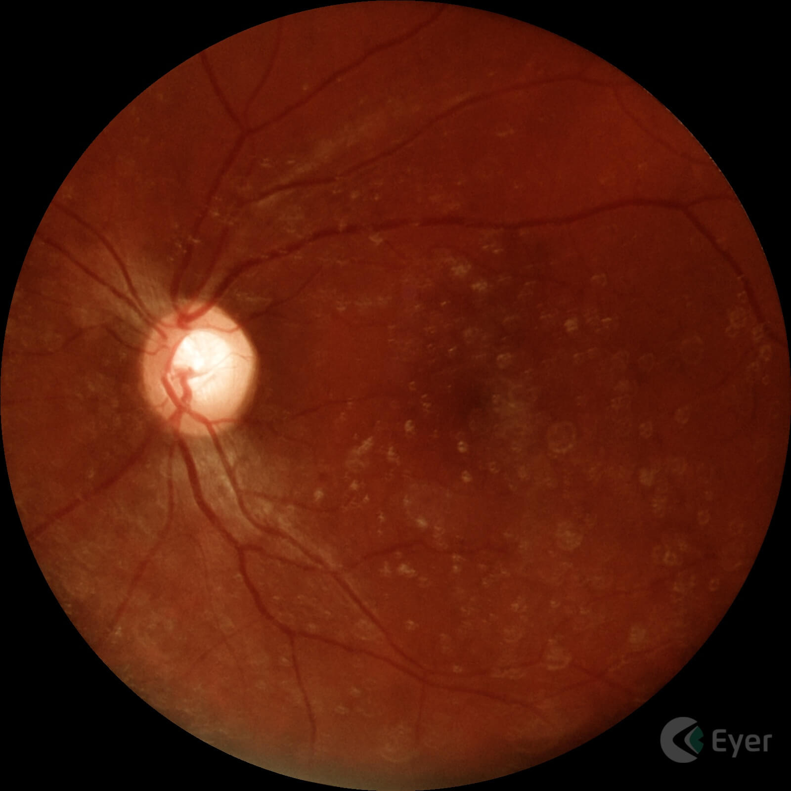
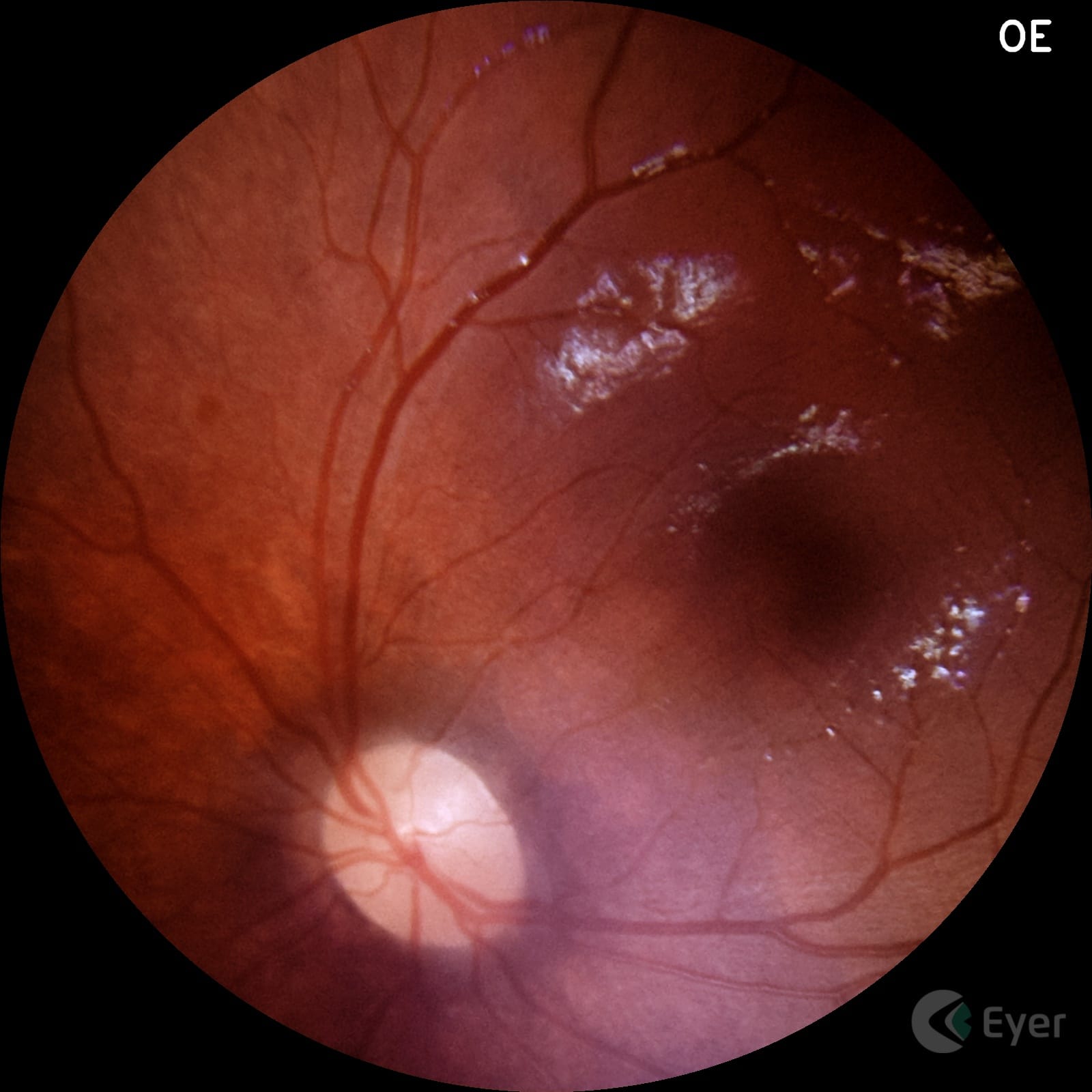
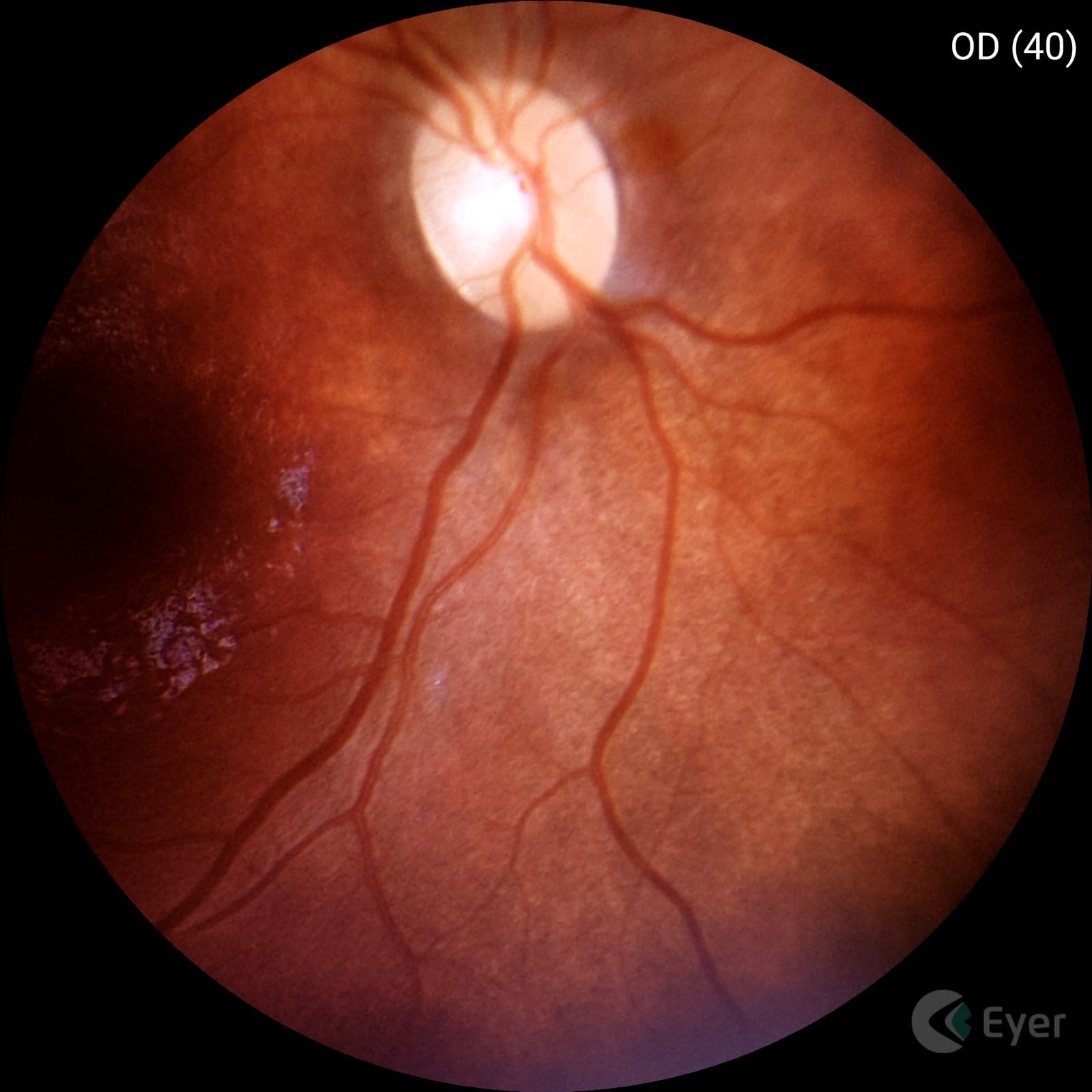
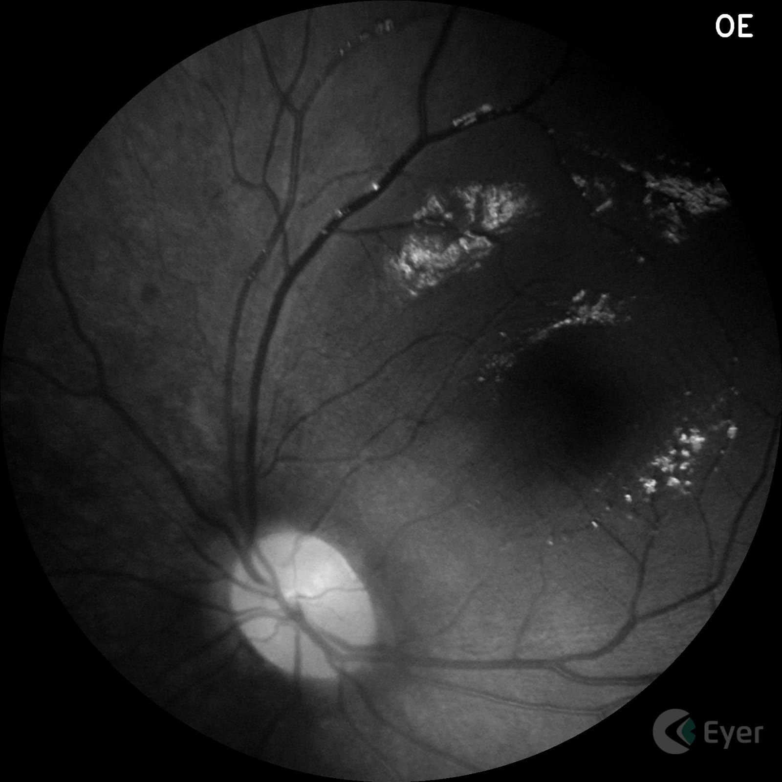
Retinal image of a patient diagnosed with glaucoma taken with Eyer.
Retinal image of a patient diagnosed with glaucoma taken with Eyer.
Lavinsky also uses the EyerMaps artificial intelligence (AI) system, embedded in Eyer, which detects potential retinal abnormalities in real time. If a suspected abnormality is identified, the AI generates a new image with a heatmap highlighting potential abnormalities in the retina. “For clinics that don’t have OCT, this can be a guideline for ordering the test.”
The ophthalmologist points out that AI does not yet accurately identify changes in the optic nerve such as cupping, however, Eyer offers tools for diagnosing the disease.
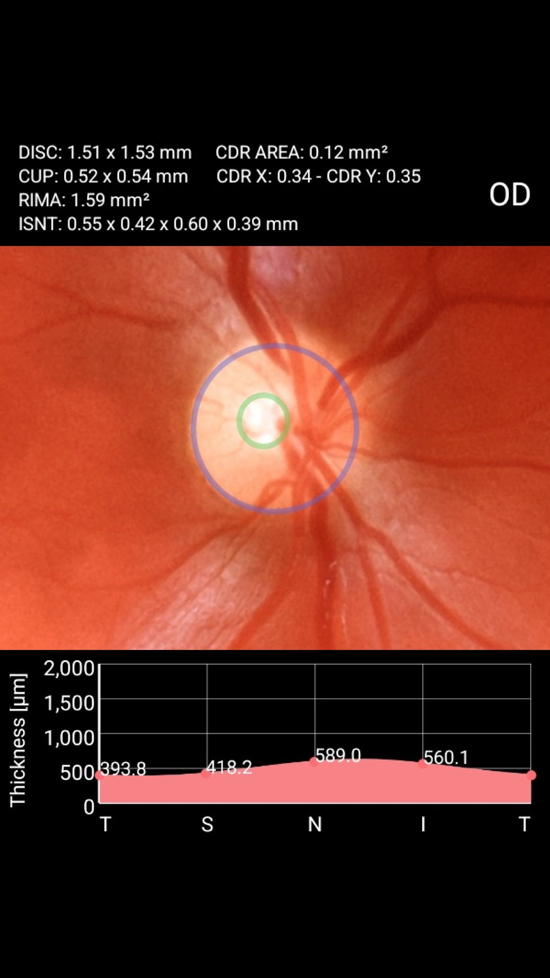
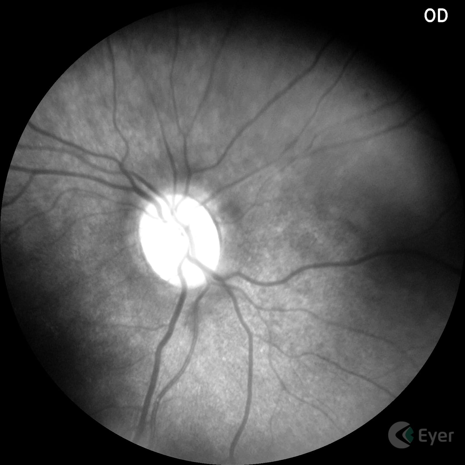
Stereophoto and CDR – Cup to Disk Ratio
The stereophoto is a video created by Eyer based on the superimposition of retinal images with the nerve and macula in the center of the image. This way, doctors can have a clearer view of the region of interest for diagnosing glaucoma.
Stereophoto taken with Eyer.
The Cup to Disk Ratio (CDR) tool shows the percentage ratio between the optic nerve and the excavation, a measurement which, together with other clinical and diagnostic findings, can indicate the presence or risk of the disease.
With just a few clicks and by selecting the circles that outline the nerve and the excavation on the device’s screen, Eyer not only calculates the CDR, but also the neural RIMA measurements and the ISNT graph, metrics that support the diagnostic interpretation of the exam quickly and more accurately.
CDR – Cup to Disk Ratio tool built into Eyer.
Education
Lavinsky is currently in the United States working as a researcher in imaging and ophthalmology at Wills Eye Hospital in Philadelphia, Pennsylvania. But he is also a member of the medical faculty at the University of Vale do Rio dos Sinos (Unisinos), in São Leopoldo (RS).
As well as bringing better clinical results for patients, he believes that Eyer can help teach future doctors. “The Eyer should be like a stethoscope for medicine. Courses should have devices in internal medicine teams and in other related specialties to take pictures of patients with systemic pathologies with ocular repercussions, in the classroom, as well as promoting discussions with students about findings”, he reflects.
Lavinsky explains that the direct ophthalmoscope has a very small field of vision. Although it has some advantages, such as three-dimensionality and showing the optic nerve clearly, the Eyer comes out on top because it has excellent optics and high-quality images that can be viewed digitally by several students. “More specifically, the Eyer should be part of the semiology course. It would be fantastic, something almost revolutionary in education.”
For the doctor, the technology should not only be used in the field of ophthalmology, but also in those where diseases have ocular manifestations, such as endocrinology, cardiology and neurology. “Using EyerCloud, ophthalmologists could provide diagnostic support to colleagues in related specialties. This would greatly speed up the diagnosis and management of important ocular complications, having a positive impact on patients’ clinical outcomes”, he says.
Fábio Lavinsky has no commercial relationship with or receives compensation from Phelcom.He was interviewed for this article because of his expertise and experience in the medical field, with the aim of sharing relevant information about glaucoma and ophthalmic technologies.
Undoubtedly, one of the obstacles in pediatric ophthalmology is being able to carry out examinations on little ones. After all, it can be difficult to keep a child still for examinations.
This difficulty is routine for ophthalmologist, Dr. Patrícia de Freitas Dotto, MD, PhD at the Dr. Jeser Amarante Faria Children’s Hospital, at the São José Municipal Hospital (HMSJ) and in her office, all in Joinville (SC), and at the Lavinsky clinic in Porto Alegre (RS).
“The main challenge is the non-mydriatic fundoscopic evaluation of children between ten months and three years old without sedation or restraint. That’s why it’s essential to create a playful and calm environment that brings peace of mind to the whole family. In this context, photographic documentation, or even fundoscopic imaging, in real time allows parents to be involved in the care, which improves the doctor-patient relationship and, consequently, has a positive impact on the child’s follow-up and/or treatment.”
Dotto says that she uses the Eyer portable fundus camera to speed up examinations on pediatric patients. The equipment, which is very suitable for examining infants and children due to its portability and high image quality, works in conjunction with a smartphone and performs retinal examinations in a few minutes. These photographs are then available on the EyerCloud online platform, making it easier to study and monitor the progression of cases.
“The field of vision is excellent. Under medicated mydriasis, it is also an excellent complementary propaedeutic tool for assessing the mid-periphery of the retina, particularly small hyperpigmented lesions of the choroid (nevus) and retina (melanocytomas), using it as an infrared scan,” she explains.
In her office, with help of the Eyer Slit Lamp Adapter, the ophthalmologist attaches the device to her slit lamp allowing exams to be performed more easily. “Although I really enjoy using it this way, it’s magical to use it in ‘mobile’ form in ICUs, operating rooms, home care and within the office itself, such as evaluations of children on their mother’s lap or in the waiting room,” she points out.
The doctor also uses the Eyer in the emergency room, in adult and child care, for documenting infectious diseases and Retinopathy of Prematurity (ROP) in neonatal ICUs severely debilitated adults in ICUs, as well as inter-consultations for neurology, neurosurgery, nephrology, cardiology, and in medical expertise for the National Civil Aviation Agency (ANAC) and the Santa Catarina Court of Justice (TJSC). “I literally carry Eyer in my bag. It makes my job a lot easier,” she says.
The ophthalmologist, Dr. Patrícia de Freitas Dotto, MD, PhDtreats infants and children at the Dr. Jeser Amarante Faria Children’s Hospital, the São José Municipal Hospital (HMSJ) and at her office, all in Joinville (SC).
Shaken Baby Syndrome
With daily use of the Eyer in various locations, Dotto has witnessed several remarkable cases, such as the care of a child suffering from Shaken Baby Syndrome. The syndrome occurs when the baby is shaken intensely, causing permanent brain damage or even death.
Here’s the doctors report on the case of shaken baby syndrome:
“The child had been followed up for a few months in hospital for convulsions and severe malnutrition. The suspicion of Shaken Baby was raised when neuroimaging alterations (hematoma) were observed during the complementary investigation of status epilepticus.
During the ophthalmological assessment, when I examined her visual acuity (grating), I found that the vision in one eye was outside the normal range for her age, despite the fact that she had no changes in ocular motility.
When I laid her on the gurney for the retinal mapping, she started crying desperately. It was a very difficult examination and I was struck by the fact that the child eventually let go, almost gave up, and then reacted again, without showing any change in her level of consciousness.
It was at one of these moments that I managed to photograph the fundus of the eye and identify retinal lesions, indicating serious damage to the central nervous system. In one particular case, the hemorrhage was very small, probably because it was being reabsorbed, and I could only be sure of the diagnosis because of the examination. It was a miracle for me and for her.
I was devastated that day, but very grateful to HaShem and Phelcom [the company that invented the Eyer]. The case was really investigated from a social point of view, the assaults were confirmed and the appropriate measures were taken.
We saved a life. And in my religion, Judaism, saving a life means saving the whole world.”
Next, see the images of this case taken by Dotto with Eyer:
More cases
Dotto performs retinography on all patients, as she considers it important the normal state of the retina. “We often look for illnesses and forget how important it is to make sure the fundus of the eye is normal, because problems happen throughout life and it’s important to make sure of the moment when the state of eye health is lost,” she says.
She highlights the case of a 22-year-old woman who was admitted to the ER with compromised visual acuity, severe hypertensive retinopathy and serous detachment of the macula. The team suspected Autoimmune Nephropathy (IgA). “Unbelievably, four or five days after pressure control (in the ICU), she progressed to 20/20 visual acuity and total resolution of the serous detachment.”
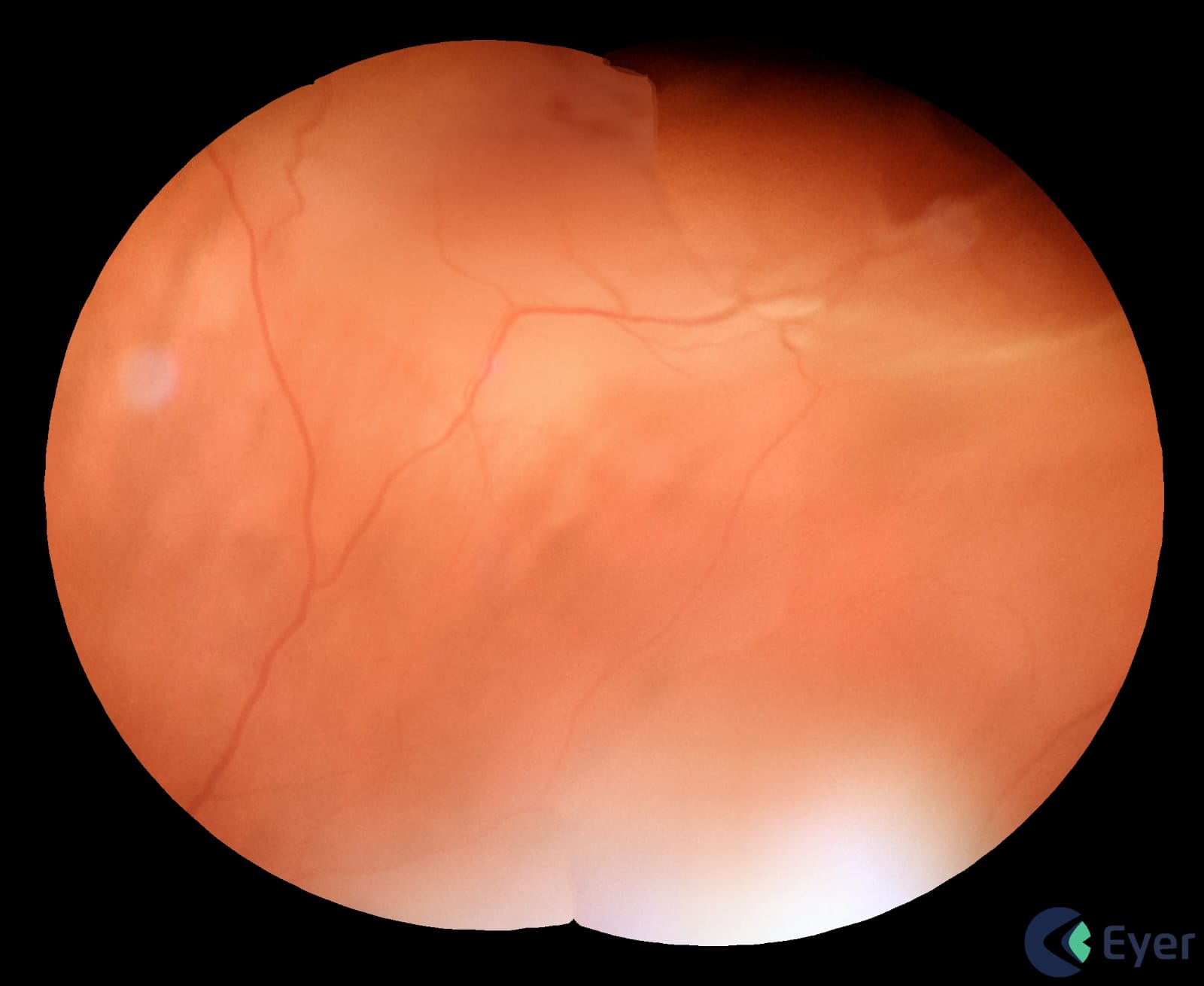
Another situation involved a patient in the ER with anterior uveitis and an apparent normal USG. After remission, when mapping the retina, the doctor noticed a peripheral blackened lesion. “Using the Eyer, I was able to document it. We redid the USG thinking it was a melanoma. And it was indeed a melanoma at the level of the ciliary body,” she says.
In children, the ophthalmologist recalls the care of two patients with slight vitreous hemorrhage: “After improving the transparency of the media, it was possible to record the presence of a slight vascular malformation at the level of the optic nerve,” she recalls.
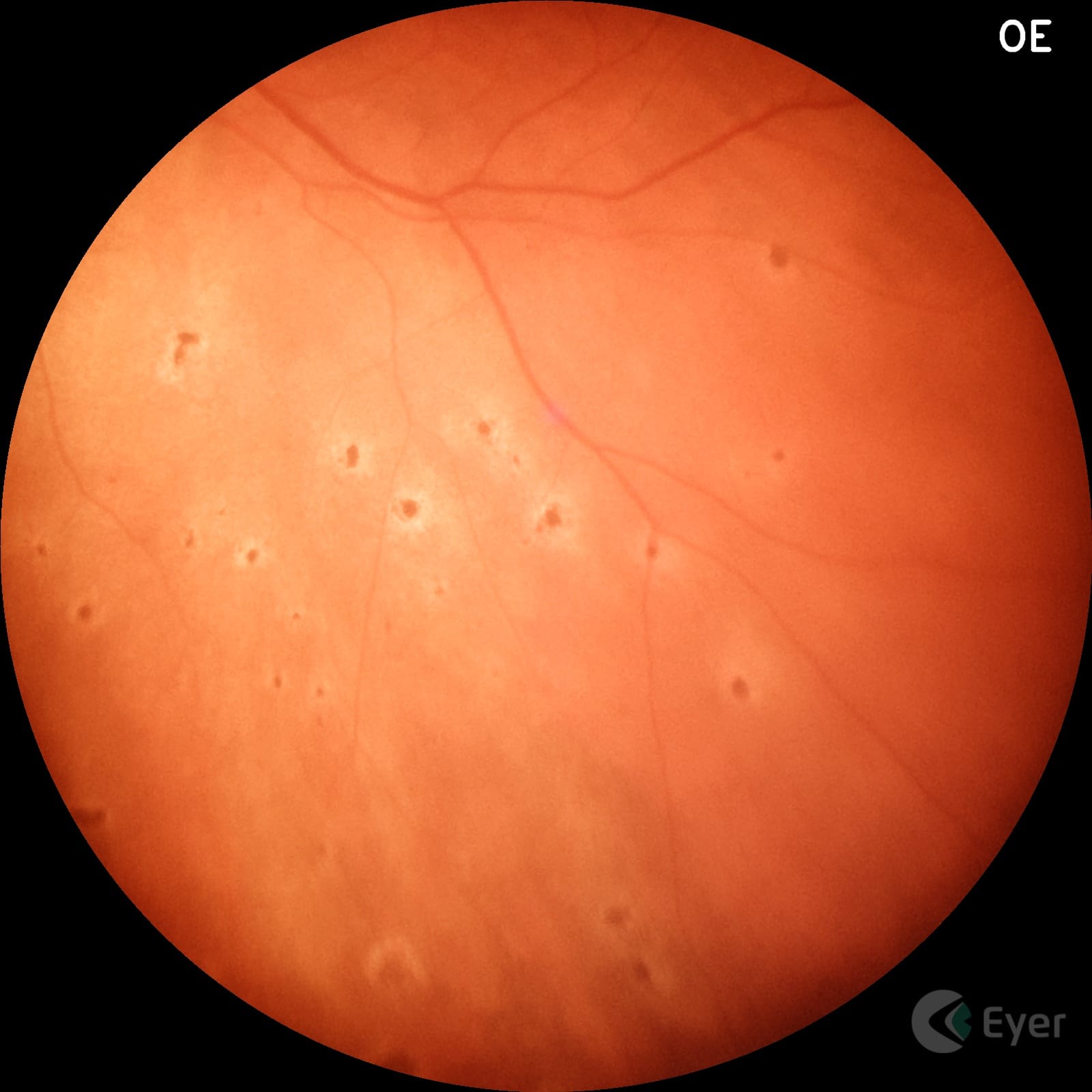
Among the most commonly diagnosed diseases in babies are ROP, congenital infections, optic nerve hypoplasia and retinal and optic nerve colobomas. “I also use the Eyer a lot to quantify the size of the optic nerve and the excavation. This helps with the differential diagnosis of suspicious glaucomas, particularly in short-sighted children,” she concludes.
Below are some images taken by the ophthalmologist with the Eyer:
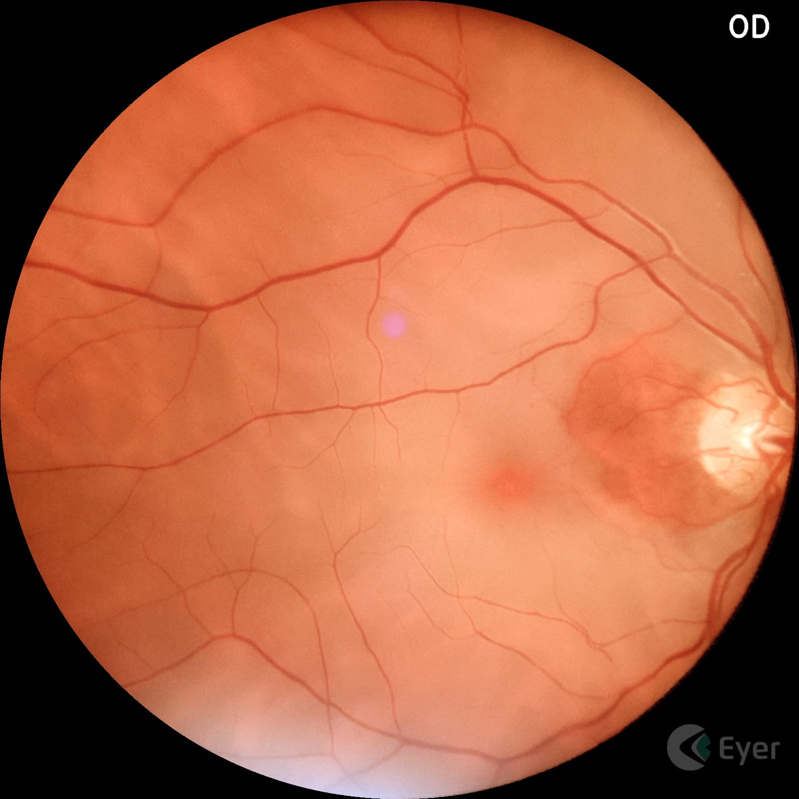
Patient with retinal displacement.
Patient with microembolization in the choroid due to covid.
Patient with arterial occlusion.
The Eyer
The Eyer is a portable fundus camera that works in conjunction with a smartphone and performs high-quality retinal examinations in a few minutes without the need for pupil dilation.
The technology supports the diagnosis of more than 50 diseases, including glaucoma, cataracts, diabetic retinopathy, AMD, ROP, retinoblastoma, hypertensive retinopathy and ocular toxoplasmosis. Currently, more than 10 million tests have been carried out in Brazil, the United States, Chile, Colombia and Japan.
It was recently approved in the United Arab Emirates and is in the regulatory process of being marketed in Mexico, Egypt and Saudi Arabia.
Portability, connectivity and integration with intelligent functions such as EyerMaps, together with the technology’s affordability, contribute to increasing access to retinal examinations.
By 2030, over half a billion people are expected to be diagnosed with diabetes. Currently, Brazil ranks as the sixth country worldwide with the highest population of diabetics.
Currently, there is no national diabetic retinopathy screening strategy by the Unified Health System (SUS). Thus, social initiatives for diagnosing the disease in communities with inadequate healthcare infrastructure are crucial.
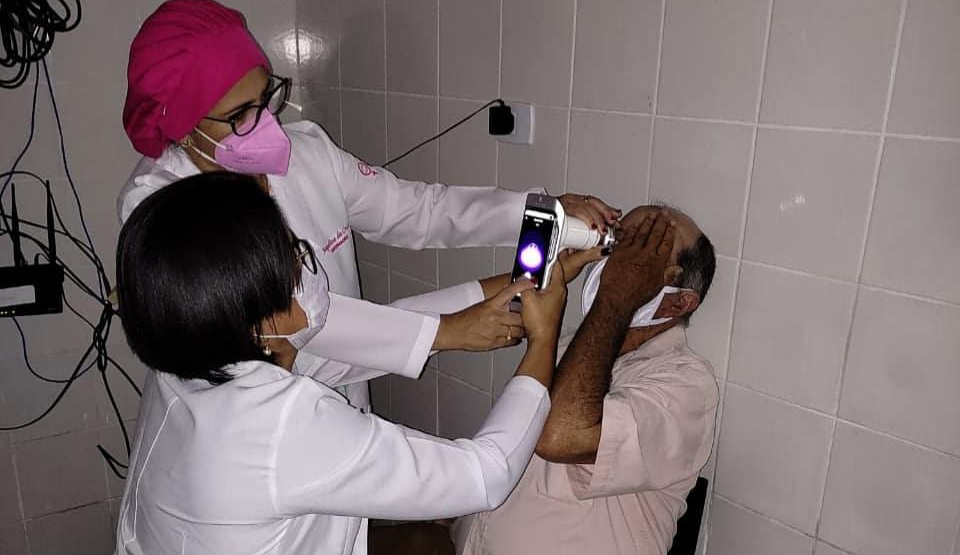
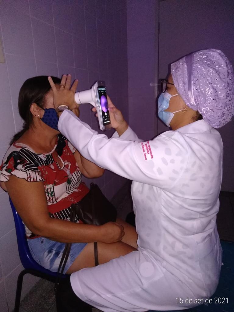
For instance, we have the “Mutirão do Diabetes” in Itabuna, Bahia, and “Iluminar” in the countryside of Sergipe, supported by the NGO Retina Global. This American institution focuses on developing sustainable solutions for managing retinal diseases in underserved areas around the world.
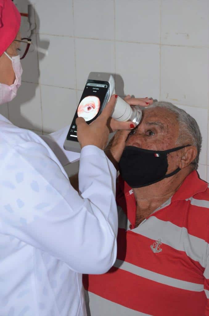
From September 2021 to March 2022, “Iluminar” used the portable retinal camera, Eyer for diabetic retinopathy screening in Itabi, Graccho Cardoso, Canindé de São Francisco, and Poço Redondo.
The device is connected to a smartphone and performs retinal exams within minutes, producing high-quality images, and uploads the images to the EyerCloud online platform, facilitating remote assessments.
The results of the pilot project were presented at ARVO 2023, one of the most renowned international ophthalmology conferences held in the United States in April.
The Study
One of the leaders of the “Iluminar” project, ophthalmologist Fernando Malerbi, explains that the aim of the retrospective observational clinical study was to evaluate the grading capacity of retina images obtained using a low-cost, non-mydriatic portable retinal camera — in this case, the Eyer — along with the use of artificial intelligence and telemedicine for diabetic retinopathy screening.
In total, 968 individuals with diabetes were evaluated:
65.9% were female;
Average age of 60.3 ± 14.2 years;
Duration of diabetes: 8.0 ± 7.2 years;
64.2% had systemic hypertension;
17.7% were using insulin;
28.5% had previously undergone fundus exams;
20.6% were illiterate;
50.6% had only completed primary education;
3.4% had health insurance.
A trained technician captured images without pupil dilation and then assessed image quality. Diabetic retinopathy requiring referral, defined as severe non-proliferative or proliferative retinopathy or the presence of diabetic maculopathy, was automatically detected by an embedded artificial intelligence system (EyerMaps). The AI was provided for clinical validation.
In a matter of seconds, EyerMaps indicated the possibility of retinopathy with a high sensitivity rate. Subsequently, all exams were evaluated by ophthalmologists.
Patients with inadequate images underwent pupil dilation and then a new assessment. Those with non-gradable images even after mydriasis, along with cases of referable diabetic retinopathy, were referred for ophthalmological evaluation.
Corneal opacities that hindered retinopathy classification were the exclusion criteria. The primary outcome measure was image gradability.
The Results
Grading was possible for 858 individuals (88.6%), with 85 of these (9.9%) showing referable diabetic retinopathy. Non-grading was associated with older age and longer diabetes duration.
Among patients with gradable images, 81% did not require pupil dilation. The need for mydriasis was associated with older age, longer diabetes duration, higher hypertension rates, and more severe retinopathy.
The strategy of utilizing a low-cost portable camera with embedded AI system and mydriasis when necessary achieved suitable images in 90% of cases within a resource-limited real-world environment. Malerbi emphasizes, “Avoiding unnecessary pupil dilation contributes to higher adherence to diabetic retinopathy screening programs.”
Enhanced Portability Facilitated Screening
This novel screening was conducted in primary care clinics located near patients’ homes to encourage participation.
Malerbi highlights that the portability of Eyer was a facilitator, along with connectivity. “We had remote experts evaluating images, sometimes in real time,” he explains. All of the exams taken were uploaded to EyerCloud.
Lastly, Malerbi emphasizes the availability of EyerMaps for use in social initiatives. “Partner ophthalmologists were instantly notified whenever EyerMaps identified a high likelihood of retinopathy. Thus, we could prioritize patients for confirmatory exams and, if necessary, treatment,” he shares.
The AI accurately detects any suspicions of retinal abnormalities. Within seconds of capturing the fundus image, if an abnormality is detected, the system generates a new image with an attention map (heatmap) highlighting potential retinal anomalies.
Synchronized with EyerCloud, it categorizes images and exams captured based on the likelihood of abnormalities using color markers:
Green: Image or exam with low likelihood of an abnormality (up to 30%);
Yellow: Image or exam with moderate likelihood of an abnormality (31 to 70%);
Red: Image or exam with high likelihood of an abnormality (71 to 100%).
All patients diagnosed with diabetic retinopathy were referred for free laser treatment.

 English
 Português