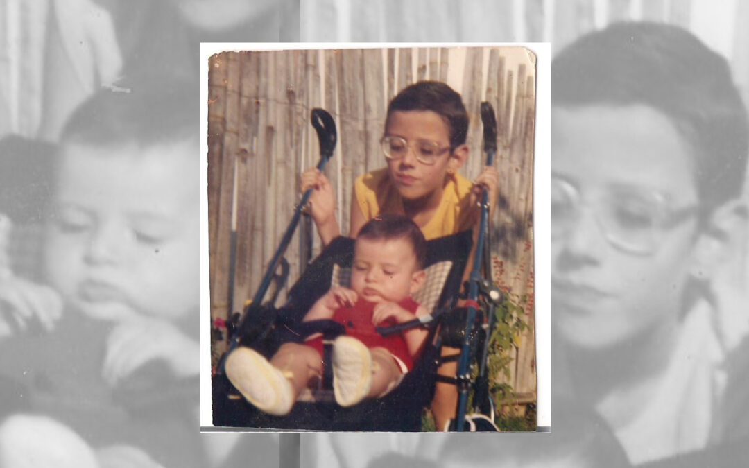
As a child, Phelcom’s Co-Founder and CTO Diego Lencione dreamed of becoming an ophthalmologist. His goal was to help people who suffered from retinal and vision impairment like his older brother, Welber Lencione.
Welber was diagnosed with high myopia when he was seven months old. During his childhood, it reached 17 degrees. The main difficulty with the disease is being able to see distant objects clearly. However, for those with high degrees of myopia, the symptoms are even more serious and have a significant impact on quality of life.
At school, seeing the small print on the blackboard was a real challenge for Welber. “For me to be able to see well, I have to get very close to my cell phone, computer or television. Without my glasses, I can’t even leave the house”, he explains.
High myopia can cause a series of eye complications and significant damage to vision. At the age of 11, Welber suffered retinal detachment in both eyes. At the time, he was admitted to the hospital for two months for treatment.
At 18 years old, he had another episode of retinal detachment, also in both eyes. However, laser surgery was available and he returned home the same day. Since then, he has had no further complications and his myopia has dropped to 11 degrees. “All this has matured me a lot. In the end, time goes by and things settle down”, he reflects.
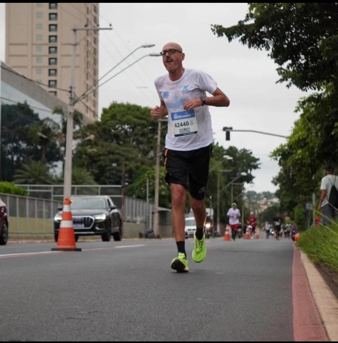
Welber currently has a productive life full of achievements. He works as a security guard at the University of São Paulo (USP) for 18 years, has a 12 year old son named Andrew, is a regular reader and recently became a marathon runner.
Welber Lencione during a competition
Running
Welber says he has always loved sports. Seven years ago, he started running. “I don’t have any difficulties related to my eyesight. I feel 100% myself when I run”.
The athlete has been taking part in competitions in and around São Carlos (SP) for three years. To prepare, he trains in his own neighborhood, day in and day out, in the company of some colleagues. In January, he achieved something he never had before: he completed a marathon in Ribeirão Preto (SP).
Welber finished the last 100 meters of the Ribeirão Preto Marathon alongside his son, Andrew.
Welber reveals that he wants to do it again, but in the famous “São Silvestre International Race”, which takes place every year on December 31st in the city of São Paulo. The obstacle? “It’s a very bad date,” he jokes.
Inspiration
During Welber’s first surgery, Diego was four years old. He had to spend days away from his beloved brother and his mother, who stayed with her firstborn for two months in the hospital.
Throughout his childhood and adolescence, Diego followed his brother’s journey: “When I grew up, life took me into other areas and I ended up studying Physics. At university, I specialized in optics and, before I knew it, I was already working in research and development of ophthalmic equipment,” Diego recalls.
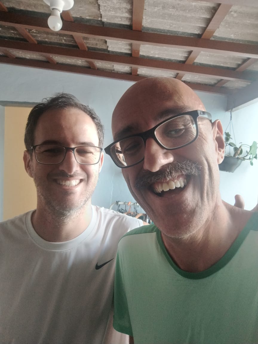
Phelcom’s Co-Founder and CTO, Diego Lencione, with his brother, Welber Lencione.
This gave rise to Phelcom and the idea of creating Eyer -a portable fundus camera that aims to help combat severe visual impairment and blindness. The first tests performed with the first prototypes were on Welber’s eyes. “It was an unforgettable moment. We created something that allowed us to clearly see the changes in his retina and that it would soon be useful for many other people as well. Many times throughout Phelcom’s history, I’ve found myself returning to this time, especially in the company’s most difficult moments”, he reveals.
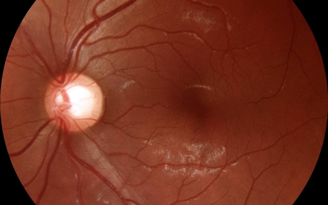
His brother continues to be a source of inspiration for Diego. Recently, the youngest brother performed a new retinal examination on Welber with the help of EyerMaps, an Artificial Intelligence (AI) system that runs on board Eyer and detects any suspected retinal abnormalities with high accuracy.
The AI pointed to the presence of something new in the back of Welber’s eye. “My brother even suspected a new retinal detachment. But with further tests, the ophthalmologist concluded that it was a kind of membrane that had grown there and that it didn’t affect his vision”, he says, relieved.
About Phelcom
Phelcom Technologies is a Brazilian medtech company based in São Carlos, in the interior area of São Paulo. The company’s story began in 2016, when three young researchers – a physicist, an electronics engineer and a computer engineer (physics, electronics, computing) – created a portable fundus camera integrated with a smartphone.
The Lavinsky Clinic in Porto Alegre (RS) uses the Eyer portable fundus camera to take images of the posterior segment. The equipment is used more frequently in one of the units where optical coherence tomography (OCT) is not an option.
“For example, in cases of glaucoma, the OCT exam provides fundamental structural information for diagnosis and progression, but only the photo shows findings such as optic disc hemorrhage. By combining the two tests, we can better diagnose and monitor the progress of the disease. In this way, retinal imaging is essential for glaucoma analysis”, says glaucoma specialist, researcher from Wills Eye, and coordinator of the Diagnostic Unit at the Lavinsky Clinic, Fábio Lavinsky.
Fábio Lavinsky is a glaucoma specialist, researcher from Wills Eye, and coordinator of the Diagnostic Unit at the Lavinsky Clinic.
The images taken with the Eyer can be accessed on the EyerCloud online platform, allowing for complete documentation, patient follow-up and remote reports.
The fundus images help to identify other retinal problems as well. “That’s why Eyer can be differential in the diagnosis of glaucoma, as it takes an exceptional picture”, he points out.
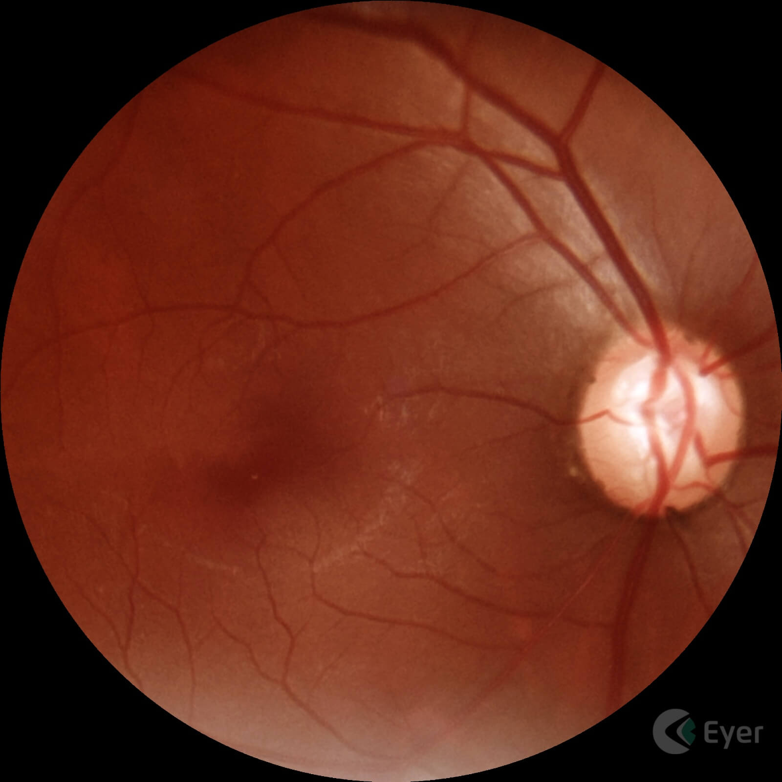
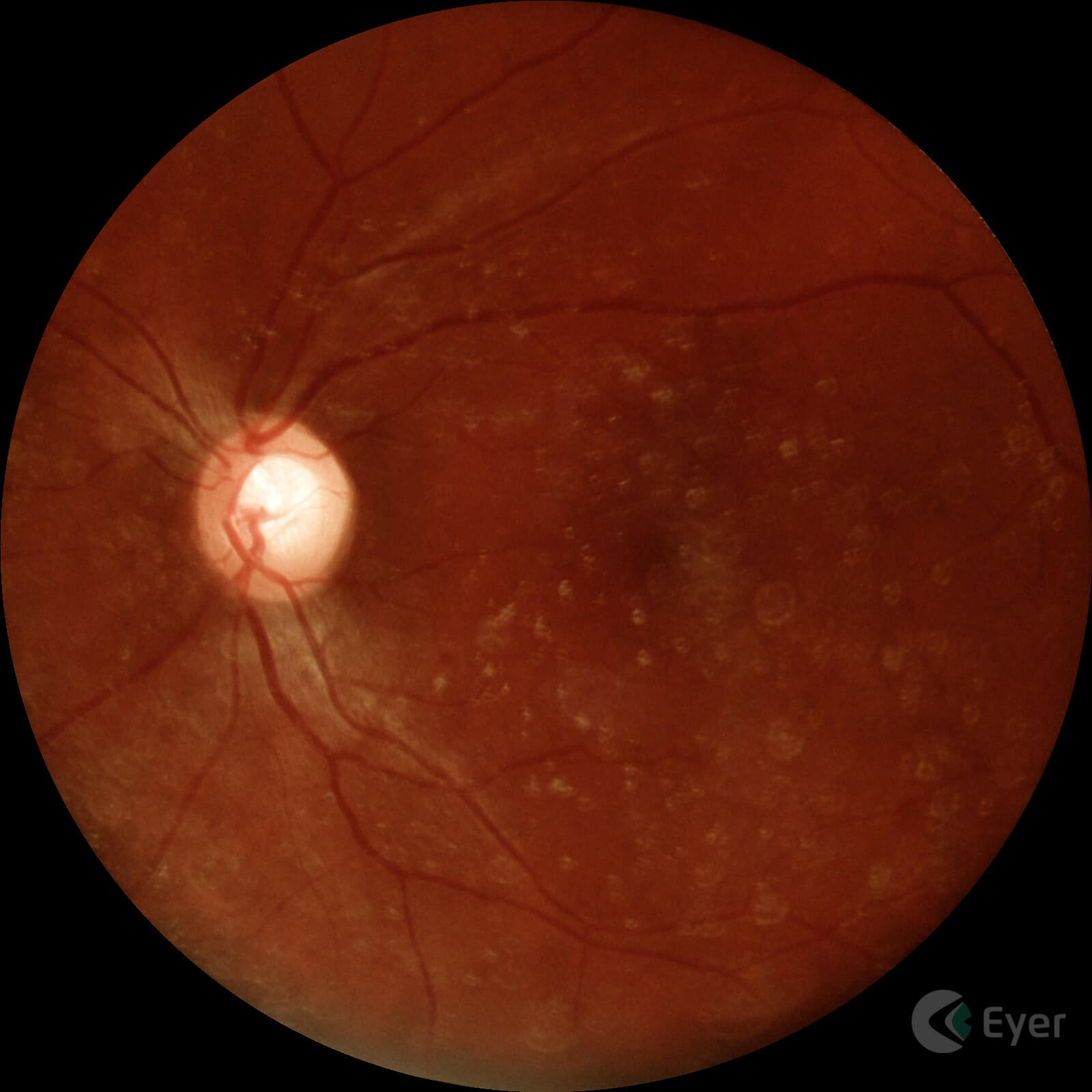
Retinal image of a patient diagnosed with glaucoma taken with Eyer.
Retinal image of a patient diagnosed with glaucoma taken with Eyer.
Lavinsky also uses the EyerMaps artificial intelligence (AI) system, embedded in Eyer, which detects potential retinal abnormalities in real time. If a suspected abnormality is identified, the AI generates a new image with a heatmap highlighting potential abnormalities in the retina. “For clinics that don’t have OCT, this can be a guideline for ordering the test.”
The ophthalmologist points out that AI does not yet accurately identify changes in the optic nerve such as cupping, however, Eyer offers tools for diagnosing the disease.
Stereophoto and CDR – Cup to Disk Ratio
The stereophoto is a video created by Eyer based on the superimposition of retinal images with the nerve and macula in the center of the image. This way, doctors can have a clearer view of the region of interest for diagnosing glaucoma.
Stereophoto taken with Eyer.
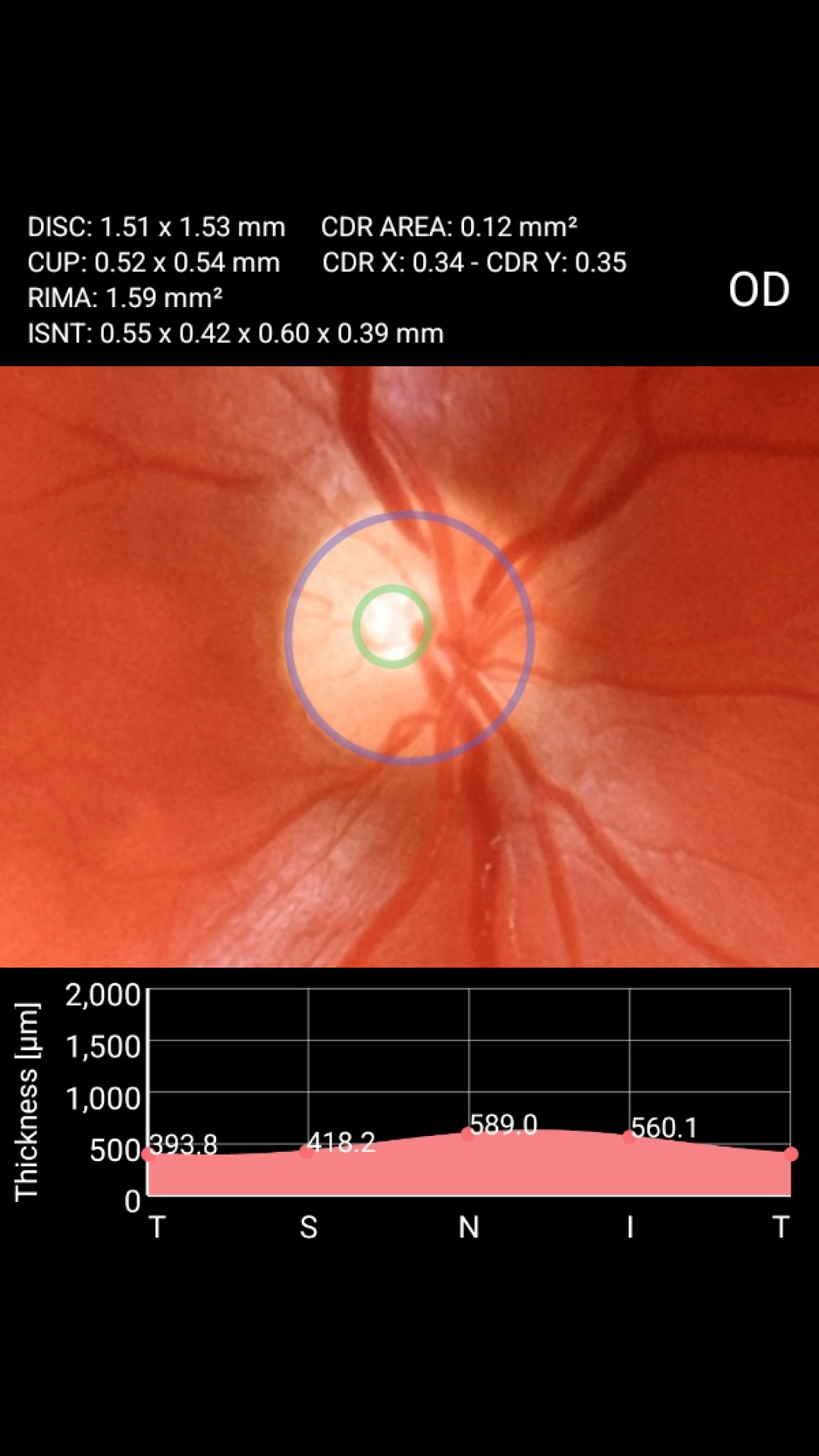
The Cup to Disk Ratio (CDR) tool shows the percentage ratio between the optic nerve and the excavation, a measurement which, together with other clinical and diagnostic findings, can indicate the presence or risk of the disease.
With just a few clicks and by selecting the circles that outline the nerve and the excavation on the device’s screen, Eyer not only calculates the CDR, but also the neural RIMA measurements and the ISNT graph, metrics that support the diagnostic interpretation of the exam quickly and more accurately.
CDR – Cup to Disk Ratio tool built into Eyer.
Education
Lavinsky is currently in the United States working as a researcher in imaging and ophthalmology at Wills Eye Hospital in Philadelphia, Pennsylvania. But he is also a member of the medical faculty at the University of Vale do Rio dos Sinos (Unisinos), in São Leopoldo (RS).
As well as bringing better clinical results for patients, he believes that Eyer can help teach future doctors. “The Eyer should be like a stethoscope for medicine. Courses should have devices in internal medicine teams and in other related specialties to take pictures of patients with systemic pathologies with ocular repercussions, in the classroom, as well as promoting discussions with students about findings”, he reflects.
Lavinsky explains that the direct ophthalmoscope has a very small field of vision. Although it has some advantages, such as three-dimensionality and showing the optic nerve clearly, the Eyer comes out on top because it has excellent optics and high-quality images that can be viewed digitally by several students. “More specifically, the Eyer should be part of the semiology course. It would be fantastic, something almost revolutionary in education.”
For the doctor, the technology should not only be used in the field of ophthalmology, but also in those where diseases have ocular manifestations, such as endocrinology, cardiology and neurology. “Using EyerCloud, ophthalmologists could provide diagnostic support to colleagues in related specialties. This would greatly speed up the diagnosis and management of important ocular complications, having a positive impact on patients’ clinical outcomes”, he says.
Fábio Lavinsky has no commercial relationship with or receives compensation from Phelcom.He was interviewed for this article because of his expertise and experience in the medical field, with the aim of sharing relevant information about glaucoma and ophthalmic technologies.
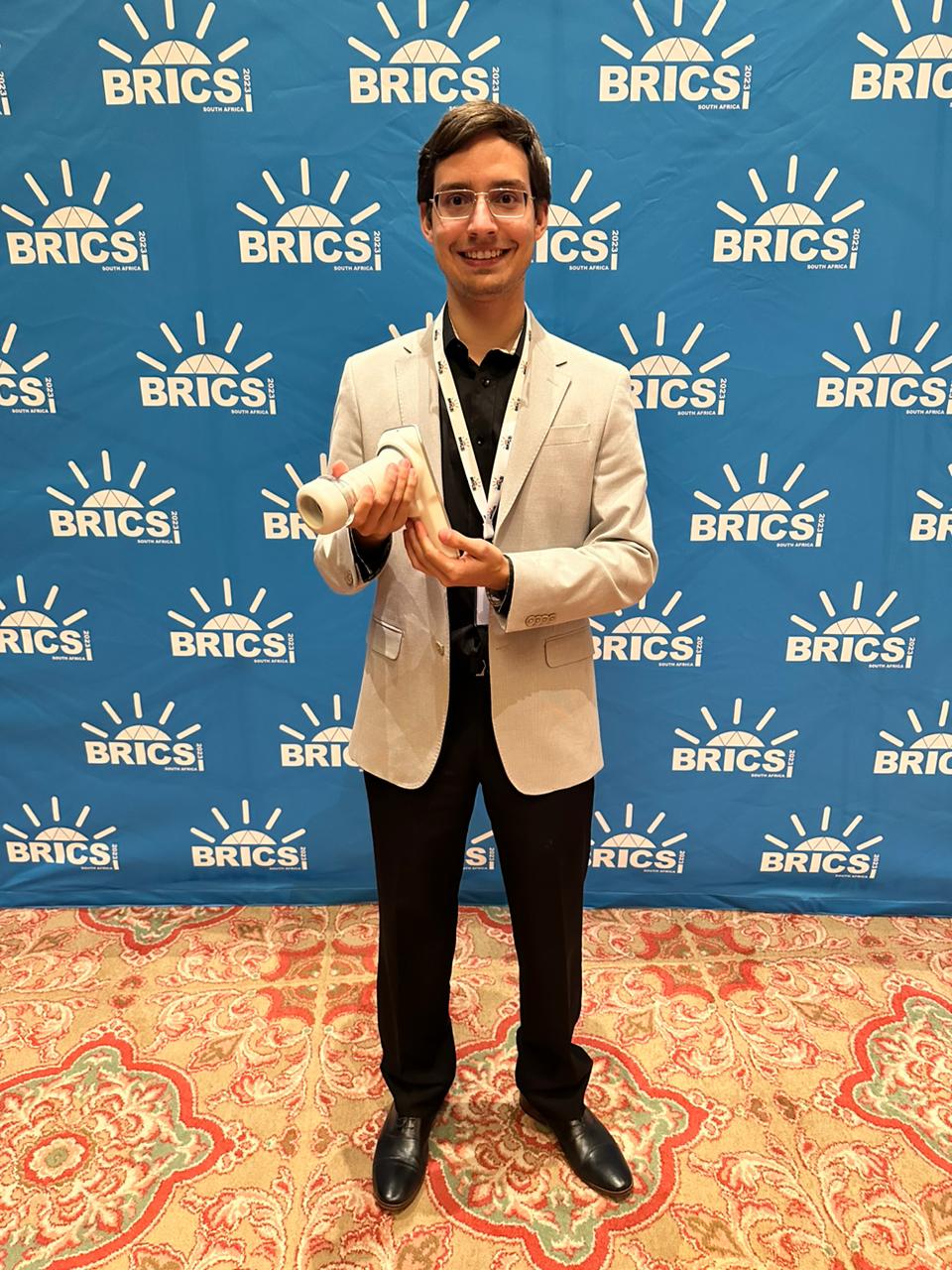
The doctor Gustavo Rosa Gameiro, a PhD student in ophthalmology at Unifesp, was selected by the Brazilian Academy of Sciences (ABC) and the Ministry of Science, Technology and Innovation (MCTI) to participate in the 8th BRICS Young Scientists Forum. The event occurred from July 31th to August 8th in Gqeberha, South Africa.
BRICS is a group of five developing countries focused on mutual economic cooperation: Brazil, Russia, India, China and South Africa.
Gameiro was one of the six Brazilian scientists nominated to take part in the panel “The future of education, skills and skill sets”. The PhD student points out that education entered the BRICS agenda with great force this year. “In my presentation, we discussed applications of basic models and the use of the Eyer portable fundus camera with artificial intelligence for teaching ophthalmology based on the results of our workshops,” says the doctor, the youngest member of the Brazilian delegation at the event at 27 years old.
Gustavo Gameiro used the Eyer portable fundus camera in his project presented at the 8th BRICS Young Scientists Forum. Photo: personal archive.
Gameiro explains that the first approach to diseases such as glaucoma, Age-Related Macular Degeneration (AMD) and diabetic retinopathy in primary care is often carried out by the newly qualified clinical doctor.
“However, studies reveal that they have a huge deficit in the teaching of ophthalmology during their degree, compromising the correct approach and prognosis of these cases. This can lead to insecurity in referring or treating patients with ophthalmic complaints,” he explains.
Workshops
The workshops took place with undergraduate students from Unifesp and the Albert Einstein Israelite Institute for Education and Research and were supported by the ophthalmologists Thiago Gonçalves Martins and Paulo Schor, Gameiro’s PhD advisor. The Eyer portable fundus camera and the EyerMaps AI system were provided free of charge by Phelcom Technologies for the project.
“With the EyerMaps AI resource, which uses a heat map to highlight areas of the retina with possible alterations caused by different pathologies, we were able to teach and correct the student’s interpretation findings at the same time as captured by the fundus of the eye,” says the doctor.
“The clarity of the images is absurdly incredible. We did exams on our colleagues and we were able to see every detail of the retina, optic nerve and vessels. Transforming large, heavy devices like a fundus camera into portable ones makes the doctor’s life much easier, because we can go to the patients and achieve better results,” says Unifesp medical student Suellyn Alves, who took part in the workshop.
Gameiro goes further: “Perhaps we need to change the paradigm of only teaching students how to acquire images using ophthalmic equipment. We need to focus on how to interpret them and manage these exams, organizing them in the cloud, for example. and with the Eyer, you can easily teach interpretation and management” he says.
With the satisfactory results of the workshops, the doctor reveals his desire to expand the project and turn it into support material for teaching ophthalmology throughout Brazil.
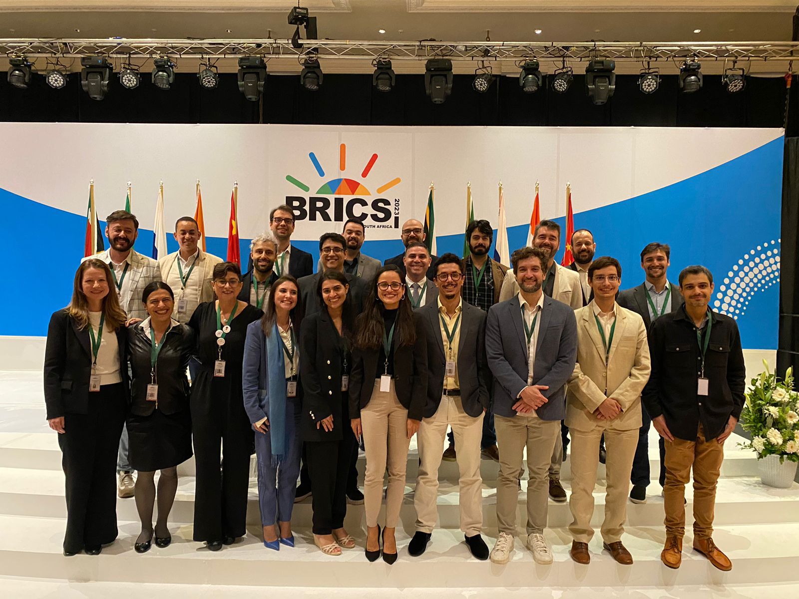
Brazilian delegation during the 8th BRICS Young Scientists Forum. Photo: personal archive.
“Eyes to the Future” contest
Gameiro is also working on a new project at the same time: evaluating the impact and follow-up of the projects presented in the “Eyes to the Future” contest, held by the Brazilian Association of Academic Ophthalmology Leagues (ABLAO), in partnership with Phelcom and with the support of the Brazilian Ophthalmology Council (CBO).
The competition seeks to teach students and encourage leagues to develop extension activities aimed at creating educational and/or assistance projects with the objective of reducing blindness due to posterior segment pathologies. To accomplish this.Phelcom provided 20 Eyer units with access to the EyerMaps feature and the EyerCloud cloud system.
The competition selected 10 projects and the top three will receive an Eyer. “It would be very interesting if we could permanently leave a portable fundus camera with each of the 10 leagues. We’re going to work on getting sponsorship to buy the seven remaining devices,” says the doctor.
“After the Academic Leagues were selected, we spoke to ABLAO’s president Luís Sabage and realized the need to evaluate and follow up on the projects submitted. Our future goal is to increase the number of ophthalmologic leagues, medical students and patients reached by the outreach projects developed,” he explains.
Furthermore, based on the results obtained in the “Eyes to the Future” contest and the points for improvement found, Gameiro intends to structure an online ophthalmology course for medical students and general practitioners, covering basic eye examination techniques, the use of artificial intelligence platforms and the interpretation of retinography images.
The doctor points out that traditional equipment for evaluating the fundus of the eye, such as fundoscopy performed with an indirect ophthalmoscope and condenser lens, is relatively difficult to handle, requires lengthy training, has a learning curve and depends on the examiner for evaluation. Besides, most of the time it doesn’t allow photographic recording of the retina for later discussion and review.
Alternatively, there is the conventional fundus camera. However, it is expensive to purchase. “Capturing images of the retina is extremely important for more accurate assessment and for monitoring the disease and treatment. It also plays a fundamental role in the training of new professionals through the presentation and discussion of findings in a group, allowing students and doctors to compare their exams and review the results,” he says.
The Eyer portable fundus camera is an extremely advantageous option in several aspects:
It makes it easy to capture high-quality images of the retina without much prior training;
Lightweight and small (it fits in the palm of your hand);
It does not require specialized labor;
Relatively more affordable than a traditional fundus camera;
It is non-mydriatic, shortening the examination time and avoiding possible adverse effects (visual discomfort, photophobia, keratitis and increased intraocular pressure);
Through telemedicine, it sends the images to the cloud, enabling remote diagnosis.
For Gameiro, the Eyer can have a significant impact on medical education. “The device can be used by medical students as a practical learning opportunity, demonstrating clinical cases and monitoring the progression of eye diseases over time, as well as stimulating interactive discussions between students and teachers, encouraging research projects and cases and facilitating access to and recording of a wide variety of cases,” he points out.
The equipment also has on-board artificial intelligence, which can be a reliable and cost-effective option for screening retinal and optic nerve pathologies using algorithms built on extensive databases.
“These algorithm models are able to predict the risk of alteration and thus notify the examiner of the need for follow-up with a more qualified specialist. In this way, the use of AI, together with deep learning and telemedicine, can represent an effective long-term solution for screening and monitoring patients in primary health care,” he concludes.
About the Eyer
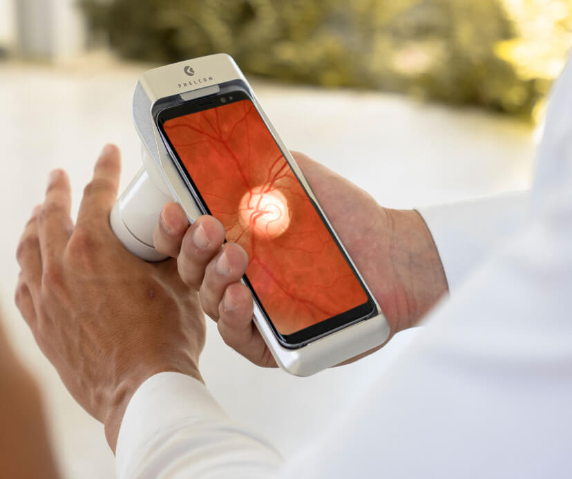
Eyer Portable Fundus Camera
The Eyer is a portable fundus camera that works in conjunction with a smartphone and performs high-quality retinal examinations in a few minutes without the need for pupil dilation.
The technology supports the diagnosis of more than 50 diseases, including glaucoma, cataracts, diabetic retinopathy, AMD, retinoblastoma, hypertensive retinopathy and ocular toxoplasmosis. Currently, more than 10 million tests have been carried out in Brazil, the United States, Chile and Colombia.
The technology’s portability and affordability democratize access to retinal examinations. It costs approximately six times less than a conventional tabletop fundus camera, which still needs to be integrated with a computer.
About Phelcom
Phelcom Technologies is a Brazilian medtech company based in São Carlos, in the interior area of São Paulo. The company’s story began in 2016, when three young researchers – a physicist, an electronics engineer and a computer engineer (physics, electronics, computing) – created a portable fundus camera integrated with a smartphone.
The idea for the first prototype was realized by Diego Lencione’s interest in visual health, as his brother has had a condition that has severely compromised his retina and vision since childhood.
In 2019, Phelcom launched its first product on the Brazilian market: the Eyer portable fundus camera. Today, the technology has reached more than two million people across Brazil and worldwide.
In four years, the company has participated in more than 100 social actions and was recently named one of the 10 most innovative companies in Brazil by Forbes.
Undoubtedly, one of the obstacles in pediatric ophthalmology is being able to carry out examinations on little ones. After all, it can be difficult to keep a child still for examinations.
This difficulty is routine for ophthalmologist, Dr. Patrícia de Freitas Dotto, MD, PhD at the Dr. Jeser Amarante Faria Children’s Hospital, at the São José Municipal Hospital (HMSJ) and in her office, all in Joinville (SC), and at the Lavinsky clinic in Porto Alegre (RS).
“The main challenge is the non-mydriatic fundoscopic evaluation of children between ten months and three years old without sedation or restraint. That’s why it’s essential to create a playful and calm environment that brings peace of mind to the whole family. In this context, photographic documentation, or even fundoscopic imaging, in real time allows parents to be involved in the care, which improves the doctor-patient relationship and, consequently, has a positive impact on the child’s follow-up and/or treatment.”
Dotto says that she uses the Eyer portable fundus camera to speed up examinations on pediatric patients. The equipment, which is very suitable for examining infants and children due to its portability and high image quality, works in conjunction with a smartphone and performs retinal examinations in a few minutes. These photographs are then available on the EyerCloud online platform, making it easier to study and monitor the progression of cases.
“The field of vision is excellent. Under medicated mydriasis, it is also an excellent complementary propaedeutic tool for assessing the mid-periphery of the retina, particularly small hyperpigmented lesions of the choroid (nevus) and retina (melanocytomas), using it as an infrared scan,” she explains.
In her office, with help of the Eyer Slit Lamp Adapter, the ophthalmologist attaches the device to her slit lamp allowing exams to be performed more easily. “Although I really enjoy using it this way, it’s magical to use it in ‘mobile’ form in ICUs, operating rooms, home care and within the office itself, such as evaluations of children on their mother’s lap or in the waiting room,” she points out.
The doctor also uses the Eyer in the emergency room, in adult and child care, for documenting infectious diseases and Retinopathy of Prematurity (ROP) in neonatal ICUs severely debilitated adults in ICUs, as well as inter-consultations for neurology, neurosurgery, nephrology, cardiology, and in medical expertise for the National Civil Aviation Agency (ANAC) and the Santa Catarina Court of Justice (TJSC). “I literally carry Eyer in my bag. It makes my job a lot easier,” she says.
The ophthalmologist, Dr. Patrícia de Freitas Dotto, MD, PhDtreats infants and children at the Dr. Jeser Amarante Faria Children’s Hospital, the São José Municipal Hospital (HMSJ) and at her office, all in Joinville (SC).
Shaken Baby Syndrome
With daily use of the Eyer in various locations, Dotto has witnessed several remarkable cases, such as the care of a child suffering from Shaken Baby Syndrome. The syndrome occurs when the baby is shaken intensely, causing permanent brain damage or even death.
Here’s the doctors report on the case of shaken baby syndrome:
“The child had been followed up for a few months in hospital for convulsions and severe malnutrition. The suspicion of Shaken Baby was raised when neuroimaging alterations (hematoma) were observed during the complementary investigation of status epilepticus.
During the ophthalmological assessment, when I examined her visual acuity (grating), I found that the vision in one eye was outside the normal range for her age, despite the fact that she had no changes in ocular motility.
When I laid her on the gurney for the retinal mapping, she started crying desperately. It was a very difficult examination and I was struck by the fact that the child eventually let go, almost gave up, and then reacted again, without showing any change in her level of consciousness.
It was at one of these moments that I managed to photograph the fundus of the eye and identify retinal lesions, indicating serious damage to the central nervous system. In one particular case, the hemorrhage was very small, probably because it was being reabsorbed, and I could only be sure of the diagnosis because of the examination. It was a miracle for me and for her.
I was devastated that day, but very grateful to HaShem and Phelcom [the company that invented the Eyer]. The case was really investigated from a social point of view, the assaults were confirmed and the appropriate measures were taken.
We saved a life. And in my religion, Judaism, saving a life means saving the whole world.”
Next, see the images of this case taken by Dotto with Eyer:
More cases
Dotto performs retinography on all patients, as she considers it important the normal state of the retina. “We often look for illnesses and forget how important it is to make sure the fundus of the eye is normal, because problems happen throughout life and it’s important to make sure of the moment when the state of eye health is lost,” she says.
She highlights the case of a 22-year-old woman who was admitted to the ER with compromised visual acuity, severe hypertensive retinopathy and serous detachment of the macula. The team suspected Autoimmune Nephropathy (IgA). “Unbelievably, four or five days after pressure control (in the ICU), she progressed to 20/20 visual acuity and total resolution of the serous detachment.”
Another situation involved a patient in the ER with anterior uveitis and an apparent normal USG. After remission, when mapping the retina, the doctor noticed a peripheral blackened lesion. “Using the Eyer, I was able to document it. We redid the USG thinking it was a melanoma. And it was indeed a melanoma at the level of the ciliary body,” she says.
In children, the ophthalmologist recalls the care of two patients with slight vitreous hemorrhage: “After improving the transparency of the media, it was possible to record the presence of a slight vascular malformation at the level of the optic nerve,” she recalls.
Among the most commonly diagnosed diseases in babies are ROP, congenital infections, optic nerve hypoplasia and retinal and optic nerve colobomas. “I also use the Eyer a lot to quantify the size of the optic nerve and the excavation. This helps with the differential diagnosis of suspicious glaucomas, particularly in short-sighted children,” she concludes.
Below are some images taken by the ophthalmologist with the Eyer:
Patient with retinal displacement.
Patient with microembolization in the choroid due to covid.
Patient with arterial occlusion.
The Eyer
The Eyer is a portable fundus camera that works in conjunction with a smartphone and performs high-quality retinal examinations in a few minutes without the need for pupil dilation.
The technology supports the diagnosis of more than 50 diseases, including glaucoma, cataracts, diabetic retinopathy, AMD, ROP, retinoblastoma, hypertensive retinopathy and ocular toxoplasmosis. Currently, more than 10 million tests have been carried out in Brazil, the United States, Chile, Colombia and Japan.
It was recently approved in the United Arab Emirates and is in the regulatory process of being marketed in Mexico, Egypt and Saudi Arabia.
Portability, connectivity and integration with intelligent functions such as EyerMaps, together with the technology’s affordability, contribute to increasing access to retinal examinations.
Three out of ten patients that ophthalmologist Juliana Barbi consults in her clinic in Belo Horizonte (MG) come from Instagram. Some of them live in other Brazilian states or even abroad.
“These patients form bonds with me even before the first consultation, because of the content I produce in my profile. They come to the office only to get to know me in person, because they already decided to begin treatment with me. So, the power of identification of Instagram is very strong”, states Barbi.
Doctor Barbi has been investing in producing social media content since 2018. She makes all the content, (script, recording, editing and posting) by herself.
And no, you do not need to dance performances to lure the right public (but you can, if you want). Barbi talks about medical procedures, shows patients’ “before and after” and shares about her professional – and sometimes personal – routine with her more than 10 thousand followers.
Ophthalmologist Juliana Barbi’s Instagram. The doctor has been investing in the social network for 5 years.
Doctor Barbi produces content on a daily basis. Usually, “Stories” videos that, many times, have space in her feed as Reels. “I look for dividing one single subject in various short videos to draw the follower’s attention and not to get boring. For example, I can divide a subject as “eyelid surgery” into post-surgery care, stitches, scar, etc.”, she explains..
When this article was written (October 5, 2023), she posted a Reel in her feed, showing a patient and demonstrating how a superior and inferior blepharoplasty ends. She shared her part in an interview on her story, a personal reel about wrinkles while smiling, and the recording of the next episode of her videocast “ENTRE(vistas)”, that goes live every other week on her Instagram.
In the videocast, the ophthalmologist invites professionals of healthcare and even other areas related to her work. For example, a tennis teacher highlighted the importance of knowing the dominant eye to make contact with the ball during a game. And a makeup artist taught tricks to improve the appearance of the eyes. “The idea is to diversify the subjects commonly addressed on my social profile with different topics related to vision in some way”, she explains.
TikTok and website
Recently, Barbi also signed in to TikTok, a social network to share short videos. A very successful platform, mainly among young people. “They may be my future patients”, she reflects.
In addition to social media, the doctor has a website where she presents her specialties, services and courses (including tips on content creation).
Barbi considers it fundamental to invest in communication technologies to reach good results with medical marketing. A professional profile on Instagram may be the first step. The second one is a website and a TikTok account. “Using websites to target an older audience, instagram to target middle aged patients and even tiktok to reach the youngest audience”, she says. However, she also emphasizes: “People who do not do it, end up falling behind”.
You do not need to be so extroverted as the ophthalmologist to go “viral” in social networks, as some fellow professionals believe, you only need to be yourself. “For the profile to be good, it is essential to be authentic. I do not show anything that I am not. I talk before the camera exactly as I work with my patient in the consultation office”, she states.
New rules for medical marketing
Recently, the Federal Council of Medicine (CFM) updated the rules for medical marketing. The new text, more aligned to the current needs of communication, including the medical class, affirms that the doctor may share their work on social networks, advertise equipment available in their workplace and, for educational purposes, to use images of patients or photo databases.
The proposal, according to the CFM is to assure the doctor has a right to show people the range of his/her services, respecting the market rules, but preserving medicine as a support activity. The new resolution authorizes advertising consultation prices and making of marketing campaigns.
“Before, I showed only a small part of the procedure and treatment results. Now I can share the ‘before and after’, of course, respecting the patients who authorized sharing their images. It is important for the patient to know better each step of a blepharoplasty, for example”, she explains.
Patient Journey
Medical marketing is one of the steps of the patient journey. In order to consolidate all investments being made to bring a patient to the office, it is fundamental to offer a great experience.
For example, Barbi searched to improve patient care by acquiring an Eyer handheld fundus camera. The equipment works coupled with a smartphone and carries out high-quality retina exams in a few minutes. Seconds after capturing the image, the device-embedded artificial intelligence, EyerMaps, identifies possible retinal alterations.
Barbi has used the Eyer handheld fundus camera since 2019.
“I explain what the optical nerve and macula are and, if it is the case, any alteration that may suggest pathologies, such as glaucoma, diabetic retinopathy, among other diagnoses primarily reachable through fundoscopy. They think it is great, get impressed with this technology and feel welcome”, she tells.
Eyer is non-mydriatic, offers the patient more comfort and speeds up the clinic service flow. “Nowadays, Eyer is my fundus examination. I do not even use a slit lamp anymore. I carry out the retinography and promptly store the patient’s image in EyerCloud for future follow-up”, she says. EyerCloud is a cloud system synchronized with the Eyer camera to manage patients’ data and exams.
Barbi examines all her patients using Eyer, except for the ones who already have a report. Using Eyer, there have been over one thousand exams since 2019.. “I already identified choroidal nevus from a routine exam in a preoperative of a preventive consultation. I sent it to a retinal specialist for evaluation. He found an extreme-peripheral retinal tear that was properly treated by laser before the surgery. Eyer was able to detect it and I felt safe to send the result to the retinal specialist before proceeding the surgery”, she recalls.
In terms of technology cost-effectiveness, Barbi believes it is worthwhile. “For doctors who serve patients with medical insurance, retinography costs may be sent to the insurance”, she ends.
About Eyer
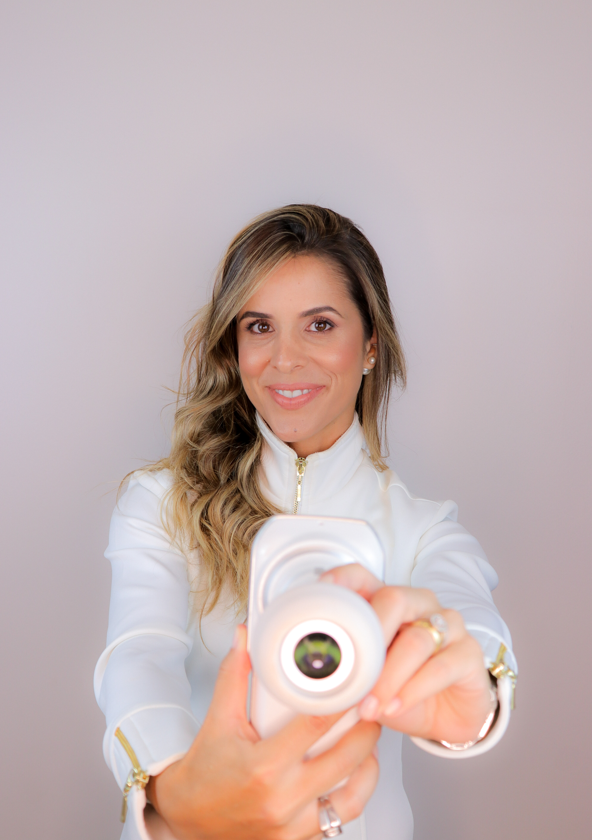
Barbi holding Eyer handheld fundus camera.
Portability, connectivity and integration with intelligent functions, as EyerMaps, along with a more accessible value, allow Eyer to contribute to increase the access to retina exams.
The equipment supports the diagnosis of more than 50 diseases, such as glaucoma, cataract, diabetic retinopathy, DMRI, retinoblastoma, hypertensive retinopathy and ocular toxoplasmosis. Currently, more than 10 million exams have been already carried out in Brazil, The United States, Chile, Colombia and Japan.
Eyer has recently been approved in the Arab Emirates and is going through the regulatory process for marketing in Mexico, Egypt and Saudi Arabia.
About Phelcom
Phelcom Technologies is a Brazilian medtech company headquartered in São Carlos, inland São Paulo. The company’s history began in 2016, when three young researchers – a physicist, an electric engineer and a computing engineer (PHysics, ELectronics, COMputing) – created a handheld fundus camera integrated with an integrated smartphone.
The project of the first prototype was born from the interest of company partner Diego Lencione on visual health, since his brother has a condition that severely compromises his retina and vision and has since childhood.
In 2019, Phelcom launched its first product in the Brazilian market: Eyer handheld fundus camera. Now, the technology has already reached more than two million people all over Brazil and the countries where it’s available.
In four years, the company has already taken part in more than 100 social actions and has recently been elected one of the 10 most innovative companies in Brazil by Forbes.
By 2030, over half a billion people are expected to be diagnosed with diabetes. Currently, Brazil ranks as the sixth country worldwide with the highest population of diabetics.
Currently, there is no national diabetic retinopathy screening strategy by the Unified Health System (SUS). Thus, social initiatives for diagnosing the disease in communities with inadequate healthcare infrastructure are crucial.
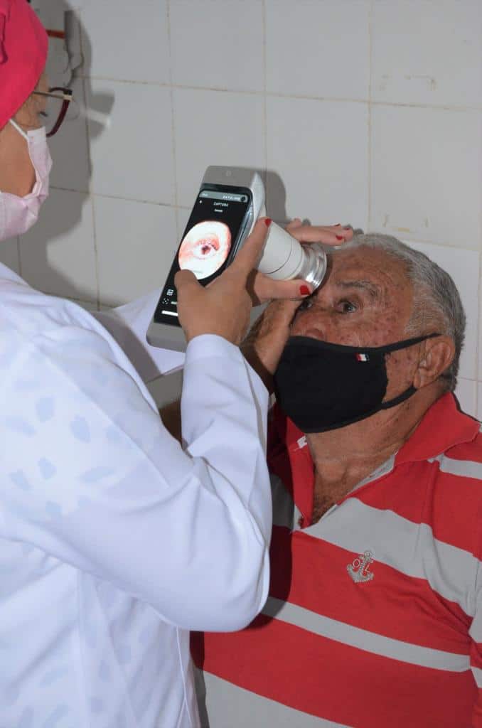
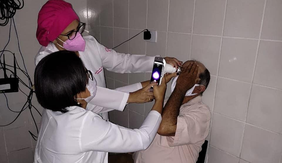
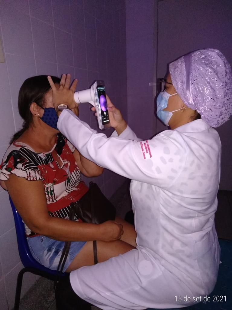
For instance, we have the “Mutirão do Diabetes” in Itabuna, Bahia, and “Iluminar” in the countryside of Sergipe, supported by the NGO Retina Global. This American institution focuses on developing sustainable solutions for managing retinal diseases in underserved areas around the world.
From September 2021 to March 2022, “Iluminar” used the portable retinal camera, Eyer for diabetic retinopathy screening in Itabi, Graccho Cardoso, Canindé de São Francisco, and Poço Redondo.
The device is connected to a smartphone and performs retinal exams within minutes, producing high-quality images, and uploads the images to the EyerCloud online platform, facilitating remote assessments.
The results of the pilot project were presented at ARVO 2023, one of the most renowned international ophthalmology conferences held in the United States in April.
The Study
One of the leaders of the “Iluminar” project, ophthalmologist Fernando Malerbi, explains that the aim of the retrospective observational clinical study was to evaluate the grading capacity of retina images obtained using a low-cost, non-mydriatic portable retinal camera — in this case, the Eyer — along with the use of artificial intelligence and telemedicine for diabetic retinopathy screening.
In total, 968 individuals with diabetes were evaluated:
65.9% were female;
Average age of 60.3 ± 14.2 years;
Duration of diabetes: 8.0 ± 7.2 years;
64.2% had systemic hypertension;
17.7% were using insulin;
28.5% had previously undergone fundus exams;
20.6% were illiterate;
50.6% had only completed primary education;
3.4% had health insurance.
A trained technician captured images without pupil dilation and then assessed image quality. Diabetic retinopathy requiring referral, defined as severe non-proliferative or proliferative retinopathy or the presence of diabetic maculopathy, was automatically detected by an embedded artificial intelligence system (EyerMaps). The AI was provided for clinical validation.
In a matter of seconds, EyerMaps indicated the possibility of retinopathy with a high sensitivity rate. Subsequently, all exams were evaluated by ophthalmologists.
Patients with inadequate images underwent pupil dilation and then a new assessment. Those with non-gradable images even after mydriasis, along with cases of referable diabetic retinopathy, were referred for ophthalmological evaluation.
Corneal opacities that hindered retinopathy classification were the exclusion criteria. The primary outcome measure was image gradability.
The Results
Grading was possible for 858 individuals (88.6%), with 85 of these (9.9%) showing referable diabetic retinopathy. Non-grading was associated with older age and longer diabetes duration.
Among patients with gradable images, 81% did not require pupil dilation. The need for mydriasis was associated with older age, longer diabetes duration, higher hypertension rates, and more severe retinopathy.
The strategy of utilizing a low-cost portable camera with embedded AI system and mydriasis when necessary achieved suitable images in 90% of cases within a resource-limited real-world environment. Malerbi emphasizes, “Avoiding unnecessary pupil dilation contributes to higher adherence to diabetic retinopathy screening programs.”
Enhanced Portability Facilitated Screening
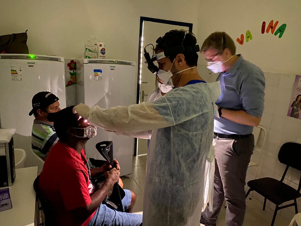
This novel screening was conducted in primary care clinics located near patients’ homes to encourage participation.
Malerbi highlights that the portability of Eyer was a facilitator, along with connectivity. “We had remote experts evaluating images, sometimes in real time,” he explains. All of the exams taken were uploaded to EyerCloud.
Lastly, Malerbi emphasizes the availability of EyerMaps for use in social initiatives. “Partner ophthalmologists were instantly notified whenever EyerMaps identified a high likelihood of retinopathy. Thus, we could prioritize patients for confirmatory exams and, if necessary, treatment,” he shares.
The AI accurately detects any suspicions of retinal abnormalities. Within seconds of capturing the fundus image, if an abnormality is detected, the system generates a new image with an attention map (heatmap) highlighting potential retinal anomalies.
Synchronized with EyerCloud, it categorizes images and exams captured based on the likelihood of abnormalities using color markers:
Green: Image or exam with low likelihood of an abnormality (up to 30%);
Yellow: Image or exam with moderate likelihood of an abnormality (31 to 70%);
Red: Image or exam with high likelihood of an abnormality (71 to 100%).
All patients diagnosed with diabetic retinopathy were referred for free laser treatment.