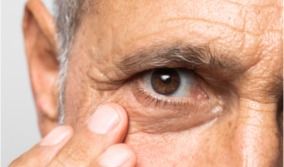
The choroidal nevus is a dark spot that occurs at the eye fundus and is only detectable through routine examinations such as retinal mapping. Usually, treatment only includes an yearly follow-up.
There are also skin nevi, which dermatologists follow-up with dermatoscopy to check for possible changes in their characteristics, such as enlargement. The same follow-up occurs with the spots at the eye fundus, for example.
If they grow, they can evolve into very advanced stages, such as choroidal melanoma, a very rare disease that affects less than 1% of patients diagnosed with the condition. This number is equivalent to five people in a million.
Melanoma (a type of cancer) is asymptomatic at the initial stage. An estimated 85% of cases arise in the uveal tract – iris, ciliary body and choroid. When not identified early, it can metastasize to the liver.
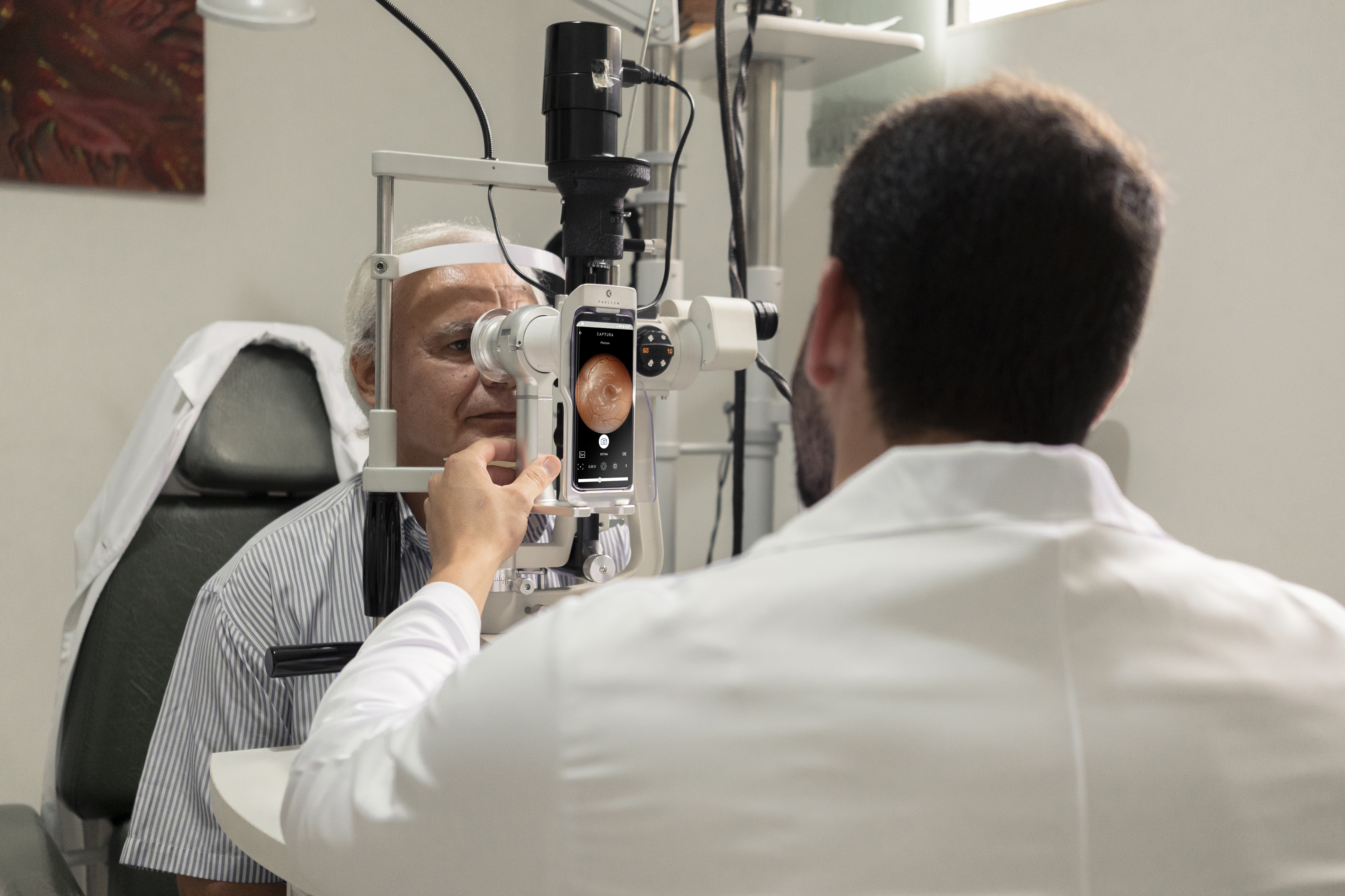
Retinography applies for checking the coroidal nevus size. Learn how this examination can help early diagnosis and disease follow-up.
Choroidal nevus – diagnosis
The ophthalmologist can only identify a coroidal nevus in a routine examination, because the disorder is not visible to the naked eye and does not usually present early symptoms.
Retinal mapping is one of them. By observing a nevus, the doctor can carry out further examinations to finish the diagnosis, such as optical coherence tomography (OCT) and retinography.
If the nevus grows, the first diagnosis may be undetermined melanocytic lesion, to which the doctor will define a protocol of examinations and follow-up. Observed new nevus increases confirm the choroidal melanoma diagnosis.
Choroidal melanoma – treatment
In fact, choroidal melanoma has no cure, but is treatable and requires lifetime monitoring. Thus, the therapy will be established according to the patient’s vision condition and age, as well as the status, location and extent of the cancer. As with all diseases, an early diagnosis determines a better prognosis.
Brachytherapy is most recommended for small and medium sizes. This surgery has a control rate of approximately 95% and maintains the eye and, in some situations, the ability to see.
An older method was removing the ocular globe. Enucleation may still occur for large tumors with symptoms as intense pain, poor vision and disorganization of internal structures. In some cases, radiotherapy, laser therapy and transpupillary thermotherapy are also indicated.
Reviewed by Paulo Schor, ophthalmologist, associate professor and director of innovation of the Federal University of São Paulo (Unifesp) and collaborator of the Faculty of Medicine of the Albert Einstein Hospital.
Follow Phelcom blog and stay on top of the main news on office management!
A patient with retinitis pigmentosa was able to recover part of the vision after undergoing optogenetic therapy and light stimulation. For the first time, this technique has achieved partial recovery of visual function, according to clinical trial researchers. The study was published in Nature Medicine journal .
Before treatment, the man could only perceive the presence of light. Now, he already finds, counts, and touches objects. Learn about the clinical trial and how optogenetic therapy works.
The research
Researchers from the Sorbonne University, Quinze-Vingts Hospital and the company GenSight Biologics, from France, in partnership with the University of Pittsburgh, from the United States, and the Institute of Molecular and Clinical Ophthalmology of Basel, from Switzerland, conducted clinical trials with optogenetic therapy in patients with retinitis pigmentosa.
That degenerative genetic disease damages the retinal photoreceptor cells, causing progressive loss of vision. The condition evolves until the patient is completely blind. The problem affects one in 3.5 thousand people, according to Orphanet database. Currently, an estimated two million cases exist worldwide.
A 58-year-old man, blind for 20 years, received an injection into one of his eyes with a gene called ChrimsonR, that encodes opsin proteins and identifies amber light. These proteins are responsible for sending visual information to the brain.
He then underwent treatment with flashes of light directly on the retina. In optogenetic therapy, light pulses control gene expression and activation of neurons. Currently, they are widely used in laboratories to unravel neural circuits and can be a potential treatment for pain, blindness and brain problems.
Results
After producing enough opsins, which occurred five months after beginning therapy, the patient was given camera glasses that project amber-colored images onto the retina.
In the first exercise, the man needed to notice, find and touch a large book and a small box of staples. In total, he managed to touch the book in 92% of evaluations, and the boxes in 36% of the time.
In the second test, the patient achieved 63% efficiency when counting glasses on a table. In the third exercise, he wore an electrode helmet that monitored if he recognized a glass on the table or not. In this one, he was successful 78% of the time.
Seven months after receiving the injection, the patient already showed signs of improvement in vision.
After two years of treatment, the man still uses the glasses to see better. In fact, images will never be the same as natural ones, but for those who have been blind for 20 years, it is life-changing.
It is the first time that optogenetic therapy has managed to partly reverse vision loss by a genetic degenerative eye disease. The trial will now advance to phase 3 to confirm the effectiveness of this therapeutic approach. However, it will still take some time to offer the technique, as it needs more studies, more patients and more longevity.
Reviewed by Paulo Schor, ophthalmologist, free professor and director of innovation of the Federal University of São Paulo (Unifesp) and collaborator of the Faculty of Medicine of the Albert Einstein Hospital.
Follow Phelcom’s blog and stay on top of the main news about coronavirus and the eyes.
Currently, it is estimated that 1.5 million people worldwide lose their vision each year due to corneal injuries and diseases. Thus, problems in this membrane are the third largest global cause of visual impairment, behind only cataracts and glaucoma.
But this scenario may change. Recently, Israeli doctors performed the world’s first successful artificial corneal transplant. The patient, a 78-year-old man, was able to regain his sight after 10 years of blindness.
In fact, synthetic corneal implants already exist, but because they required more complex surgeries, they were used only as a last resort, such as rejection in corneal transplants. The new technology, on the other hand, can be implanted in a relatively simple way, with minimal cutting and suturing.
In the following, understand how the artificial cornea transplant occurred, how it acts inside the organism, the next steps, and how the result can change the reality of millions of people waiting for a cornea transplant to see again.
The artificial cornea transplant
Israeli startup CorNeat Vision has developed the KPro artificial implant to replace a patient’s deformed cornea. The procedure was performed at the Rabin Medical Center hospital in Israel.
The device has a non-degradable synthetic nano-tissue, which is placed under a membrane that lines the surface of the eyelid and the sclera (white part of the eyeball). When implanted, it unifies with the living tissue and encourages cell proliferation within the eye.
The synthetic cornea is only indicated in cases where the tissue is deformed, opaque, or scarred.
In an interview with the Israel Hayom website, the doctor and creator of the technology, Gilad Litvin, said that the surgery was relatively simple and lasted less than an hour.
Elderly Man Recognizes Relatives After Surgery
Patient Jamal Furani was able to regain his sight already the day after the artificial cornea transplant. The elderly man says that light was the first thing he could see. Afterwards, he was able to recognize relatives and even read texts.
“The result exceeded all our expectations,” says physician Irit Bahar, head of the Department of Ophthalmology at Rabin Medical Center.
Next Steps
The expectation is that the procedure will become viable and end the waiting line for donors around the world. “This technology was key to turning the tide against global blindness. It is very exciting to be at the forefront of this project that will undoubtedly impact millions of lives,” Bahar believes.
“We hope this will enable millions of blind patients around the world, in areas where there is no corneal practice or organ donation culture, to regain their sight,” says Gilad Litvin, medical director of CorNeat Vision. However, the company has not yet announced a market launch date.
Now the clinical trials continue. A further 10 approved Israeli patients are awaiting artificial corneal transplantation at Rabin Medical Center hospital. In addition, countries like Canada, France, the United States, and the Netherlands also have patients eligible for clinical trials.
Eyes are an open door.Contaminated tears as a possible source of contagion.Conjuntctivitis.Retina alterations.Glaucoma.
From symptoms as conjunctivitis to possible sequelae as retinopathy and glaucoma, diverse studies and reports point to the relation of the new coronavirus and the eyes.
As it is a new disease, we sill do not know for sure how itreaches this organ.Therefore, nowadays, researches serve mainly as an alert.
Now, a new study identified small nodules in the eyes of patients with severe covid-19. The work of the French Radiology Society (SFR) was published in the scientific journal Radiology.
Know more about the research, results and how data is important to better understand and encourage investigation of the possible sequelae of the new coronavirus to the eyes.
Study
From March to May 2020, researchers submitted 129 severe covid-19 patients, 43 women and 86 men, to brain MRI exams.They aimed to detect possible anomalies in the eyes.
Results
Nine patients presented one or more small nodules in the posterior macular area:
From them, eight had anomalies in both eyes;
Eight were hospitalized in the ICU;
Seven remained prone for an extended time;
Six were obese;
Two had diabetes;
Two suffered from hypertension.
The team could not identify the reason for the nodules to appear.However, they raised some hypotheses:
Virus-caused inflamation;
Poor blood circulation in the ocular veins in patients intubated in the ICU, such as damage and blockage;
Small eye hemorrhages;
Disruption of nerve fibers.
According to scientists, it is the first time there is documentation of this kind of sequelae through MRI.
Next steps
The volunteers remain under monitoring to observe possible changes of the ocular nodules or vision impairment. In addition, new severe patients from the second and third waves are under evaluation.
In fact, the study needs to deepen, in order to prove the formation of nodules in the eyes due to the new coronavirus. However, the study highlights it is important to capture images of the eyes in severe covid-19 cases and follow the patient post-treatment evolution.
Brain MRI, fundoscopy and optical coherence tomography figure as useful exams.
“Our study advocates triage for all patients hospitalized in ICU with severe covid-19. We believe they must receive specific eye protection treatments”, said ina note the main author of the study, Augustin Lecles, associated professor at the University of Paris, and neuroradiologist* of the Department of Neuroradiology of the Adolphe de Rothschild Foundation Hospital in Paris.
Conclusion
In fact, there is still a long way to prove the correlation between coronavirus and the eyes.However, studies are essential to encourage more attention to the eyes of infected patients, by means of exams and monitoring after the cure.
More than that, this research highlights that patients with pre-existing diseases, such as diabetes and hipertension, have a higher risk factor. Therefore, they also must undergo eye investigations.
Learn first-hand information on the main researches about the coronavirus and the eyes. Follow Phelcom’s blog.
Undoubtedly, diagnosing Parkinson’s disease is a challenge. Some procedures to identify signs of the disease have high costs, such as computed tomography and magnetic resonance imaging. In addition, millions of people worldwide do not have access to these technologies. According to the World Health Organization (WHO), currently, about 6.3 million suffer from the problem.
Since the first symptoms only manifest with the progression of the disease, early detection is another challenge. A new research from the United States has developed a cheaper way to diagnose the disorder: artificial intelligence applied to the retinal exam. The results were presented at the last annual meeting of the Radiological Society of North America (RSNA).
Learn about the study, how it was done and how the results achieved help to democratize access to healthcare.
Research
Researchers at the University of Florida (USA) used the machine learning principleto create an artificial intelligence tool that learns to detect signs of Parkinson’s disease in retinal examinations. They trained the “support vector machine” with eye fundus images of patients with the disease and control participants without the disorder.
As the problem deteriorates nerve cells and, consequently, thins the retinal walls, a simple fundus examination can already diagnose it. The disease also damages the retinal microvasculature.
Results
The results show that Parkinson’s disease can be diagnosed from changes in the retina. Currently, several studies prove that damage to the brain can be observed through the eyes .
“The most important finding was that a brain disease was diagnosed with a simple eye image. The diagnosis can be made in less than a minute and the equipment costs much lower than a computed tomography or magnetic resonance imaging ”, says Maximillian Diaz, the researcher in charge.
Conclusion
Undoubtedly, by applying machine learning techniques to the artificial intelligence system used in the retinal scan, scientists can diagnose Parkinson’s disease faster, more assertively, cheaply and accessibly.
Early detection, even before the first symptoms, allows the treatment to start as soon as possible and provide the patient more life-quality.
The results of the research can also help in a better understanding of the disease in the search for a cure and in ways to slow the evolution.
In addition, the researchers say the new artificial intelligence tool can be used to identify other diseases that damage the brain, such as Alzheimer’s disease and multiple sclerosis.
Follow the main researches related to the eyes on Phelcom’s blog.
Cataract is one of the most frequent side effects on patients who undergo macular hole surgery. For some time now, specialists already indicate that multiple surgical procedure to correct the diseases.
Now, the simultaneous treatment of macular hole and cataract has received scientific approval after a study by the University of São Paulo (USP), campus of Ribeirão Preto (SP), which proved the effectiveness of the technique.
Details on research results and the benefits of this approval – for doctors, health institutions, the Brazilian Unified Health System, and the patient – follow below.
The research
Researchers divided 65 patients with macular hole in two groups. The first one, of 33 people, underwent the multiple surgery with both techniques. The other group, with 32 patients, underwent a single procedure of cataract correction after the first surgery.
This is the first prospective study in the world to evaluate the multiple surgeries in a single intervention.
Results
Twenty-seven patients who underwent only the macula correction surgery had cataract afterwards. Because of that, they needed a new surgery in less than one year. In general, they presented significant worsening in vision.
Patients from the first group improved their visual acuity, similar to those who underwent sequential surgery.
The results demonstrate effectiveness of joining both techniques in a single procedure.
Benefits
In most cases, cataract occurs only a few months after the macula surgery. Therefore, the multiple procedure, joining both techniques, is advantageous to doctors, hospitals, the Brazilian Unified Health System and the patient.
Offering a safer treatment, cost and time reduction and less suffering of the patient are some remarkable advantages.
Follow the main healthcare news on Phelcom’s blog.