The search for a cure for diabetes is one of the main aspects of the health research sector. According to the World Health Organization (WHO), 422 million people suffer from the disease worldwide. It is also responsible for 1.6 million annual deaths.
Scientists at the University of Alberta, Canada, recently announced that they had discovered a possible solution for the problem. With a new stem cell process the team was able to transform the patient’s own blood into insulin-producing cells. The clinical research was carried out on mice. Learn more about the study, the results and next steps.
Research
Researchers at the University of Alberta in Canada are working with scientists from all around the world on a new stem cell technique.
The study basically turns the blood of patients with type 1 diabetes into insulin-producing cells .
To do this the team adapted the technique of the 2012 Nobel Prize winner in Physiology or Medicine, Shinya Yamanaka, for the treatment of diabetes. Yamanaka discovered how to reprogram skin cells, through the use of hormones and other growth factors, in induced pluripotent stem cells (iPSCs). These could be induced to become any type of cell.
The Canadian researchers took blood samples from patients and treated the cells with a cocktail of hormones and other growth factors to “go back in time” and induce them to become insulin-producing cells. Then, they transplanted into mice with type 1 diabetes.
Results
The new technique achieved a cure for diabetes in mice. If successful blood cell transplantation would overcome the challenges of islet transplantation, such as the need for anti-rejection drugs and lifelong insulin applications. The project leader is physician James Shapiro, who made history 20 years ago with the “Edmonton Protocol“. The procedure places new insulin-producing cells in the patient by transplanting islets harvested from pancreases of organ donors.
However, they have significant side effects, such as an increased risk of cancer and potentially fatal infections.
But this new research believes that using the patient’s own cells should eliminate the problem.
Now the hope is that it will also generate results in humans. However, there is still a long road ahead. Undoubtedly, further evaluations in animals are still needed to demonstrate that the procedure can be possible, safe and effective. Only then should the research test people.
Obstacles
Financing is one of the current biggest obstacles today to continue the work. But in an interview withCTV News Edmonton, Shapiro said there are volunteers working on raising $ 22 million by 2022.
Conclusion
Undoubtedly, if it works, the results of the research can be considered the next major advance in the treatment of diabetes. Even if the technique is successful there is a lot of work to be done in the areas of robotic engineering, artificial intelligence and stem cell science to improve the process and make it less laborious. Mass production of iPSC and personalized medications may occur for the patient to cure diabetes in the future.
Stay in the main news about eye healthcare. Follow the Phelcom’s blog.
Have you ever thought that having an eyer handheld fundus camera in your clinic can be a real advantage?
After all, having quicker access to your patients’ fundus images can assist in early diagnosis, treatment initiation and monitoring various diseases: glaucoma, diabetic retinopathy and age-related macular degeneration (AMD).
However, the equipment is expensive. There are models up to BRL 100,000 on the market. In addition to the other devices your business needs, the investment can burden your budget. Even more for professionals beginning their careers. Given that scenario, a new alternative is available: Eyer handheld fundus camera. Connected to a smartphone, the technology captures high-quality images of the fundus and sends them to an online platform. It allows reporting the exam and filing the patients’ history, among other features.
Another advantage is the price: the equipment is up to 4 times cheaper than the conventional handheld fundus camera.
The technology is national and developed by the startup Phelcom Technologies headquartered in the countryside of Sao Paulo.
Next, learn more about Phelcom Eyer handheld fundus camera, its advantages, how the Eyer Cloud platform works and how the device has been used in research projects and social actions.
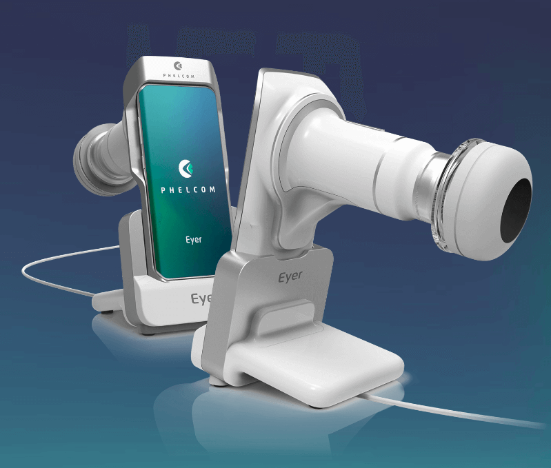
Phelcom Eyer handheld fundus camera
Released in 2019, Phelcom Eyer is a handheld fundus camera that works coupled to a smartphone with a high-resolution camera. The device captures high-quality images of the fundus, in a few minutes and without the need for pupil dilation. After that, the data is automatically sent to the Eyer Cloud online platform. It allows reporting the exam and storing patients’ history with total data security.
Check out all the features of the Eyer:
High quality
Phelcom’s patented technology allows high quality exams to be performed on a portable device integrated with the smartphone.
Telemedicine
Carried out exams are automatically synchronized with the internet and made available in the cloud, enabling remote diagnosis.
Embedded artificial intelligence
Eyer has intelligent functions to assist medical diagnosis and capture retinal exams.
Connectivity
The device is naturally connected because it is integrated with the smartphone. It eases sharing and accessing exam data in the cloud through the Eyer Cloud system.
Non-mydriatic
Eyers carries out retinal examinations at any location without the need to use eye drops for pupil dilation. It is more comfortable to the patient and provides a faster exam.
Autofocus
With the Autofocus function it is possible to compensate refractive errors in the range of -20D to + 20D. This allows retinal examinations with a high level of details.
Accessible
Eyer allows the democratization of access to retinal examination technology through innovative and more accessible business models.
Easy operation
Any minimally trained healthcare professional can use the equipment to perform high-quality retinal exams in less than 1 minute. It guarantees faster and more accurate diagnoses.
Panoramic
The Eyer generates panoramic exams with a more than 100 degree visual field. The device has internal fixation points that assist capturing and generating panoramics.
Portability
The portable device allows performing exams anywhere with remotely-issued diagnosis. This feature helps in the democratization of health, since 85% of Brazilian cities do not have access to specialists and devices that diagnose eye diseases.
Low cost
Portability and small size allow Eyer to have a much lower cost compared to traditional fundus cameras. Even with cutting-edge technologies applied to the device production.
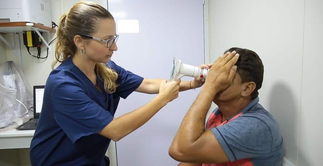
Recently Phelcom also released a slit lamp holder to allow Eyer attachment to a tabletop. This way, it provides the experience of a tabletop retinograph by easing image capture without movement.
In addition, it helps to maintain social distance in care, since the professional does not need to touch the patient’s forehead as occurs in the traditional exam.
Eyer handheld fundus camera – advantages
Eyer offers state-of-the-art technology which makes the device one of the most modern portable fundus cameras for the prevention and diagnosis of vision-related diseases.
Learn about the advantages of the equipment below:
High-quality eye exam through a smartphone;
Accurate and quick diagnosis;
Lower cost compared to traditional retinographs;
Exams are possible in several locations due to portability;
Democratization of retinal exams, especially in places with low infrastructure of quality services in the area, such as doctors, health professionals, equipment, medicines etc;
Faster service through computerized systems integrated to an online platform with access via computers, smartphones and tablets;
Tests are easy to carry out in clinics and health centers;
Diagnosis made by specialists and reference professionals located anywhere in the world;
Reduction of attendance time and operational costs;
Decrease in the displacement of patients to hospitals and large urban centers;
Improvement in the quality of the reports issued;
Increase in prevention and early diagnosis of diseases such as diabetic retinopathy, glaucoma, cataract, macular degeneration, retinoblastoma, retinal detachment, premature retinopathy and blindness, inter alia.
Eyer handheld fundus camera – Eyer Cloud
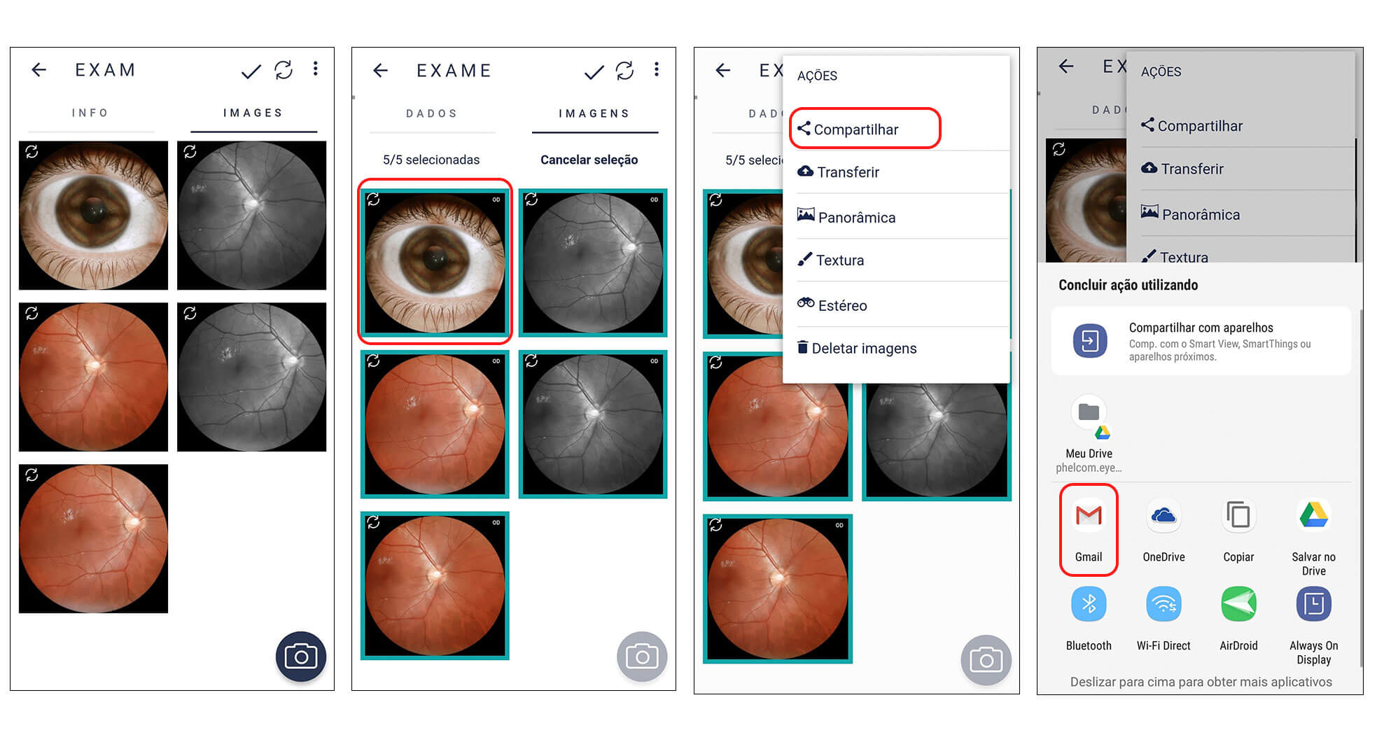
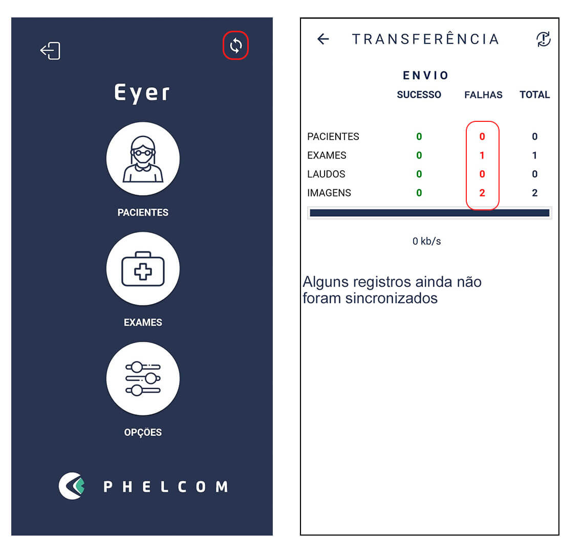
Eyer Cloudis an online platform integrated with the Phelcom Eyer handheld fundus camera that allows you to store and manage patient exams. All data captured by the equipment is automatically synchronized with the system, allowing them to upload to the cloud with complete security.
Any health professional – without necessarily being a specialist – can carry out the exam in the field after a brief training. With the information available on the web, a doctor from anywhere in the world can diagnose.
In addition to ensuring data backup on a secure server, the user has all data organized in a friendly, functional and intuitive interface. Moreover, the platform can be accessed on the device itself or in a smartphone, tablet and computer.
If there is no internet access at the time of the exam the images are saved on the device and are sent to the cloud as soon as a connection is available.
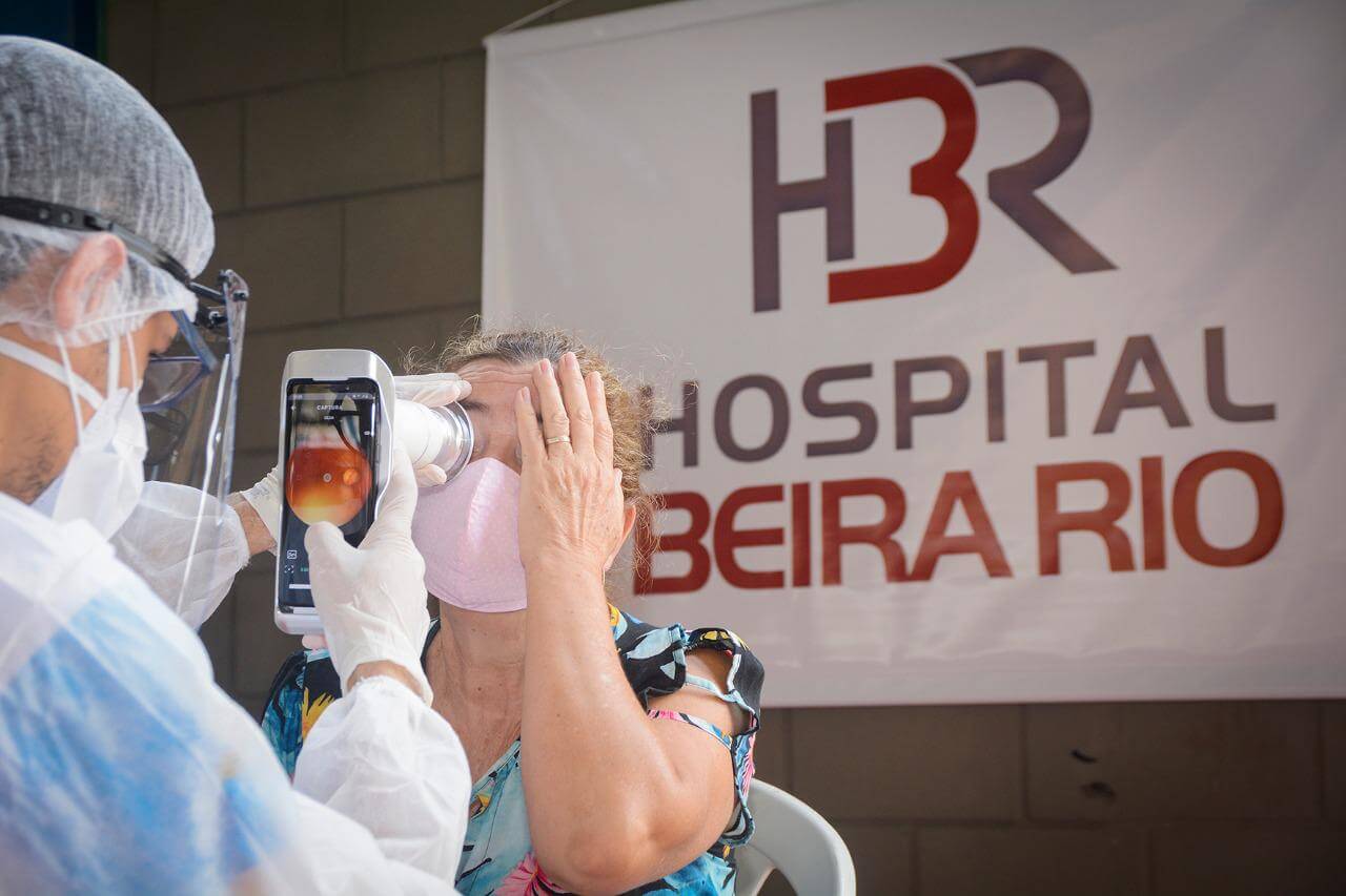
Eyer handheld fundus camera – social actions
Currently there are several social actions that use the device to bring more health to the whole country.
This is the case of the project “Unidos pelo Diabetes em Ação”, in Itabuna (BA). The event gave rise to the collective task force “Unidos pelo Diabetes”that needed to be cancelled due to the pandemic. On the whole, 400 patients participated in the first phase. 100 of these had more severe diabetic retinopathy or macular edema.
The project used advanced technologies to ensure screening without agglomerations. Eyer was the main screening tool. “Phelcom was fundamental to the success of the project as it made the devices available, provided prior training, helped in the strategy of assembling reports using its Eyer Cloud program and made its team of engineers available for support during the action”, says Andrade. “In addition, in an innovative way in Brazil, it created an artificial intelligence algorithm to help sorting patients who would probably be serious and those without changes, easing the logistics through distance-reporting”, adds Andrade.
Expeditions
The expedition carried out by Barco Hospital São Francisco, in the region of the municipality of Terra Branca (PA),at the end of last year, also featured the Eyer. Ophthalmologist Mariana Lafetá, one of the volunteers on this trip, says that the equipment helped in the diagnosis of diseases such as cataracts, glaucoma and diabetic retinopathy, inter alia.
“It is easy to carry out the exams, take the photos, find them in the files and store them later. We can also send or print the images, which I think is very interesting, in addition to the easy access from anywhere with the internet connection ”, she analyzes. Ophthalmologist Fernando Korn Malerbi also used the Eyer on an expedition to three indigenous communities in the state of Mato Grossoearlier this year. The doctor evaluated 193 Indians. Diabetic retinopathy and cataracts figure among the main diseases found.
“The experience with the equipment was very good, mainly due to portability and ease of use”, he evaluates. He recalls that he has been involved in other projects with the Eyer handheld fundus camera for the diagnosis of diabetic retinopathy. “I believe that the Eyer is very relevant for this type of action, representing an important alternative for tracking populations that live in remote areas”, he concludes.
Do you want to know more about how technology helps with eye exams? Follow the Phelcom’s blog.
Can you imagine regenerating the damaged optic nerve with gene therapy and being able to recover patients with glaucoma?
A research from the University of Cambridge (United Kingdom) has achieved promising results in this regard. Through gene therapy, scientists have rehabilitated damaged eye nerve fibers. The study was published recently in Nature Communications Journal.
Learn about details on the research: methodology, results and how important it may be, in the future, to treat glaucoma.
However, results are still initial and require further tests to determine an effective therapy for humans.
Research
Researchers from the University of Cambridge (UK) used a gene therapy technique to stimulate, in vitro, the increase and activity of the protruding protein in the eye and optic nerve.
First, the scientists cultivated the cells in laboratory. Then, laser-injured the nerve cells (axons). Finally, they introduced the gene and followed the changes with a microscope.
The study was carried out in mice.
Results
The scientists noticed that the more the protruding protein in these cells increased in number, the greater was the retinal neuron regeneration. More than improving the protein growth, it decreased the chance of nerve cell death.
The investigation showed an almost complete “protection” of the nerve cells growing in a cell culture of one of the guinea pigs.
This protein can help healing or, at least, reducing optic nerve damage. Thus, it may assist protection against glaucoma in the future.
It is worth mentioning that further tests are needed to determine an effective treatment for humans.
Conclusion
In fact, the gene therapy study results are promising. Especially because it seemed impossible to find a way to regenerate injured optic nerve in the past.
This research gives hope, however it still requires many tests on mice, primates and humans. Only then will an effective treatment be defined to reach the population within a few years. Nevertheless, the study is still a major breakthrough against glaucoma and other diseases that affect neurons such as Alzheimer.
Stay in the main news about eye care. Follow Phelcom’s blog.
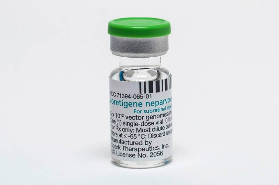
Voretigene neparvopeque (trade name: Luxturna®) is a special kind of advanced therapy medicine. The enzyme it produces improves functioning of retina cells, decreasing the disease progress.
Learn more about this gene therapy, how it acts in the organism and its results to patients.
Gene therapy
In brief, gene therapy is based on inserting exogenous genetic material in a person’s cells for therapeutic purposes. Therefore, the transference aims to recover the function of a gene, provide a new gene function or intensify the functioning of active genes.
Objectively, gene therapy fixes defective genes by inserting healthy genes in patients with different diseases.
Hereditary retinal dystrophy
Hereditary Retinal Dystrophy (HRD) is a rare disease provoked by the mutation of human gene RPE65. Leber congenital amaurosis, retinitis pigmentosa, Usher syndrome and Stargardt disease are among the disorders it causes.
This gene mutation provokes gradual rupture of the cells that form the retina, located at the back of the eye. Therefore, it causes gradual loss of vision, culminating in blindness.
Some of the symptoms are difficulty or lack of adaptation to dark, loss of peripheral vision, abnormal color vision, photophobia and visual acuity loss, evolving to complete blindness.
How the medicine works
Elaborated through genetic engineering, the product consists of a virus with an inserted copy of the human gene RPE65, responsible for producing an enzyme necessary for the retina proper functioning.
Such enzyme allows a better performance of retina cells, thus decreasing the disease progress. During the study phase, it demonstrated gain of functional vision and autonomy in adults and children.
It is worth mentioning the virus used in manufacturing that medicine does not cause diseases in humans.
It is possible to treat children from 12 months old on and adults with vision loss due to hereditary retinal dystrophy. Novartis Biociências S.A. produces the medicine.
So far, there was no therapeutic alternative for the disturbance.
Another important remark: it is a hospital use product and must be used under expert medical supervision, via sub-retinal injection.
The physician also must take precautions, such as having the patient undergo mandatory tests able to prove his/her vision loss was due to mutations of RPE65 gene, over which the Luxturna’s active substance works.
Moreover, the doctor must use other lab and clinical evaluation criteria. For example, a certain amount of still functional retina cells is necessary for a successful treatment. If not, results may be low.
Conclusion
Gene therapy, as a whole, has enormous potential to treat diseases provoked by a single imperfect gene. That happens because, by fixing part of the defective genetic code, it solves or decreases the problem. It thus increases patient’s life quality.
Undoubtedly, this treatment brings up positive expectations towards rare diseases caused by genetic disorders.
Follow the main news on eye health on Phelcom blog.