Everyone already knows that cigarettes are bad for health. This includes the eyes. For example, smoke is a risk factor for various diseases such as dry eye syndrome, glaucoma, cataract, and age-related macular degeneration (AMD).
Learn more about the work that correlated cigarette and eyes, its results and next steps.
Cigarettes and eyes – the research
Scientists at Gifu Pharmaceutical University, in Japan, created cultures of cells from the epithelium of the human cornea and exposed part of them to an extract of cigarette smoke and PTA aerosol, which contained most of the ingredients inhaled by smokers.
After 24 hours, the number of dead cells in cultures exposed to smoke and aerosols was higher than compared to those that did not interact with the substances. Upon contact with cigarette components, the ferritin inside eye cells decomposes, releasing the stored iron.
Results
Exposure to the components of cigarette smoke accumulates iron, which kills cornea epithelium cells. The same reaction was observed with the aerosol produced by heated tobacco products (PTA). Although different from electronic cigarettes, these also require an electronic device for use and do not always come with nicotine.
Generally, cigarette smoke does not cause permanent problems. However, continuous exposure can cause corneal injury such as leukoma and even lead to blindness.
Despite the important results of the study on cigarettes and eyes, more research is still needed, especially in humans, to confirm the findings.
Information from Einstein Agency.
Reviewed by Paulo Schor, ophthalmologist, associate professor and director of innovation of the Federal University of São Paulo (Unifesp) and collaborator of the Faculty of Medicine of the Albert Einstein Hospital.
Follow Phelcom blog and stay on top of the top of main research about cigarettes and eyes.
Retinal neurons were first identified more than 100 years ago. But now scientists at the University of Utah in the United States have discovered a new type of retinal cell.
Published in the Proceedings of National Academy of Sciences, United States, the research found a hitherto unknown type of interneuron in the retina of mammals.
Learn more about the study, how it was conducted, and the next steps.
The research
In the central nervous system, a complex circuit of neurons communicates with each other to transmit sensory and motor information. “Interneurons” act as intermediaries in the communication chain.
Researchers at John A. Moran Eye Center of the University of Utah (USA) have identified a new type of interneuron in the mammalian retina. The new cell does not fit into the current five classes of retinal neurons: photoreceptors, horizontal cells, bipolar cells, amacrine cells and endogenous cells. This is due to differences presented in their morphology, physiology and genetic properties.
Thus, the scientists responsible for the discovery propose that this new type of cell should belong to a new class of retinal neurons.
The team dubbed the discovery a “bell cell” because of the hand-bell-like shape. The discovery unites two cell types, cones and rods, and does extra processing in the cells. Thus, they relay visual signals of both types of photoreceptor rods and photoreceptor cones in the retina, but their precise purpose is subject of ongoing research.
Experiments have shown that bell cells remain activated for an unusually long time – up to 30 seconds – in response to a 10-millisecond flashlight stimulus.
“In the brain, persistent firing cells are believed to be involved in memory and learning. Once campana cells have a similar behavior, we theorize that they could play a role in requesting a temporal ‘memory’ of a recent stimulation,” said research leader Ning Tian.
Undoubtedly, it is a great discovery that contributes directly to the search for a better understanding of the central nervous system, since it detects all classes of neurons and their connections.
Source: Medical Xpress
Reviewed by Paulo Schor, ophthalmologist, associate professor and director of innovation of the Federal University of São Paulo (Unifesp) and collaborator of the Faculty of Medicine of the Albert Einstein Hospital.
Follow Phelcom’s blog and stay on top of the main health news.
The World Health Organization (WHO) has long warned about the danger of diabetes. The disease grows year by year around the world, and in the past 40 years the number of cases has quadrupled.
According to the 10th edition of the Diabetes Atlas, published by the International Diabetes Federation (IDF) and recently released, 537 million people aged 20 to 79 have diabetes worldwide. A growth of 16% compared to 2019.
This equals one diabetic out of ten people. The scenario gets even worse: almost half (44.7%) do not even imagine that they face the disease. The projection for next years are 643 million diabetics in 2030 and 784 million in 2045.
Lifestyle, lack of access to healthcare in developing countries and the present pandemics – which increased sedentary lifestyles, poor diets and postponed medical care – are the main factors for these numbers. Learn more about preliminary data IDF presented.
Diabetes around the world
According to the survey, done every two years, 10.5% of the world’s population have diabetes. Thus, the number proportionally exceeds the global population growth. Until then, one person out of 11 was diabetic.
More than that, 44.7% don’t even know they are sick. That can greatly aggravate diabetes, since people only seek help when symptoms arise. Undoubtedly, figures are worrying. Lack of control can lead to other serious problems, such as blindness, kidney damage, changes in the heart and even death.
The disease is also one of the most deadly: 6.7 million people have lost their lives due to diabetes. That is, every five seconds a person dies from this condition. This account does not yet include deaths resulting from complications of other diseases that have been aggravated due to diabetes, such as covid-19.
The presence of the disease is much higher in developing countries: 81% of sick adults live in these localities. That is, 4 out of 5 diabetics. According to atlas, 32 million diabetics are from Latin America and Central America.
So many sick people cost a lot of money: USD 966 billion were spent worldwide with healthcare, a rise of 316% in the last 15 years, according to IDF.
Diabetes in Brazil
In the 2019 edition, there were 16.8 million diabetics in Brazil. In the world ranking, we are in 5th place behind only China, India, the United States and Pakistan.
Among Brazilian Capitals, Rio de Janeiro stands out with the highest rate of diagnoses in the country: 11.2%. Then there is Maceió (11%) and Porto Alegre (10%). The disease also affects more women (9%) than men (7.3%) here.
Data are from the research “Vigilância de Fatores de Risco e Proteção para Doenças Crônicas por Inquérito Telefônico” (Vigitel), 2020, a telephone survey research of the Ministry of Health.
With regards to the costs invested in the treatment of Brazilian diabetics aged 20 to 79, Atlas estimates USD 52.3 billion per year. This equals to USD 3 thousand per adult.
More data on Brazil should be published in the full edition, with a release preview for December 6.
Causes
Experts claim that diabetes is increasingly out of control and that there is a lack of information and awareness for prevention. Current lifestyle is one of the main factors for the increasingly high number of the disease cases. Sedentary lifestyle and poor diets, rich in fats and carbohydrates, have brought problems such as hypercholesterolemia, hypertension, overweight, obesity and pre-diabetes, among others.
In low-and middle-income countries, which have the largest number of diabetics, there is a lack of access to healthcare, delaying diagnoses, treatments and even guidance for a balanced diet.
Diabetes and the eye
One of the possible complications of diabetes is in the eyes. According to a study by the Brazilian Society of Ophthalmology (SBO), 40% of people who suffer from diabetes present ophthalmic changes.
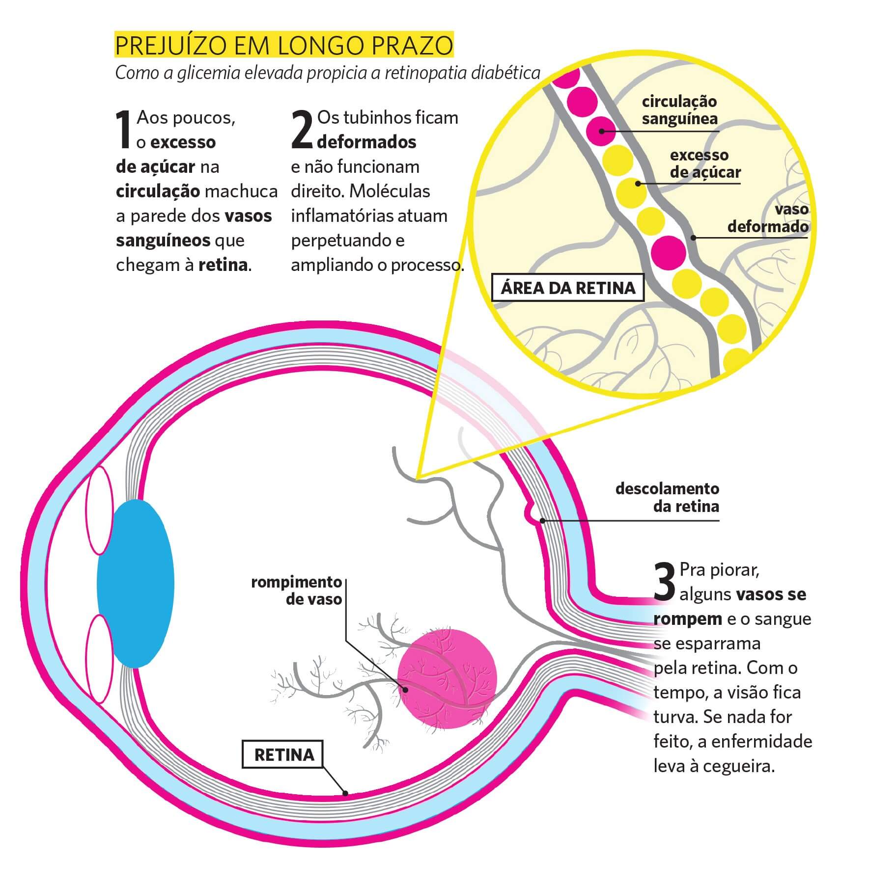
Diabetic retinopathy figures among them. Currently, about 40% of the 4 million Brazilians diagnosed with retinopathy have diabetes. In fact, the duration of diabetes and the uncontrolled blood glucose have a direct relationship with retinopathy.
Source: Infographic on diabetic retinopathy – Saúde magazine
This disease subdivides into two types: pre-proliferative, which does not require laser treatment, and proliferative, in which neovases occur and also demand therapy.
For the diagnosis of the correct type, one must evaluate the fundus with examinations.
The disease can also cause glaucoma. The Ministry of Health estimates that people with diabetes are 40% more likely to develop the problem.
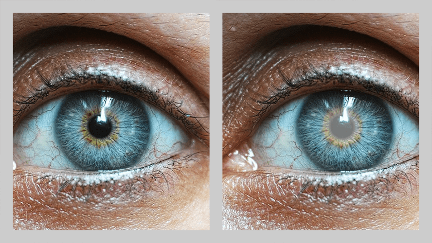
In addition, diabetics are 60% more likely to have cataract earlier. In this situation, the problem appears earlier and progresses faster than senile cataracts. Therefore, it is the main cause of vision loss in diabetic patients. However, it is reversible through surgery.
Comparison between a healthy eye and one with cataracts.
It is surely essential for diabetics to be attentive to any changes in their vision. Therefore, even if everything is fine, it is important to undergo periodic examinations with an ophthalmologist.
Prevention and early diagnosis are the keys to avoid serious damage, such as severe visual impairment and even blindness, in case of complications from diabetes.
Reviewed by Paulo Schor, ophthalmologist, associate professor and director of innovation of the Federal University of São Paulo (Unifesp) and collaborator of the Faculty of Medicine of the Albert Einstein Hospital.
Follow Phelcom blog and stay on top of the main health news.
For some time now, researchers have been investigating whether eye diseases can be a risk factor to manifest other health problems. For example, scientists at Sun Yat-sen University in China found that one in four people with eye disorders also develops depression.
Now, a new study links age-related macular degeneration (AMD), cataract and eye disorders brought on by diabetes to the increased risk of dementia. The researchwas recently published in the British Journal of Ophthalmology.
Understand the work, its results and how eye diseases can be directly linked to dementia cases.
Research and results
Researchers at the Guangdong Academy of Medical Sciences, in China, evaluated data from 12,364 adults with AMD, cataract or glaucoma, aged 55 to 73, from 2006 to 2010. Participants had follow-up until 2021.
The risk of cognitive decline was 26% higher in patients with AMD, 11% higher in those with cataracts and 61% more in diabetics compared to those who did not have eye diseases at the beginning of the study. Glaucoma was not considered one of the risk factors.
The scientists also looked at eye and systemic diseases alongside the incidence of dementia. Patients with cataract and a systemic condition were 1.19 to 2.29 times more likely to develop dementia compared to those without these problems. Regarding eye diseases related to diabetes and systemic diseases, such as diabetic retinopathy, this figure was 1.50 to 3.24 higher.
From the beginning, the study detected that diabetes, heart diseases, strokes and depression associate with increased risk of dementia. Hypertension joined the list until the research ended. All mediated the association of cataract and incipient dementia, as well as other eye diseases related to diabetes incipent dementia.
Despite the expressive results, it is worth noting that the research is observational. However, the scientists state in the article that “AMD, cataract and diabetes-related eye diseases associate with increased risk of dementia. Individuals with ophthalmic and systemic diseases have an even greater risk.”
Reviewed by Paulo Schor, ophthalmologist, associate professor and director of innovation of the Federal University of São Paulo (Unifesp) and collaborator of the Faculty of Medicine of the Albert Einstein Hospital.
Follow Phelcom blog and stay on top of the main news on health research!
With the pandemic and the need for social isolation, children around the world began to stay longer at home. A daily life with longer time in front of screens and less outdoor activities in leisure moments. The “new normal”, experienced a year and a half ago, is already paying its price: the growth of myopia among children aged 6 to 8 years in China.
Learn about the research, the data raised and what are the recommendations of experts to slow the growth of the disease among young people.
The research
Researchers examined 123 thousand children and teenagers, from 6 to 13 years old, in schools in Feicheng, China, in 2020. The evaluation technique used was photoscreening, a camera that analyzes the eyes and does not require pupil dilation.
Children aged 6 years were the ones who suffered the most from the increase in myopia: from 5.7%, between 2015 and 2019, to 21.5% in 2020. The 7-year-olds, in the same period, showed a raise from 16.2% to 26.2% and the 8-year-olds, from 27.7% to 37.2%. The increased degree of myopia also drew attention: 1.5-2 degrees.
In the 9 – 13-year group, there was no significant evolution.
Another interesting result is that girls developed myopia earlier than boys.
With this, researchers concluded that the social isolation caused by the new coronavirus pandemic can influence myopia in children. Especially among those aged six to eight years because they are at a stage more sensitive to the problem.
Does increased myopia also occur here as overseas?
In Brazil, there are no concrete data on the increase in myopia in children and teenagers during the pandemic. But in a recent survey conducted by the Brazilian Council of Ophthalmology (CBO), 72% of ophthalmologists reported an increase in diagnoses in patients from zero to 19 years old.
295 ophthalmologists, specialized in various areas, such as retina, cornea, glaucoma and pediatrics, were heard between April and June this year. 76% of doctors believe excessive exposure to electronic devices may directly relate to the explosion of cases. 22% believe only smartphones and tablets are to blame. On the other hand, a small percentage of experts believe there is no link between the two events.
Less screen, more outdoor action
The increase of myopia in young people during the pandemic is influenced by genetic and environmental factors. The disease can be hereditary, passing from parents to sons. In relation to external conditions, the problem lies in the longer period focused on objects very close to the eyes, not resting nr being exposed to sunlight.
Looking at things too closely, less than 33 centimeters from the eyes, without intervals, causes the release of chemical agents inside the eye, which can grow the eyeball larger and increase myopia.
Another aggravating factor is the progression to severe myopia, which seriously affects vision. Currently, this untreated disease is the leading cause of mild and moderate visual impairment and the second largest cause of blindness in the world, according to the World Health Organization (WHO). Besides this, it can cause more serious problems in the future, such as glaucoma, cataracts and retinal detachment.
The Brazilian Society of Pediatrics (SBP) has recommendations on the use of screens by children and teenagers. One of the main is not exposing children up to two years to screens, even if passively. From two to five years, only one hour a day. From six to ten years, two hours a day. Other guidelines are to avoid screens during meals and two hours before bedtime. And, when using, take periodic breaks every 30 minutes or 1 hour in a row.
At the same time, it is critical to increase outdoor activities so that cases decrease. Sunlight releases neurotransmitters that reduce eye enlargement.
Myopia: the epidemic of the century
It has been a few years since the WHO warns of a worldwide myopia epidemic. The entity estimates that the disease currently affects 35% of the population and may reach more than half (52%) by 2050. Only in Brazil, the organization believes that there are 59 million short-sighted people.
Regular visits to the ophthalmologist
How to slow the increase in myopia among children and teenagers taking other actions than reducing close focus without intervals and having more outdoor activities? It is advisable for parents or legal guardians not to only take youngsters to the ophthalmologist after a visual issue. It is essential to keep a routine of visits to the specialist, mainly because at this age it is possible to prevent and early diagnose eye disorders.
Reviewed by Paulo Schor, ophthalmologist, free professor and director of innovation of the Federal University of São Paulo (Unifesp) and collaborator of the Faculty of Medicine of the Albert Einstein Hospital.
Follow Phelcom’s blog and stay on top of the main news about coronavirus and the eyes.
Ocular syphilis is a manifestation of syphilis that can arise when the disease is not treated properly. This stage occurs years after infection and has a challenging diagnosis. Despite directing lesions, we call the treponema palidum (etiological agent of the disease) “the great copycat”. The agent can simulate several different manifestations. At this stage, the problem can even cause blindness.
But, a new study pointed out that Optical Coherence Tomography (OCT), A common ophthalmological examination in SUS, can help in the early identification of ocular syphilis. The University of São Paulo (USP) carried out the research and published it recently in the journal Ocular Immunology and Inflammation.
Learn about the research, results and what should be the next steps for the use of OCT to diagnose the disease.
The research
Researchers from the Faculty of Medicine of Ribeirão Preto (FMRP), from USP, evaluated one of the eyes of 54 patients with ocular syphilis admitted to the FMRP clinical hospital (HCFMRP). After part of them received the treatment, scientists still analyzed 31 eyes.
Through Optical Coherence Tomography (OCT), researchers found retinal lesions that may aid in early diagnosis of the disease.
Results
The ophthalmological exam identified round spots, irregularities, elevations and detachment in the retinas studied. According to the authors of the work, it is the first time that OCT checks for frequent changes in the retina in a large series of cases of ocular syphilis. These modifications are imperceptible on clinical exams.
Undoubtedly, the findings of OCT have diagnostic value in ocular syphilis, but do not predict the prognosis. However, the examination – common both in the Brazilian Unified Health System (SUS) and private clinics – can help visualize signs of the disease even in early stages. After confirming the diagnosis with serology and referring to the indicated treatment, the patient has a good chance of not having permanent sequelae in vision.
Photo: Eduardo Paulino Eye Institute.
Reviewed by Paulo Schor, ophthalmologist, free professor and director of innovation of the Federal University of São Paulo (Unifesp) and collaborator of the Faculty of Medicine of the Albert Einstein Hospital.
Follow Phelcom’s blog and stay on top of the main news about coronavirus and the eyes.