Phelcom Technologies is proud to expand its mission of transforming eye care globally. The company’s presence in the United States is a key part of that journey, led by a passionate team based in Cambridge, Massachusetts.
Located in the heart of Kendall Square, Phelcom is housed at the Cambridge Innovation Center (CIC), a vibrant innovation hub that fosters collaboration, growth, and innovation.
“Being located in such a vibrant healthcare ecosystem like Cambridge is important because it helps us build relationships with world-class doctors and the best innovators in the world,” says Gabriel Finch, Sales and Business Development Supervisor for the US Market.
It’s a natural fit for Phelcom, whose mission is to make visual health more accessible through innovative tools like the Eyer2 — a portable, versatile fundus camera transforming the way photo documentation is performed.
Cambridge Innovation Center (CIC), Cambridge, Massachusetts.
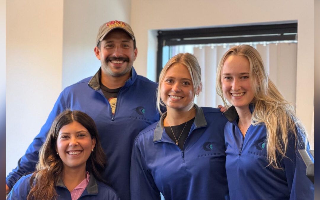
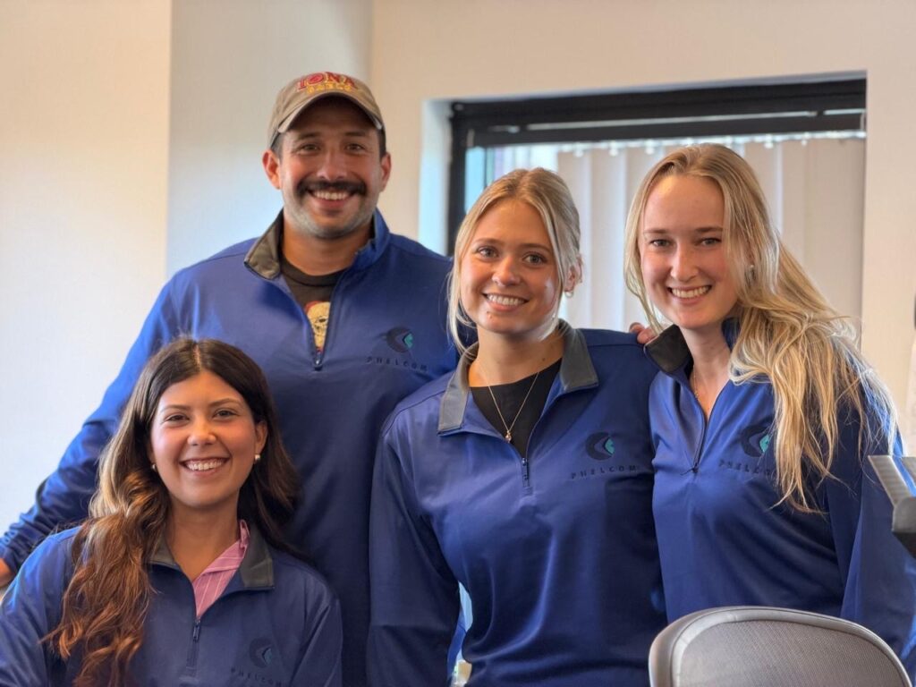
US Team
The US team is composed of four professionals focused on sales, business development, marketing and customer success.
Beyond the technology, the company culture is a key driver. “I love the team spirit, even across countries I always feel supported and connected,” shares Marcela Martins, whose main focus is prospecting.
For Finch, what stands out is the global aspect of Phelcom’s work. “Getting to work with clients all over the world, whether it be from South America to Japan, to the Middle East, to Europe, it’s a wide breadth of types of doctors that we get to talk to and all from different parts of the globe”.
In addition to supporting the growing US customer base, the team collaborates closely with colleagues in Brazil, reflecting Phelcom’s strong international spirit.
“My favorite thing about Phelcom is being able to work with our international team in Brazil as well as our team in the Cambridge office,” says Emily Homiller, the fourth sales professional that composes the US Team.
Phelcom US team members, from left to right: Marcela Martins, Gabriel Finch, Emily Jakacki and Emily Homiller.
Eyer2: An improved way to deliver eye care
At the heart of Phelcom’s innovation is the Eyer2 — a device designed to simplify workflows and expand access to high-quality eye care by combining portability, ease of use, and advanced imaging capabilities for both the posterior and anterior segments.
“With the Eyer2 camera, you bring the device to the patient, making workflow more efficient and patient-friendly. What many OD and MD practices love about it is that anyone in the clinic can easily capture high-quality images — and there’s no need to move patients from room to room,” explains Finch
The device’s panoramic imaging function is another key highlight. “It enables you to create a panoramic image with up to 120 degrees field of view — giving you wider retinal coverage with excellent detail,” adds Hulsander.
For Martins, two things stand out: “The image quality and how easy it is to use.” This accessibility is crucial for clinics of all sizes.
Portability is another key advantage, as Homiller points out: “My favorite thing about Eyer2 is its portability so it can reach underserved communities”.
By combining advanced technology with real-world usability, the Eyer2 is helping improve eye care on a global scale — making it easier for clinics to capture faster, more accurate, and more efficient eye images.
About Phelcom
Phelcom Technologies is an American-Brazilian company that employs Physics, Electronics, and Computing to make visual health simpler, more connected, and smart.
Founded in 2016 by three young researchers, the company developed a portable retinal camera with an integrated smartphone. Its first prototype was inspired by co-founder Diego Lencione’s personal experience, as his brother had struggled with a severe vision condition since childhood.
In 2019, Phelcom launched its first product, the Eyer portable retinal camera. Five years later, the company introduced the Eyer2, a photo documentation platform capable of capturing high-quality images of both the posterior and anterior segments.
To date, Phelcom’s technology has benefited over two million people across multiple countries, including the United States, Japan, Chile, Colombia, the United Arab Emirates and Brazil. It has also been used in over 100 social outreach initiatives.
Fundus imaging plays a crucial role in diagnosing and monitoring various eye diseases, including diabetic retinopathy. However, in many low- and middle-income countries, access to conventional retinal imaging equipment remains limited due to high costs and infrastructure requirements. Additionally, publicly available image datasets for doctors, researchers, and clinicians remain scarce, particularly for images captured with portable fundus cameras.
In this context, portable fundus cameras like the Eyer have emerged as a more accessible and cost-effective solution for eye screening and disease management. Mounted onto a smartphone, the Eyer captures high-quality retinal images in minutes—without requiring pupil dilation.
These devices can be used in a variety of settings beyond hospitals, such as community health screenings and remote telemedicine consultations. Additionally, they record detailed metadata typically unavailable in other datasets, including patient age, sex, diabetes duration, treatments, and comorbidities.
Recently, Scientific Data, a journal from the Nature portfolio, published the article “A portable retina fundus photos dataset for clinical, demographic, and diabetic retinopathy prediction,” introducing mBRSET: the first publicly available diabetic retinopathy dataset captured using handheld fundus cameras in real-world, high-burden environments. Among the authors of the study are ophthalmologist Fernando Korn Malerbi and Phelcom’s CEO, José Augusto Stuchi.
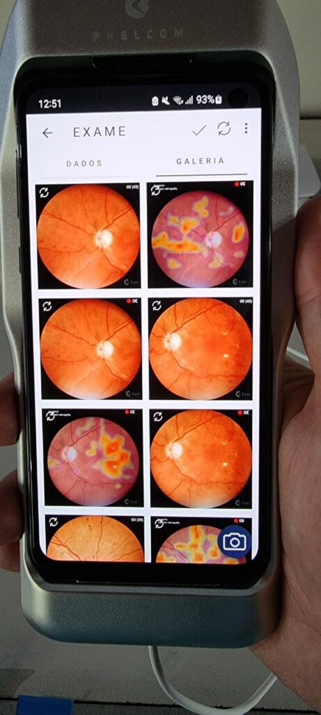
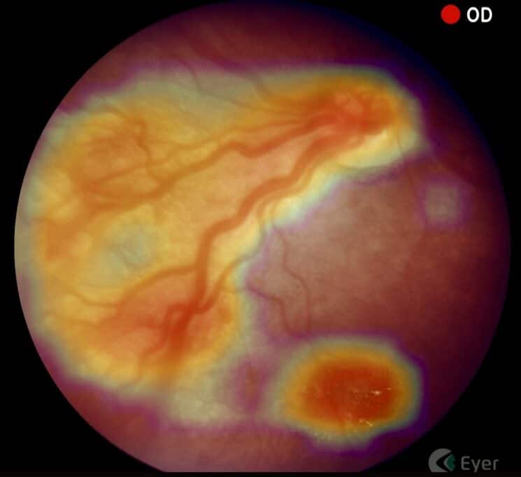
Fundus images captured with Eyer during the Itabuna Diabetes Campaign, held in 2022, highlighting the attention map generated by EyerMaps, indicating possible anomalies.
mBRSET: A Groundbreaking Dataset
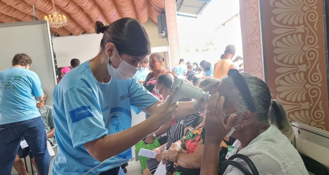
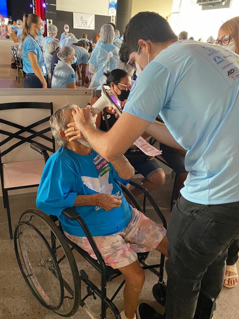
mBRSET consists of 5,164 from 1,291 patients of diverse backgrounds, all captured with the Eyer device during the 2022 Itabuna Diabetes Campaign in Bahia. Recognized as one of the world’s largest diabetes prevention and treatment initiatives, this campaign provides hundreds of patients with vital screenings—such as fundus examinations—and referrals for specialized treatment.
Exam performed with Eyer during the Itabuna Diabetes Campaign, held in 2022.
To validate the utility of mBRSET, state-of-the-art deep learning models were trained for benchmarking, demonstrating high accuracy in diagnosing diabetic retinopathy and macular edema, as well as predicting demographic data.
An analysis of 4,885 assessed images revealed that 3,759 images (76.79%) showed no signs of diabetic retinopathy (DR), 272 images (5.56%) indicated mild non-proliferative DR, 570 images (11.64%) exhibited moderate non-proliferative DR, 82 images (1.67%) showed severe non-proliferative DR, 427 images (8.69%) displayed signs of macular edema.
A Milestone for Ocular Health and Scientific Research
The significance of the mBRSET dataset can be outlined in 5 key aspects:
Representing Brazil’s Diverse Population
mBRSET helps reduce the underrepresentation of low- and middle-income country (LMIC) populations in ophthalmological datasets by including individuals from various ethnic and socioeconomic backgrounds in Brazil.
The First Public Dataset with Portable Camera Images
This is the first publicly available dataset featuring images captured with portable fundus cameras, reflecting the increasing adoption of this technology in resource-limited settings.
Data Collection in Real-World, High-Demand Environments
Images were captured in high-volume clinical settings, ensuring the dataset accurately represents real-world challenges in eye disease screening and management.
Inclusion of Detailed Demographic Data
mBRSET goes beyond retinal images, incorporating information such as gender, education level, and health insurance status. This enables researchers to evaluate AI algorithm performance across different subpopulations.
A Foundation for AI Development in Ophthalmology
This dataset serves as a critical resource for training and validating AI algorithms, fostering advancements in automated screening, diagnosis, and monitoring of diabetic retinopathy and other eye conditions.
Phelcom CEO José Augusto Stuchi emphasizes the dataset’s impact on the scientific and medical communities:
“The creation of this mBRSET marks a significant milestone in ocular health, particularly for regions with limited resources. By providing high-quality images captured with portable devices, we expand research opportunities and accelerate the development of AI-driven solutions that can revolutionize the diagnosis and treatment of eye diseases.”
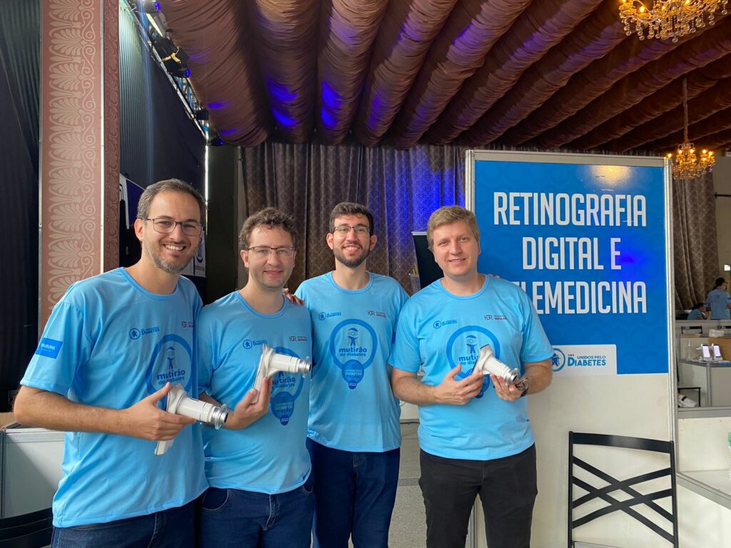
Diego Lencione, co-founder and CTO of Phelcom, Flavio Pascoal Vieira, co-founder and COO of Phelcom, Paulo Prado, coordinator of Mobile Software and AI at Phelcom, and José Augusto Stuchi, co-founder and CEO of Phelcom, during the Itabuna Diabetes Campaign, held in 2022.
Eyer
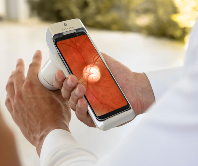
The Eyer is a portable fundus camera that attaches to a smartphone, enabling high-quality retina exams in just minutes—without the need for pupil dilation.
The technology supports the diagnosis of more than 50 diseases, including: glaucoma, cataracts, diabectic retinopathy, retinoblastoma, hypertensive retinopathy, retinopathy of prematurity, ocular toxoplasmosis.
Recently, Phelcom launched Eyer2, an enhanced version of the device featuring new built-in tools for expanded diagnostic capabilities. In addition to posterior eye imaging, Eyer2 enables the detection of anterior segment conditions such as: blepharitis and other eyelash abnormalities, meibomian gland dysfunction, styes, conjunctival and eyelid tumors, advanced cataracts, foreign bodies and burns, corneal injuries, keratitis caused by dry eye, contact lenses, infections and ulcers.
About Phelcom
Phelcom Technologies is a Brazilian medtech company based in São Carlos, São Paulo. Founded in 2016 by three young researches—a physicist, an electronics engineer, and a computer engineer—the company developed a portable retinal camera integrated with a smart phone.
The first prototype was inspired by co-founder Diego Lencione’s personal experience, as his brother struggled with a severe vision condition from childhood.
In 2019, Phelcom launched its first product, the Eyer portable retinal camera, in Brazil. Five years later, the company introduced the Eyer2, a platform capable of capturing high-quality images of both the posterior and anterior segments.
To date, Phelcom’s technology has benefited over two million people across Brazil and multiple countries, including the United States, Japan, Chile, Colombia and the United Arab Emirates. It has also been used in over 100 social outreach initiatives.
Doctor Iddi Ndyabawe, ophthalmologist and a passionate ROP specialist in Uganda, East Africa, was responsible for the very first ROP study in the country, published by BMC Ophthalmology in 2023. He’s currently involved in Adult Retina services as well, working in a screening project for diagnosing CMV Retinitis in HIV patients with meningitis admitted at Kiruddu National Referral Hospital and Mulago National Referral Hospital. He also offers Diabetic Retinopathy screening services to private patients at Kisubi Hospital.
Dr Iddi in the Aravind Eye Hospital Library, Coimbatore India
Dr. Iddi got to know about the Eyer Retinal Camera in February 2023, when Nurse Nancy Maria Douat Dietrich from Santa Catarina, Brazil came along with the high quality images device and joined the ROP screening activities at Kawempe National Referral Hospital.
“When Nancy saw my passion for ROP in Uganda, she communicated with Mr. Jose Augusto, CEO of Phelcom, who then sent me an Eyer Fundus camera by June 2023 as a support for my humble efforts in ROP “, recalls Dr. Iddi.
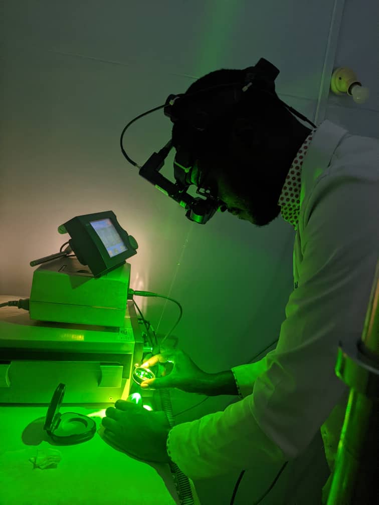
Dr Iddi training in laser therapy for ROP on an eye model
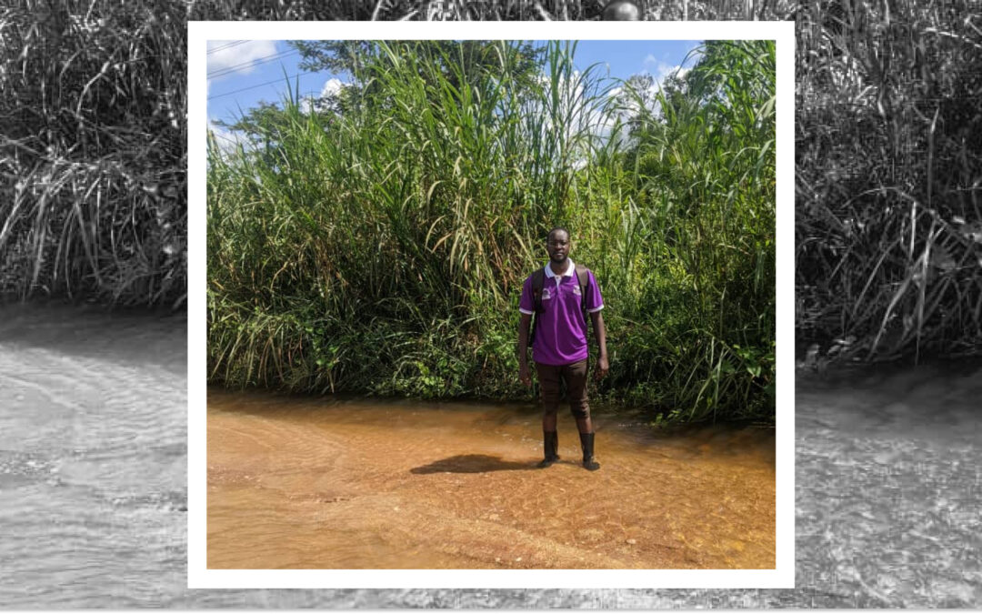
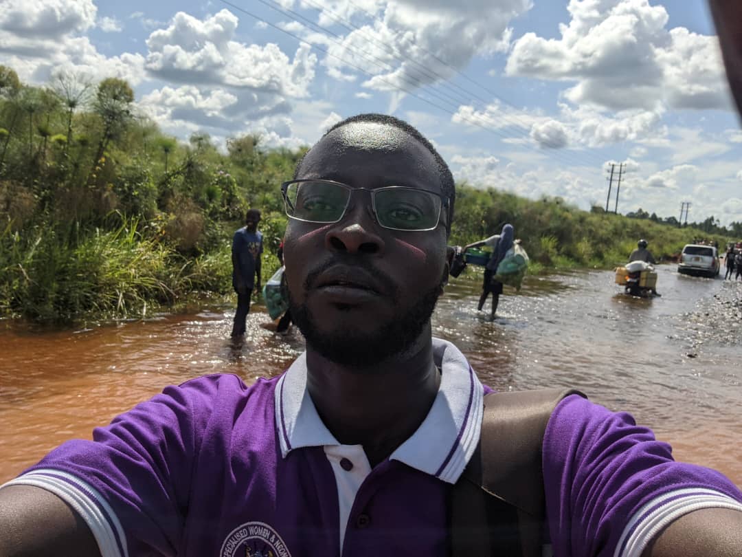
Dr. Iddi crossing the flooded roads to reach the village NICU to conduct ROP screening
According to Dr. Iddi, the main advantages of the Eyer Fundus Camera for his medical practice are:
Eyer takes clear resolution images for best diagnosis of posterior disease;
It is perfect at CDR assessment;
Eyer can easily pick out the hyperemic disc in optic neuritis that could easily be missed;
EyerMaps heat map is perfect in pointing where the clinician should pay more attention.
Dr. Iddi mentions as well that the portability and non-mydriatic use of Eyer Fundus Camera, which allows him to perform examinations in different scenarios. He recalls one particular Sunday when a relative came in complaining of reduced vision. He was able to discover a diagnosis of toxoplasmosis right there in his own home.
About the Eyer
Eyer Portable Fundus Camera
The Eyer is a portable fundus camera that works in conjunction with a smartphone and performs high-quality retinal examinations in a few minutes without the need for pupil dilation.
The technology supports the diagnosis of more than 50 diseases, including glaucoma, cataracts, diabetic retinopathy, AMD, retinoblastoma, hypertensive retinopathy and ocular toxoplasmosis. Currently, more than 10 million tests have been carried out in Brazil, the United States, Chile and Colombia.
The technology’s portability and affordability democratize access to retinal examinations. It costs approximately six times less than a conventional tabletop fundus camera, which still needs to be integrated with a computer.
About Phelcom
Phelcom Technologies is a Brazilian medtech company based in São Carlos, in the interior area of São Paulo. The company’s story began in 2016, when three young researchers – a physicist, an electronics engineer and a computer engineer (physics, electronics, computing) – created a portable fundus camera integrated with a smartphone.
The idea for the first prototype was realized by Diego Lencione’s interest in visual health, as his brother has had a condition that has severely compromised his retina and vision since childhood.
In 2019, Phelcom launched its first product on the Brazilian market: the Eyer portable fundus camera. Today, the technology has reached more than two million people across Brazil and worldwide.
In four years, the company has participated in more than 100 social actions and was recently named one of the 10 most innovative companies in Brazil by Forbes.
Premature Retinopathy (ROP) is an ocular condition that affects premature infants. According to retina disease and premature retinopathy specialist Samuel Montenegro, ROP is one of the primary causes of preventable childhood blindness. In Brazil, an estimated 13,500 cases occur annually, and among these cases, 1,000 infants may need treatment.
Therefore, identifying newborns in need of treatment early is crucial to reduce ROP-related blindness. Premature babies weighing up to 1.5 kilograms and/or born before 32 weeks’ gestation are a high risk group for ROP.
This period might extend to 35 weeks if the child experiences sepsis, intraventricular hemorrhage, respiratory distress syndrome, requires blood transfusions, or if the mother had a multiple pregnancy, even if the baby’s weight is above 1.5 kilograms.
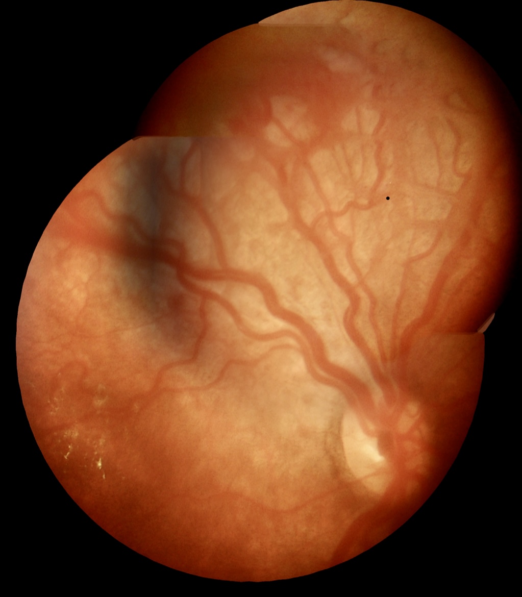
This is because premature birth can disrupt the development of the baby’s retinal blood vessels. In these cases, vascularization might expand, twist, or even rupture. In advanced stages, this can lead to the formation of retinal scars or even retinal detachment, resulting in permanent vision loss.
The International Classification of ROP (ICROP) defines the disease by severity (stages 1-5), location (zones I-III), and extension in analog hours (1-12 h), with or without additional disease (arteriolar dilation and venous tortuosity), and the presence of which would indicate disease activity (4-5).
Check the table below:
Premature Retinopathy Classification
Stage 1
White line separating vascular from avascular retina
Stage 2
Elevated ridge
Stage 3
Fibrovascular proliferation from ridge
Stage 4
Proliferation leading to subtotal retinal detachment (4a, extrafoveal; 4b, total detachment, including fovea)
Stage 5
Total retinal detachment (open or closed funnel)
Threshold disease (defined by CRYO-ROP) (untreated cases show poor anatomical outcomes in 50% of cases)
Stage 3 retinopathy, zone I or II, with at least five contiguous hours or eight cumulative hours of extension, with an additional disease disease (arteriolar dilation and venodilation).
Type 1 pre-threshold disease (defined by ET-ROP)
Any ROP in zone I with an additional disease (aggressive posterior disease) Stage 3, zone I, without plus disease Stage 2 or 3 in zone II, with additional disease(s).
Type 2 pre-threshold disease (defined by ET-ROP)
Stage 1 or 2, zone I, without additional disease(s) Stage 3, zone 2, without additional disease(s).
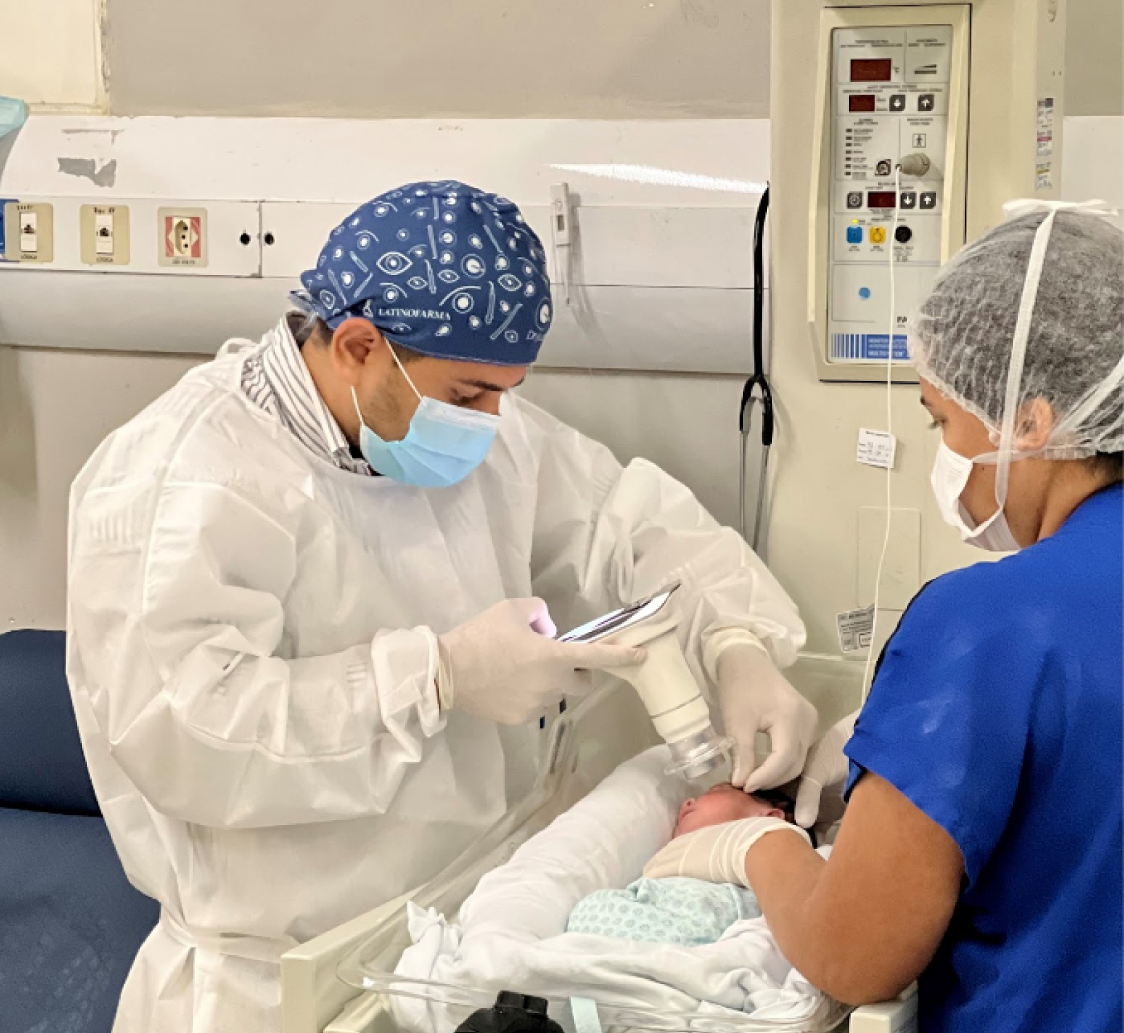
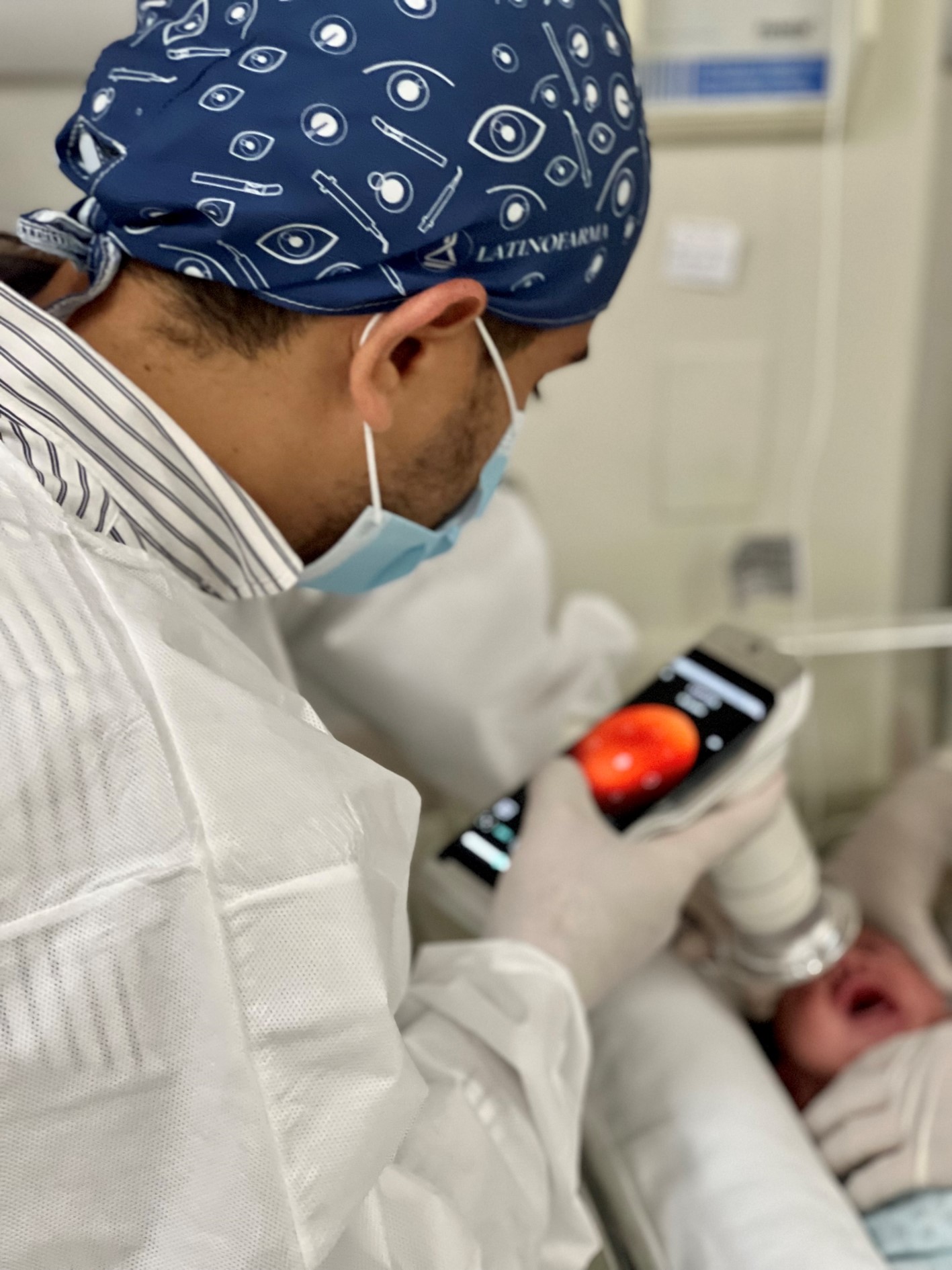
Image caption: Samuel Montenegro examining a newborn with the Eyer. Photo: personal archive.
ROP bears two dangerous characteristics: it’s silent, showing no visible symptoms, and it progresses rapidly. Therefore, adhering to international and national protocols for early diagnosis and treatment is crucial.
Montenegro explains that routine eye examinations for premature babies should be conducted four weeks after birth. “The child isn’t born with the disease, so it’s essential to assess during this period.”
The examination should be performed by an ophthalmologist experienced in evaluating preemies and knowledgeable about the disease to identify location and sequential retinal changes.
Image caption: Image taken with the Eyer. Photo: Samuel Montenegro.
Subsequent examination scheduling will be determined by findings from the initial examination.
After identifying ROP, Montenegro tracks and documents patients using a portable retinal camera, Eyer. This equipment, highly recommended for infant and child examinations due to its portability and high image quality, attaches to a smartphone and conducts retinal exams within minutes. It also makes images available on the online platform, EyerCloud, facilitating study and case progression monitoring for physicians.
Image caption: Image taken with the Eyer. Photo: Samuel Montenegro.
“The device has been a game-changer as it greatly assists me in capturing the patient’s retina at that exact moment, in a practical, quick, and high-quality manner,” he states. Previously, the specialist used a retina mapping lens with smartphone assistance for videos. “Then I’d freeze the image, take a screenshot, and store it on the computer. It was quite labor-intensive,” he recalls.
Premature Retinopathy Treatment
Montenegro explains that treatment is most effective when ROP is identified early. “The secret to managing this disease lies in early diagnosis and immediate treatment when necessary.”
Currently, retinal ablation with a laser is the gold-standard treatment. Depending on the stage, there are alternatives, such as anti-VEGF injections and cryotherapy. “In this disease, we’re fighting blindness. Therefore, we apply laser therapy to prevent blindness in cases where it’s the best indication. However, this may permanently restrict the field of vision,” he points out.
Children with ROP receive follow-up care from a multidisciplinary team: pediatric ophthalmologist, retina specialist, occupational therapist, and physiotherapist. This follow-up extends beyond retinal ablation, aiming to achieve early visual stimulation.
“Newborns diagnosed with ROP are at a higher risk of developing ophthalmological issues in the future, such as strabismus, amblyopia, and refractive errors. Therefore, ophthalmological follow-up after discharge is recommended,” he emphasizes.
Image caption: Samuel Montenegro examining a newborn with the Eyer. Photo: personal archive.
ROP Brazil
Montenegro is part of a project called ROP Brazil, which aims to share knowledge and further study premature retinopathy.
Various surveys indicate that the proportion of blindness caused by ROP is greatly influenced by the level of neonatal care (availability of human resources, equipment, access, and quality of care), as well as the presence of effective screening and treatment programs. Consequently, there’s significant variability in disease occurrence between developed and developing countries.
“That’s why, understanding more about the disease is essential to decrease cases of preventable childhood blindness in the country,” he states.
Request a Quote
Fechar
Later
Request a quote
Fill out the form below and we will contact you shortly.
Request a Quote
Our team will contact you shortly.
Close
…complete suas informações
Depois
Obrigado por completar suas informações
FECHAR
Depois
Solicite e comece SEU TEST DRIVE
Por favor, preencha o formulário abaixo que entraremos em contato.
Obrigado!
Nosso time comercial logo entrará em contato para finalizar o processo.
FECHAR
Later
Request more information
Please fill out the form below and we will contact you.
Thank you!
Our commercial team will contact you shortly to finalize the process.Our commercial team will contact you shortly to finalize the process.