The use of robotics in health has grown increasingly in recent years. By 2025, global investment in this sector is expected to increase approximately 20%, according to a report from Zion Market Research.
The technology generates several advantages for doctors and health institutions, such as more accurate and reproducible procedures. In fact, doctors’ knowledge allied to robotics make the procedure safer, faster and reduces pain and trauma to the patient.
The technology is employed in robot surgeons and even medical software. Learn about the health benefits of robotics and how it works in areas such as surgery, healthcare and even office management.
Robots X doctors
First, robotics does not replace doctors in the healthcare field. In fact, robots only work when guided by humans.
Equipments work as an extension of the surgeon’s hand, for example. This way, they follow commands and avoid the natural tremor of a person, which is a great benefit to delicate surgeries that demand millimeter movements, mainly.
Another approach is planning of the entire procedure and monitoring the execution. Therefore, if an unforeseen event occurs, the professional can adjust at the same time.
1. Robotics in surgery
It seems like something new, but the health field started employing robotics many years ago. First surgeries with such equipments occurred in 1988, in Paris.
Ophthalmology is one of the areas in which the use of robotics has been done for the longest time. One of the first uses was treating diabetic retinopathy with laser. The technology measures the duration, size and power of each pulse. For this, the device is pre-programmed so that each laser pulse is the same as the previous one.
Nowadays, the tool is also used in refractive surgeries to correct myopia. Robots guide all the programming of the procedure of laser pulsing in the patient’s, always monitored by a specialist.
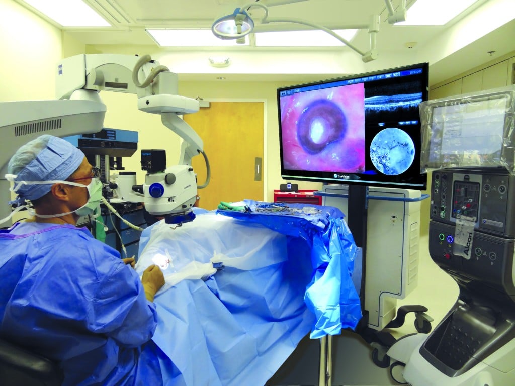
The robotics system NGENUITY uses 3D technology in high definition that greatly increases the surgeon’s view of the eye. It turns the ophthalmological surgeries more accurate. The doctor operates by looking at a high-definition 3D screen, which allows a more proper posture and reduces fatigue during the procedure.
Another ease is using digital filters to customize visualization during the procedure, increasing the image of eye structures and tissue layers.
There are also robots to carry out cataract surgery and corneal transplantation, such as the Ziemer and LensX. However, they are less used in Brazil.
Da Vinci
Several technologies have this goal nowadays. Undoubtedly, the most known is Da Vinci, the robot surgeon most used in the world. Currently, more than 5 million patients are treated. Even in Brazil.
These systems carry out minimally invasive surgeries in different procedures. One of its tools is a console – inspired by flight simulators – in which doctors visualize the high-definition 3D images and make the operative movements with their own hands, through a joystick, which are transmitted to the robot.
The doctor can move the axes for 360 degrees, reaching angles that human hands would only reach with great difficulty.
It also applies to train and requalify students, residents and specialists in attendance or within the surgical center. Thus, it is possible to analyze ethnographically the performance of the professional – who is alone during the procedure – by capturing images from a local camera.
The technology can also be operated remotely via telemedicine.
2. Robotics in healthcare
Several robots already improve medical care. Such as robot Laura, which identifies potentially dangerous generalized infections in patients and informs the medical staff. For this, the robot is connected to electronic medical records and monitors the health reports and clinical information of each patient. When it identifies any worsening or abnormality, it generates an alert. It also uses the principle of machine learning for this purpose.
Since its invention in 2016, it has helped reduce the overall mortality rate by 25%, saving 18 patients a day in institutions that work with the technology.
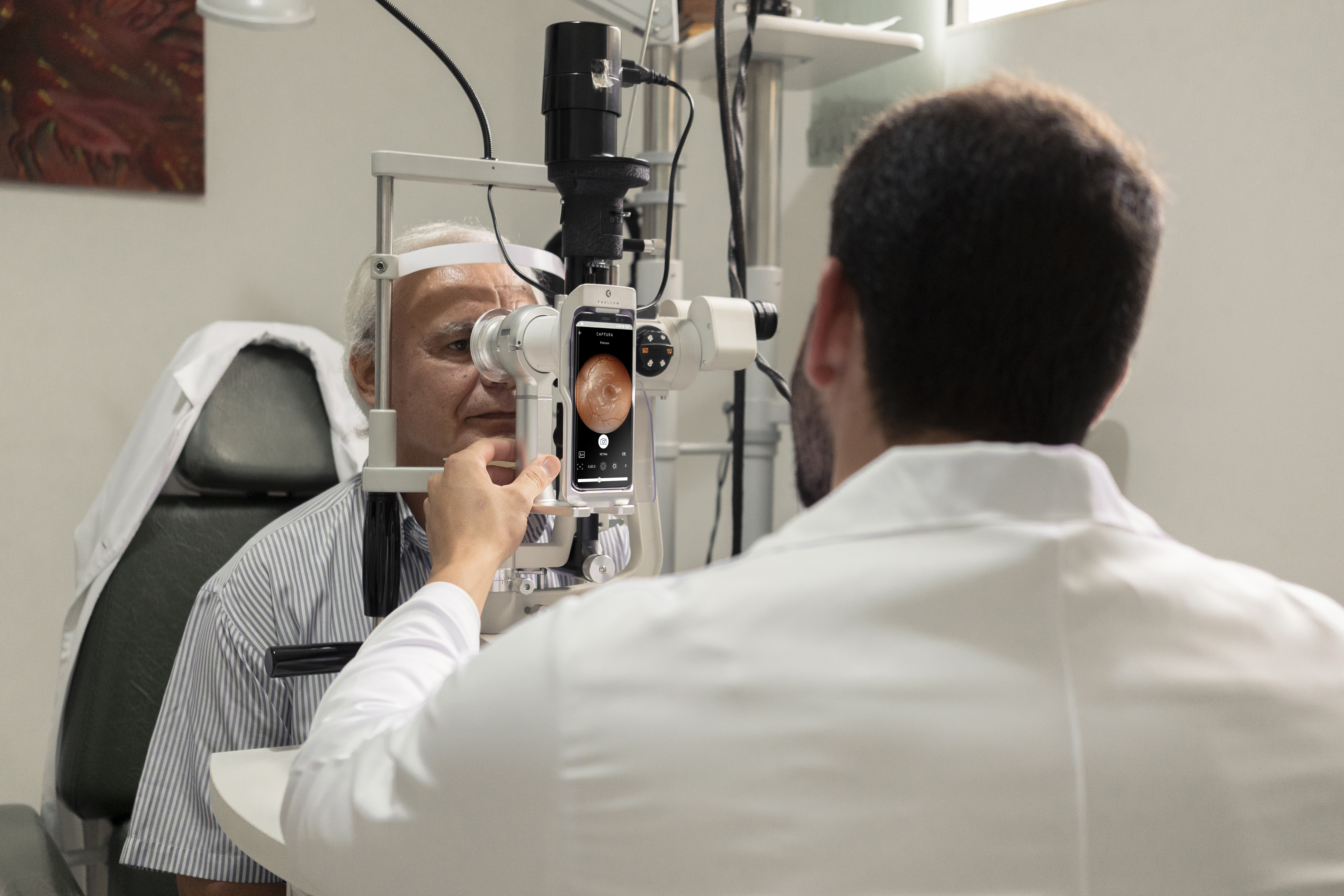
In ophthalmology, one of the proposals is the Adam robot, which can assist in checking primary visual acuity. This is because it identifies visual difficulty levels. In up to five minutes, it can detect diseases such as myopia, astigmatism, hyperopia and presbyopia.
Even for procedures that involve needle placement, such as taking blood and biopsy, there are robotic systems. This is the case of Veebot. It uses a pouch similar to blood pressure gauges, making the veins more visible. It then uses an infrared light and a camera to find the best vein through an image analysis software.
The depth with which the needle is pricked is pre-calculated and the whole process takes about a minute. The system succeeds in choosing the vein in 83% of cases. In addition, the procedure is less painful to the patient.
3. Robots in office management
Imagine receiving your patient with the help of a robot? Invented in Japan, Pepper is used in several countries for the reception of shops, exhibitions, public places and even in medical offices.
The robot has a human appearance: giant eyes, child’s face, arms, hands, 1.20 meters high, 30 kilos and a screen attached to the chest. One of its main features is the ability to analyze people’s emotions through facial expression and tone of voice.
It uses voice recognition technology, cameras and sensors and evaluates all this data in a system based on artificial neural networks. It is able to modify the voice, the color of the eyes, move the arms and show images on the screen according to the emotions of the person.
In fact, neither Pepper nor any other robot can replace professionals in the field. However, it helps greatly to improve care, reduce costs, carry out more precise and less invasive procedures, increasing agility and reducing pain in the patient.
Reviewed by Paulo Schor, ophthalmologist, free professor and director of innovation of the Federal University of São Paulo (Unifesp) and collaborator of the Faculty of Medicine of the Albert Einstein Hospital.
Follow Phelcom’s blog and stay on top of the main news about coronavirus and the eyes.
Ocular syphilis is a manifestation of syphilis that can arise when the disease is not treated properly. This stage occurs years after infection and has a challenging diagnosis. Despite directing lesions, we call the treponema palidum (etiological agent of the disease) “the great copycat”. The agent can simulate several different manifestations. At this stage, the problem can even cause blindness.
But, a new study pointed out that Optical Coherence Tomography (OCT), A common ophthalmological examination in SUS, can help in the early identification of ocular syphilis. The University of São Paulo (USP) carried out the research and published it recently in the journal Ocular Immunology and Inflammation.
Learn about the research, results and what should be the next steps for the use of OCT to diagnose the disease.
The research
Researchers from the Faculty of Medicine of Ribeirão Preto (FMRP), from USP, evaluated one of the eyes of 54 patients with ocular syphilis admitted to the FMRP clinical hospital (HCFMRP). After part of them received the treatment, scientists still analyzed 31 eyes.
Through Optical Coherence Tomography (OCT), researchers found retinal lesions that may aid in early diagnosis of the disease.
Results
The ophthalmological exam identified round spots, irregularities, elevations and detachment in the retinas studied. According to the authors of the work, it is the first time that OCT checks for frequent changes in the retina in a large series of cases of ocular syphilis. These modifications are imperceptible on clinical exams.
Undoubtedly, the findings of OCT have diagnostic value in ocular syphilis, but do not predict the prognosis. However, the examination – common both in the Brazilian Unified Health System (SUS) and private clinics – can help visualize signs of the disease even in early stages. After confirming the diagnosis with serology and referring to the indicated treatment, the patient has a good chance of not having permanent sequelae in vision.
Photo: Eduardo Paulino Eye Institute.
Reviewed by Paulo Schor, ophthalmologist, free professor and director of innovation of the Federal University of São Paulo (Unifesp) and collaborator of the Faculty of Medicine of the Albert Einstein Hospital.
Follow Phelcom’s blog and stay on top of the main news about coronavirus and the eyes.
The choroidal nevus is a dark spot that occurs at the eye fundus and is only detectable through routine examinations such as retinal mapping. Usually, treatment only includes an yearly follow-up.
There are also skin nevi, which dermatologists follow-up with dermatoscopy to check for possible changes in their characteristics, such as enlargement. The same follow-up occurs with the spots at the eye fundus, for example.
If they grow, they can evolve into very advanced stages, such as choroidal melanoma, a very rare disease that affects less than 1% of patients diagnosed with the condition. This number is equivalent to five people in a million.
Melanoma (a type of cancer) is asymptomatic at the initial stage. An estimated 85% of cases arise in the uveal tract – iris, ciliary body and choroid. When not identified early, it can metastasize to the liver.
Retinography applies for checking the coroidal nevus size. Learn how this examination can help early diagnosis and disease follow-up.
Choroidal nevus – diagnosis
The ophthalmologist can only identify a coroidal nevus in a routine examination, because the disorder is not visible to the naked eye and does not usually present early symptoms.
Retinal mapping is one of them. By observing a nevus, the doctor can carry out further examinations to finish the diagnosis, such as optical coherence tomography (OCT) and retinography.
If the nevus grows, the first diagnosis may be undetermined melanocytic lesion, to which the doctor will define a protocol of examinations and follow-up. Observed new nevus increases confirm the choroidal melanoma diagnosis.
Choroidal melanoma – treatment
In fact, choroidal melanoma has no cure, but is treatable and requires lifetime monitoring. Thus, the therapy will be established according to the patient’s vision condition and age, as well as the status, location and extent of the cancer. As with all diseases, an early diagnosis determines a better prognosis.
Brachytherapy is most recommended for small and medium sizes. This surgery has a control rate of approximately 95% and maintains the eye and, in some situations, the ability to see.
An older method was removing the ocular globe. Enucleation may still occur for large tumors with symptoms as intense pain, poor vision and disorganization of internal structures. In some cases, radiotherapy, laser therapy and transpupillary thermotherapy are also indicated.
Reviewed by Paulo Schor, ophthalmologist, associate professor and director of innovation of the Federal University of São Paulo (Unifesp) and collaborator of the Faculty of Medicine of the Albert Einstein Hospital.
Follow Phelcom blog and stay on top of the main news on office management!
A patient with retinitis pigmentosa was able to recover part of the vision after undergoing optogenetic therapy and light stimulation. For the first time, this technique has achieved partial recovery of visual function, according to clinical trial researchers. The study was published in Nature Medicine journal .
Before treatment, the man could only perceive the presence of light. Now, he already finds, counts, and touches objects. Learn about the clinical trial and how optogenetic therapy works.
The research
Researchers from the Sorbonne University, Quinze-Vingts Hospital and the company GenSight Biologics, from France, in partnership with the University of Pittsburgh, from the United States, and the Institute of Molecular and Clinical Ophthalmology of Basel, from Switzerland, conducted clinical trials with optogenetic therapy in patients with retinitis pigmentosa.
That degenerative genetic disease damages the retinal photoreceptor cells, causing progressive loss of vision. The condition evolves until the patient is completely blind. The problem affects one in 3.5 thousand people, according to Orphanet database. Currently, an estimated two million cases exist worldwide.
A 58-year-old man, blind for 20 years, received an injection into one of his eyes with a gene called ChrimsonR, that encodes opsin proteins and identifies amber light. These proteins are responsible for sending visual information to the brain.
He then underwent treatment with flashes of light directly on the retina. In optogenetic therapy, light pulses control gene expression and activation of neurons. Currently, they are widely used in laboratories to unravel neural circuits and can be a potential treatment for pain, blindness and brain problems.
Results
After producing enough opsins, which occurred five months after beginning therapy, the patient was given camera glasses that project amber-colored images onto the retina.
In the first exercise, the man needed to notice, find and touch a large book and a small box of staples. In total, he managed to touch the book in 92% of evaluations, and the boxes in 36% of the time.
In the second test, the patient achieved 63% efficiency when counting glasses on a table. In the third exercise, he wore an electrode helmet that monitored if he recognized a glass on the table or not. In this one, he was successful 78% of the time.
Seven months after receiving the injection, the patient already showed signs of improvement in vision.
After two years of treatment, the man still uses the glasses to see better. In fact, images will never be the same as natural ones, but for those who have been blind for 20 years, it is life-changing.
It is the first time that optogenetic therapy has managed to partly reverse vision loss by a genetic degenerative eye disease. The trial will now advance to phase 3 to confirm the effectiveness of this therapeutic approach. However, it will still take some time to offer the technique, as it needs more studies, more patients and more longevity.
Reviewed by Paulo Schor, ophthalmologist, free professor and director of innovation of the Federal University of São Paulo (Unifesp) and collaborator of the Faculty of Medicine of the Albert Einstein Hospital.
Follow Phelcom’s blog and stay on top of the main news about coronavirus and the eyes.
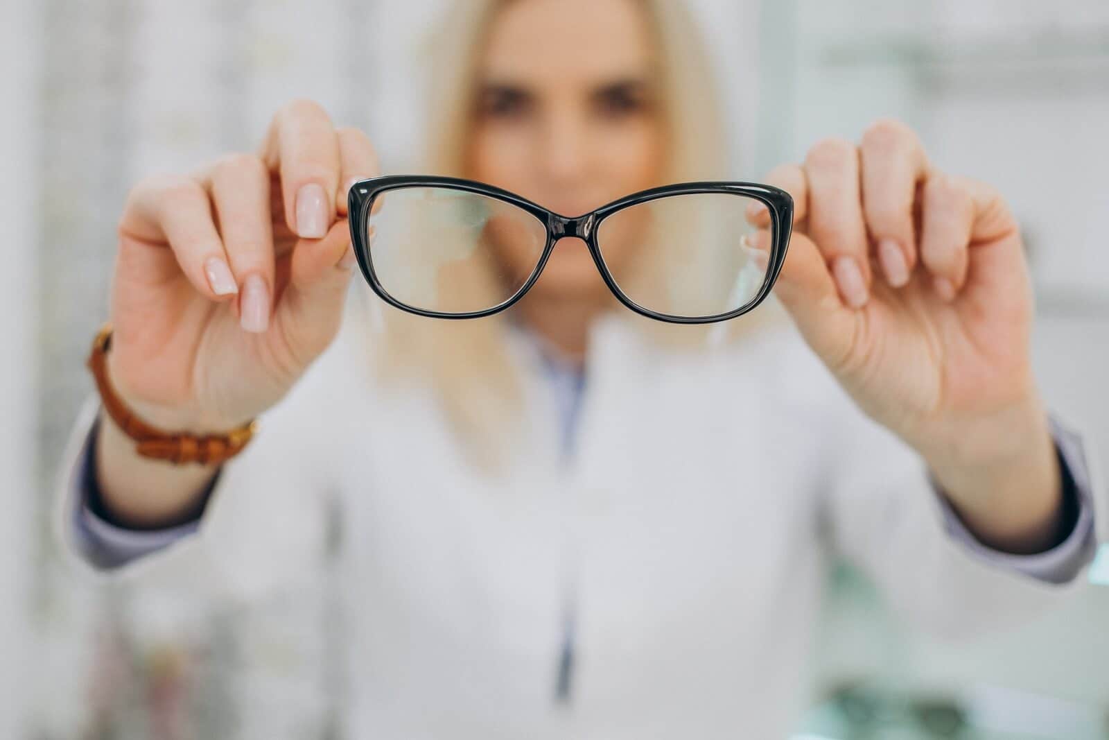
Since last year, scientists raised the hypothesis that glasses help to bar the contamination by the new coronavirus (SARS-CoV-2) through the eyes. This is because this region is also an open door for the virus, as well as a source of contagion.
Now, a recent study found out that the risk of infection by covid-19 in people who wear glasses may be twice to three times lower.The research, conducted in India, was published in the website medRxiv.
Learn more about the preliminary work, how it was done, th results and next steps. It is worth noting the new investigations are necessary, but, as the disease is new, it is important to follow studies in the area.
Glasses and coronavirus: the research
The research evaluated 304 people, 223 men and 81 women, aging from 10 to 80, residents in Northern India, during two weeks.Everyone presented symptoms of covid-19.
58 patients (19%) reported they frequently wore eyeglasses or sunglasses when exposed to sunlight.Through a questionnaire, volunteers stated they touched the face 23 times and the eyes 3 times per hour, in average.
Glasses and coronavirus: results
According to the scientists, the results related to extended use of sunglasses were significant.The current contamination rate of 1,35 of the population, in general, dropped to 0,48 among those who regularly wore glasses.The calculated risk ratio, according to the study, was 0,36%.
That is, the chance of covid-19 infection dropped three times.
Researches believe that the low transmission ratio is due to two factors: glasses protect against contaminated droplets in the air and less frequent touches in the eye while wearing them.
Scientists evaluated 276 positive covid-19 patients.Factors are the same: less touches in the eyes and protection against airborne droplets.
Conclusion
In fact, scientists remark that the relation between wearing eyeglasses and the new coronavirus is still inconclusive.This is because the study has not been revised by peers yet. Sampling was small and there was also difficulty to measure how long people wore glasses during the pandemic.
New investigations are necessary to evaluate the effects of glasses, such as protection against covid-19. However, as the disease is new, it is important to follow studies in the area.
Follow Phelcom’s blog and stay on top of main news in the area of eye care.
Currently, it is estimated that 1.5 million people worldwide lose their vision each year due to corneal injuries and diseases. Thus, problems in this membrane are the third largest global cause of visual impairment, behind only cataracts and glaucoma.
But this scenario may change. Recently, Israeli doctors performed the world’s first successful artificial corneal transplant. The patient, a 78-year-old man, was able to regain his sight after 10 years of blindness.
In fact, synthetic corneal implants already exist, but because they required more complex surgeries, they were used only as a last resort, such as rejection in corneal transplants. The new technology, on the other hand, can be implanted in a relatively simple way, with minimal cutting and suturing.
In the following, understand how the artificial cornea transplant occurred, how it acts inside the organism, the next steps, and how the result can change the reality of millions of people waiting for a cornea transplant to see again.
The artificial cornea transplant
Israeli startup CorNeat Vision has developed the KPro artificial implant to replace a patient’s deformed cornea. The procedure was performed at the Rabin Medical Center hospital in Israel.
The device has a non-degradable synthetic nano-tissue, which is placed under a membrane that lines the surface of the eyelid and the sclera (white part of the eyeball). When implanted, it unifies with the living tissue and encourages cell proliferation within the eye.
The synthetic cornea is only indicated in cases where the tissue is deformed, opaque, or scarred.
In an interview with the Israel Hayom website, the doctor and creator of the technology, Gilad Litvin, said that the surgery was relatively simple and lasted less than an hour.
Elderly Man Recognizes Relatives After Surgery
Patient Jamal Furani was able to regain his sight already the day after the artificial cornea transplant. The elderly man says that light was the first thing he could see. Afterwards, he was able to recognize relatives and even read texts.
“The result exceeded all our expectations,” says physician Irit Bahar, head of the Department of Ophthalmology at Rabin Medical Center.
Next Steps
The expectation is that the procedure will become viable and end the waiting line for donors around the world. “This technology was key to turning the tide against global blindness. It is very exciting to be at the forefront of this project that will undoubtedly impact millions of lives,” Bahar believes.
“We hope this will enable millions of blind patients around the world, in areas where there is no corneal practice or organ donation culture, to regain their sight,” says Gilad Litvin, medical director of CorNeat Vision. However, the company has not yet announced a market launch date.
Now the clinical trials continue. A further 10 approved Israeli patients are awaiting artificial corneal transplantation at Rabin Medical Center hospital. In addition, countries like Canada, France, the United States, and the Netherlands also have patients eligible for clinical trials.