Known as a reference in innovation, the country has one of the most challenging regulatory processes for foreign technologies in the world. The handheld fundus camera, developed and produced by Phelcom Technologies, is now commercialized in five countries.
Phelcom Technologies has conquered one more international market. Now, Japan took its turn to receive Eyer, a non-mydriatic handheld fundus camera, coupled to a smartphone, that carries out high-quality retinal exams in a few minutes.
“We are very proud to have our equipment sold in a country known as a reference in technology and innovation”, states the company CEO, José Augusto Stuchi.
The Japanese regulatory process for entrance of foreign technologies is considered one of the most challenging in the world. This is due to the various players accessed in the procedure, such as DMAH – Designated Marketing Authorization Holder (legal representative), RCB (Regional Certifying Body), PMDA (regulatory agency); as well as specific certification workflows for each product class. The language barrier was another difficulty, since the whole reference documentation was in Japanese.
José Roberto Santiciolli Filho, Product Development coordinator at Phelcom Technologies, explains that the certification process started by the end of 2021, when the company was approved by the Ministry of Health, Labor and Welfare of Japan (MHLW) as a Foreign Manufacturer. All the manufacturing infrastructure and the Quality Management system were evaluated by then.
“After that, we developed a technical dossier of the product, according to PDMA standards, and submitted all the technical and quality documentation to a third-party unbiased evaluation to receive the certificate”, explains Santiciolli.
During this process, Phelcom counted on the support and partnership of Allm Inc., an investor based in Japan, for interlocution with local agents. Last November, Eyer received the medical device certification no. 304AIBZI00005000.
Eyer in the USA, Colombia and Chile
More than Brazil and Japan, Eyer is present in Chile, Colombia and the United States – where Phelcom also has an office in Boston. “This new unit confirms the internationalization movement of the company, started a year ago, and shall support the offer of equipment in the North-American market”, observes Stuchi.
The CEO highlights that physical presence in the United States confirms Phelcom’s availability to work for making eye exams simpler, connected and intelligent, without borders. “In 2022, we took part, as expositors, of the main ophthalmology congresses of the United States, as the American Society of Retina Specialists Annual Meeting, in New York, and the American Academy of Ophthalmology, based at Chicago. American professionals have accepted Eyer very positively”, states Stuchi.
In March 2023, the company has also been at Vison Expo East, a commercial exposition in New York. This month, it is taking part of ARVO (April 23 to 27), in New Orleans, and ASCRS (May 5 to 8), in San Diego.
For Stuchi, being a Brazilian company which operates in important markets worldwide is a pride and great responsibility to Phelcom. “It is certain that internationalization widens our potential to act. However, it also increases our commitment to offering excellent quality products that really contribute to the professional practice”, he complements.
Phelcom Eyer
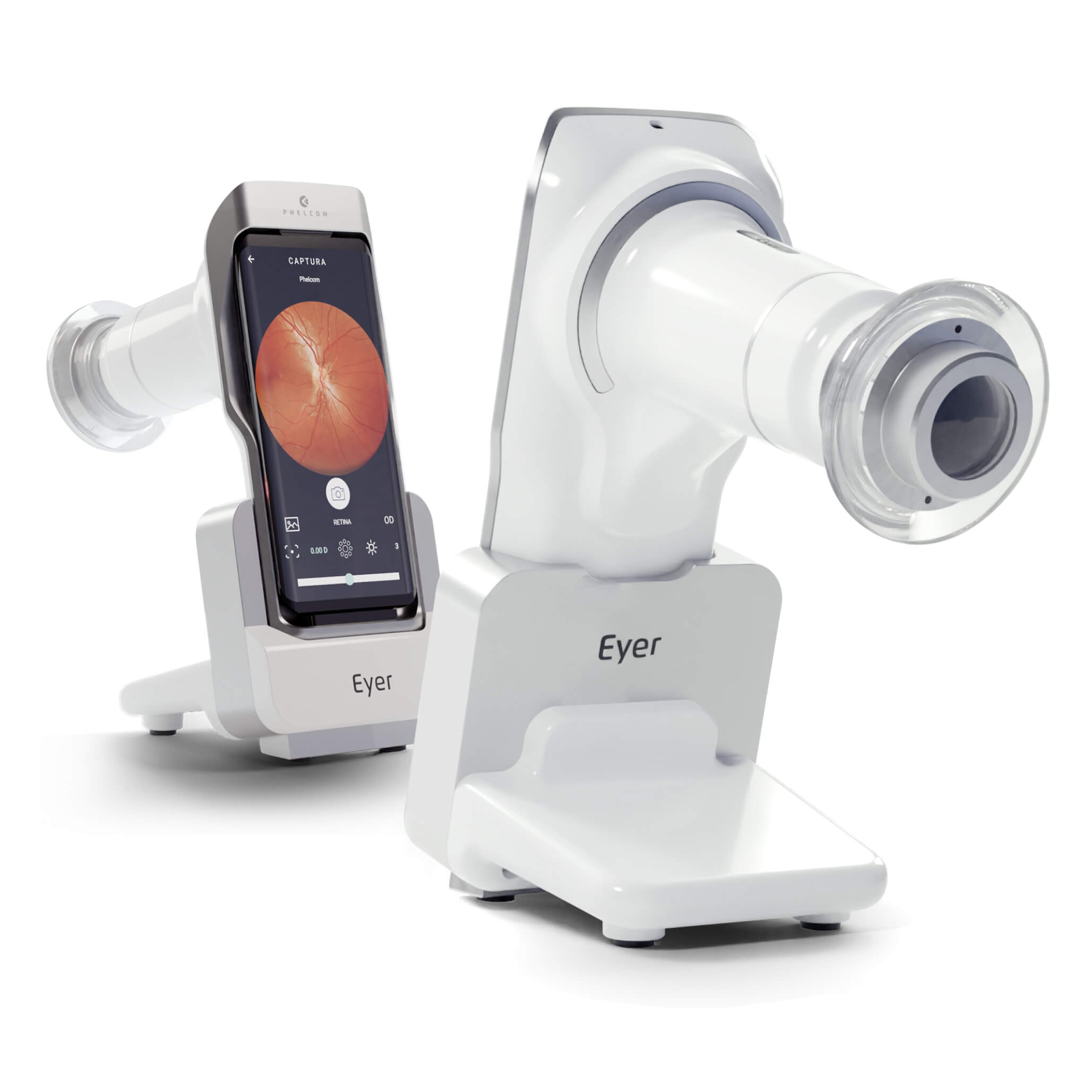
Phelcom Eyer is a handheld fundus camera that works coupled to a smartphone and carries out high-quality retina exams, in a few minutes, without need of pupil dilation.
The technology supports the diagnosis of over 50 diseases, such as glaucoma, cataract, diabetic retinopathy, DMRI, retinoblastoma, hypertensive retinopathy and ocular toxoplasmosis. Currently, more than 10 million exams have been made in Brazil, United States, Chile and Colombia. The portability and the more accessible value of the technology democratize the access to retinal exams. Because it costs approximately six times less than a conventional tabletop fundus camera, which still needs to be integrated into a computer.
About Phelcom
Phelcom Technologies is a Brazilian medtech, headquartered in São Carlos, São Paulo. Its history started in 2016, when three young researchers – a physicist, an electric engineer and a computing engineer (PHysics, ELetronics, COMputing) – created a handheld fundus camera based in technology embedded in a smartphone.The interest of Diego Lencione, a company partner, on visual health gave birth to the first equipment project. His brother has a condition that severely compromised his retina and vision since childhood.
In 2018, Phelcom launched its first product in the market: Eyer handheld fundus camera. Nowadays, the technology has already reached more than 1.2 million people all over Brazil and other countries where it is present.
Ophthalmologist Balamurali Krishna Ambati, known as “Bala” Ambati, is the youngest doctor in the world, according to the Guiness Book of Records. At the age of 17, he graduated in medicine in Mount Sinai School of Medicine, in New York (USA), in 1995.
After that, he did residency in ophthalmology at Harvard University, where he developed strategies to reverse corneal angiogenesis. Since then, he has helped various non-profit organizations all over the world, such as ORBIS – Cooperation and Development and Sight for the Sightless, which acts to combat blindness in the Indian countryside.
He received various important awards for his work, such as the Gold Humanism Award and the Troutman-Veronneau Prize. Nowadays, he leads the clinic Pacific Clear Vision Institute, in Eugene, in the state of Oregon (USA).
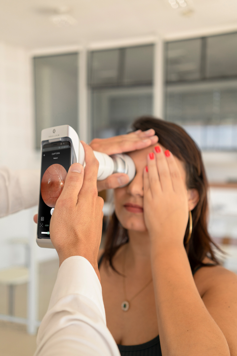
For six months, the ophthalmologist has been using the Eyer Portable Fundus Camera in the clinic, on a daily basis. The technology works coupled to a smartphone, to carry out high-quality retinal exams in a few minutes, without need of pupil dilation.
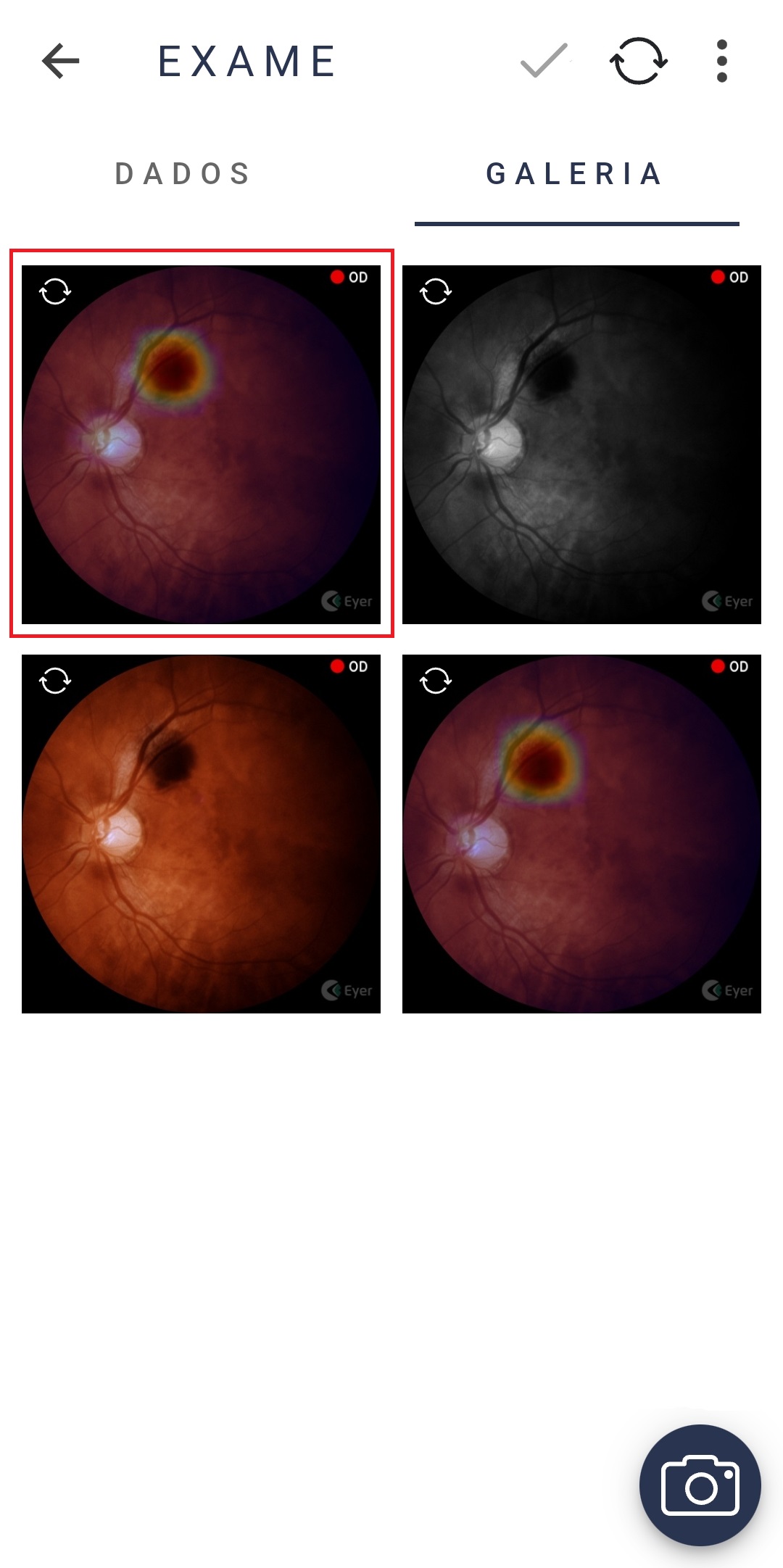
“We acquired the Phelcom Eyer camera about 6 months ago and have been incredibly pleased with it. Its image quality, easy to use and easy to learn form factor, panoramic image stitching, and heat maps make photo acquisition and image transfer to the EMR seamless”, highlights Ambati.
The doctor remarks that using the equipment does not break the clinic workflow and points out the capacity to acquire images through a 2,5mm-wide pupil as “impressive”.
Furthermore, the ease of taking photos and making the documentation simplifies the doctors’ work. The exams are automatically sent to EyerCloud, a platform for remote diagnosis.
Another pro from Eyer is its portability: one can handle it manually or stabilize it on a slit lamp.
The patient becomes more engaged in the treatment, according to Ambati
As it makes device imaging available in real time, Ambati says the patients appreciate the chance to learn about their condition and what goes on with their retinas.
The ophthalmologist tells that both them and the medical team were impressed by the way the heat maps of retinal elevation reveal the pathology. “It is a factor that motivates patients to be involved with the treatment and take diabetes or DMRI seriously”, he says.
Phelcom Eyer
Phelcom Eyer is a portable fundus camera that works coupled to a smartphone and carries out high-quality retina exams, in a few minutes and without the need of pupil dilation.
The technology can identify more than 50 diseases, such as glaucoma, cataract, diabetic retinopathy, DMRI, retinoblastoma, hypertensive retinopathy and ocular toxoplasmosis. Currently, more than 10 million exams have already been carried out.
The device integrates to an online platform, Eyer Cloud, to which data is sent automatically. A specialist can analyze them remotely from anywhere in the world.
More than that, the embedded artificial intelligence provides intelligent functions to help the medical diagnosis and the capture of retinal exams. On the other hand, portability and the technology lower prices democratize the access to retina exams.
Phelcom Technologies
Eyer is developed by Phelcom Technologies, a startup that invests in innovation and technology to democratize visual health. The company is headquartered in São Carlos-SP, Brazil and has an operation in Boston, USA.
Launched in April 2019, the technology has already reached more than 1,2 million people all over Brazil and in countries such as the United States, Chile, Japan and Colombia.
The startup creates portable devices with embedded artificial intelligence, for immediate diagnoses. It aims to revert the fact that 75% of the 250 million cases of blindness or severe visual deficiency happen due to lack of prevention or wrong treatment.
Brazil, United States, Chile, Colombia and, now, Japan. Eyer portable fundus camera, developed and produced by Phelcom, has just been approved in the country.
The Japanese regulatory process for entry of foreign technologies is considered one of the most challenging in the world. This is due to the various players accessed during the procedure, such as the DMAH – Designated Marketing Authorization Holder (legal representative), RCB (third-party certification organization), Foreigner Manufacturer and PMDA (regulatory agency), besides the specific certification flows for each product class. The language barrier was another difficulty, since all the reference documentation was in Japanese.
José Roberto Santiciolli Filho, coordinator of Product Development in Phelcom Technologies, explains that the certification process started in the end of 2021, when the Japanese Ministry of Health, Labour and Welfare approved the company as a Foreign Manufacturer. At this stage, all the manufacturing infrastructure and Quality Management system were evaluated.
“After that, we developed the technical dossier of the product, according to PMDA standards and submitted all the technical and quality documentation to third-party impartial evaluation, for then receiving the certificate”, explains Santiciolli.
During the certification process, Phelcom counted on the support of its investor, Allm Inc., headquartered in Japan, for interlocution with local agents. In November this year, Eyer received the medical device certification with the number 304AIBZI00005000.
Phelcom Eyer
Phelcom Eyer is a handheld fundus camera that works coupled to a smartphone. It carries out high-quality fundus exams in a few minutes, without the need of pupil dilation.
Integrated to an online platform, EyerCloud, data are automatically sent and can be analyzed by a specialist from anywhere in the world. That is, it makes remote diagnosis possible.
More than that, the embedded artificial intelligence provides intelligent functions to help medical diagnosis and capture of retinal exams. On the other hand, the portable technology of accessible value democratizes the access to retinal exams. The device is around six times cheaper than a conventional tabletop fundus camera, which still demands integration to a computer.
Learn about the advantages:
CONNECTIVITY
The device is naturally connected, since it is attached to the smartphone. Therefore, it eases cloud sharing and access of exam data in the Eyer Cloud system.
NON-MYDRIATIC
Eyer allows to carry out retinal exams anywhere, without needing eye drops for pupil dilation. Thus, the patient feels more comfortable and the exam is faster.
LOW COST
Portability and reduced size grant Eyer a lower cost in relation to traditional fundus cameras. Even with the cutting-edge technology applied in the device production.
Currently, more than 1.2 million people have been examined and around 10 million exams were carried out in Brazil, United States, Chile and Colombia.
Eyer is also in the certification process in Mexico. Furthermore, a new Phelcom product is being approved in Brazil and in the United States.
“The favorable result of the process in Japan demonstrates we are on the right track to democratize access to ophthalmology. We are very happy to make available to one more country an innovative solution that will make a difference in the struggle against blindness in the world”, highlights Santiciolli.
Retina Global is an international, non-profit organization that seeks to enable sustainable solutions for the care of retinal diseases in underserved areas around the world.
Currently, the institution has projects in Central and South America and in Asia. It has already implemented programs in Tanzania, Kenya, Bolivia, Belize, Bahamas, Burundi, Ethiopia, Haiti and Brazil.
The NGO, in partnership with the project Iluminar, tracked and treated diabetic retinopathy in 13 municipalities in Sergipe hinterlands.
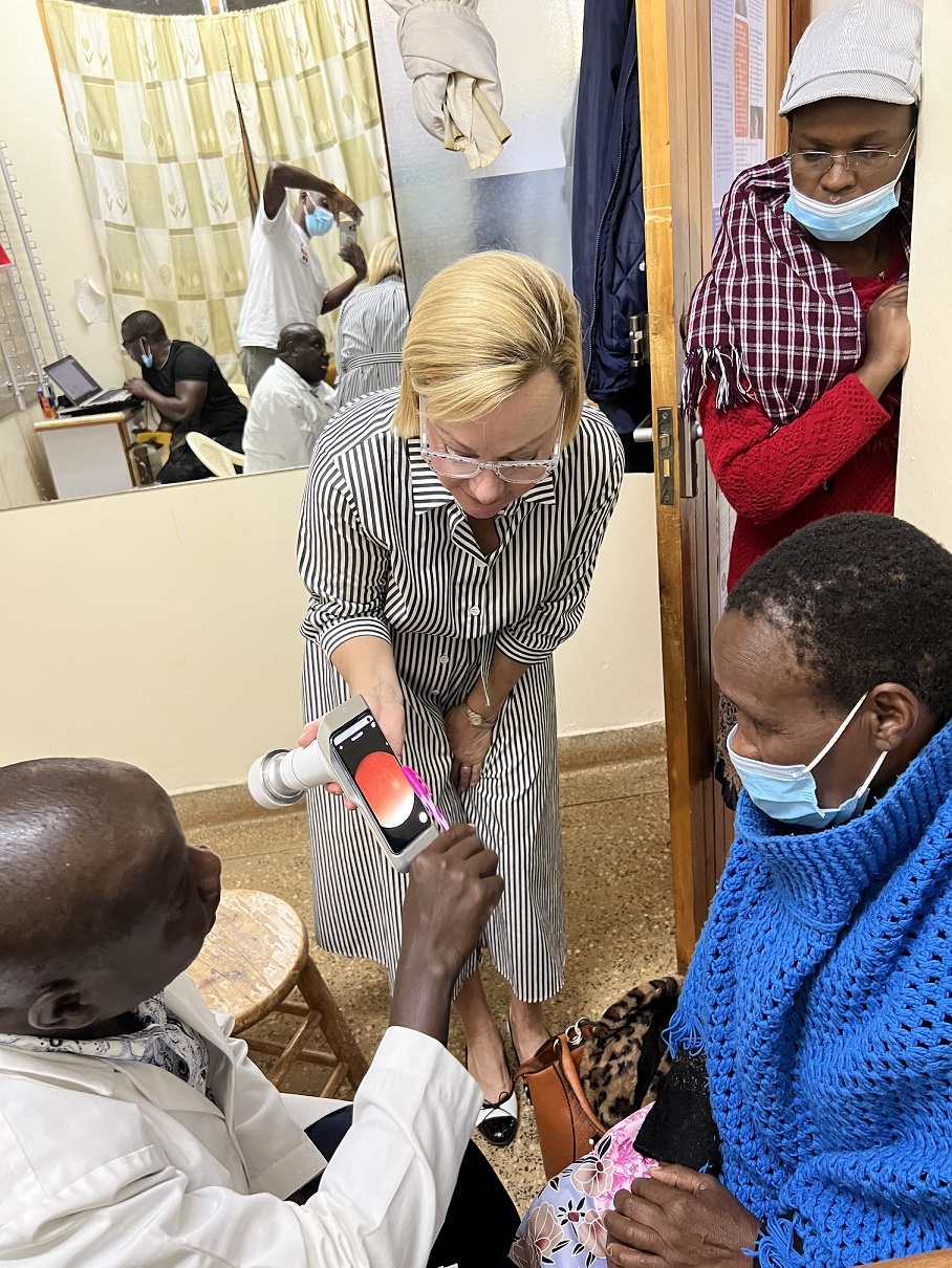
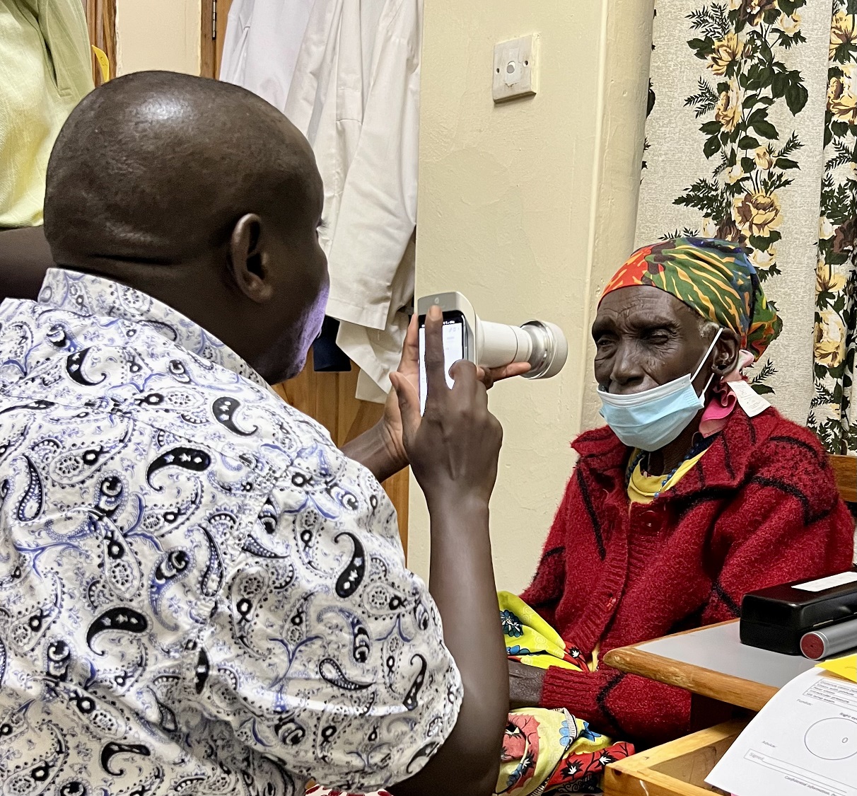
The team used the Phelcom Eyer, a smart, medical device, to carry out high-quality retinal exams (without having to dilate pupils) and make images available for remote diagnosis.
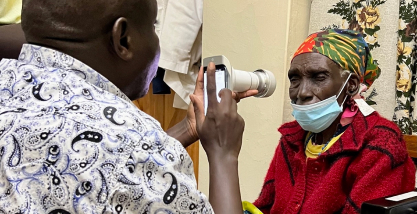
Recently, the organization has used the equipment again to track retinal diseases in Kericho, a town in rural Kenya, Africa.
Learn more about the project of Retina Global in Kenya, with the support of Eyer.
Retina Global in Kenya
For two days, the volunteer team from Retina Global made an initial evaluation, and then referred the patients for fundus exams with Eyer. Both the images and patients’ histories were
immediately available in the Eyer Cloud online platform.
Specialists in retina, based in the United States, made the reports. In total, 26 people were found to have retinal abnormalities, including a child and one young adult. “With that, we could diagnose quickly and start early treatment”, said the leader of Retina Global project in Kenya, Diane Steinhilber.
“We will keep using the equipment to identify and select the patients in need of specialized retinal care”, she highlights.
Lack of access to eye health
Steinhilber explains that the great challenge of this project is the lack of a retina specialist in this rural area of Kenya. Patients who need specialized treatment are referred to hospitals in Nairobi, the country’s capital, which is located roughly five hours away.
“To see a doctor, they face an arduous and long trip, as well as the time away from their jobs and families. Screening through Eyer allows us to quickly refer patients that are in real need of care in specialized centers”, she states.
As some patients have difficulties to get to the hospital, the project plans to send a trained team on community missions. Moreover, the project searches for a portable machine for laser treatment, as well as tools to carry out other kinds of therapies, such as intraocular injections and surgeries.
“Our team evaluated in detail the best way to implement a project that is able to provide exams and information on retinal care and also one that is sustainable in the long-term”, she says. Therefore, the project in Kenya was divided in phases to be implemented over five years.
Eyer use in Kenya
Retina Global team in Kenya took part on a remote training to learn how to handle the technology. “Eyer is extremely easy to handle. It is possible to promptly identify the images, quickly create patient database and send the photos to the doctor in the United States for review and reporting”, she says.
The project leader also highlights another advantage of Eyer: it not only has data for patient follow-up and continuous care, but also is a way to obtain information on the prevalence of retinal diseases in the region.
“Without Eyer, Retina Global would not have even started in Kenya. Further than using it in more local communities, we plan to include it in other projects to take place around the world”, she finishes.
Phelcom Eyer
Phelcom Eyer is a portable fundus camera that works to carry out high-quality fundus exams in a few minutes without the need of pupil dilation.
Integrated to an online platform, Eyer Cloud, the data is automatically sent and can be analyzed by a specialist anywhere in the world. That is, it allows remote diagnosis.
More than that, its embedded artificial intelligence provides smart features to help medical diagnosis and the capture of retinal scans. On the other hand, the portability and affordability of the technology democratize the access to retinal exams. The Eyer is about 10 times more affordable than a desktop fundus camera, which still has to be integrated into a computer.
Phelcom Technologies developed the technology, which is currently present all over Brazil and in countries such as the United States, Japan and Chile.
Google ads for doctors is a tool that helps you bring new patients to your office. We all know that people search on Google for what to do, where to go, what to buy and even which doctor to choose. This resource allows your ad to be shown exactly when someone is looking for services as yours.Thus, you can increase site visits, appointments and, consequently, the number of patients. But, how to attract more customers with Google ads for doctors? Learn how to in this article.
Google ADS for doctors: how it works
Google Ads is a Google Ads platform. It allows you to create online ads with sponsored links, which will appear to the user according to the search keywords.By this, your advertising may appear on YouTube videos, in Google search engine, on sites related to your area of activity, applications, etc. All depends on the strategy your team sets.The position of your ad on Google will depend on three factors: the relevance of the keyword to the subject searched, as well as the quality of the ad and the landing page; the maximum CPC (cost per click) bid, a comparison of the advertiser with the highest value per click; and the Ad rank, an average value between the previous two.The tool also delivers the metrics of the carried out campaigns. This way you can evaluate the number of impressions and ad clicks, as well as identify the users who clicked. Thus, it allows you to find out which type of ad performed best and better control the return on your online investments.By the way, the charge for the ad only occurs when you receive clicks. In addition, you can also limit the amount spent for each click received.
Google ADS for doctors: how it works
Check a step-by-step guide to create online campaigns with Google ADS:1.Set the target audienceWhich patients do you want to attract? Information such as age, job, location, level of education and income help create your target audience.2.Create the personasPersonas are ideal, detailed profiles of the patient you want to attract. In addition to the information above, one must analyze lifestyle, habits, behaviors, frustrations, difficulties, leisure, etc.3. Create the campaignWith personas in hand, you can define which type of media is most consumed and make ads there. Example: blogs and YouTube.4. Choose keywordsThis step is critical for your ad to appear or not to your persona. Here, you need to define the keywords that are most related to your specialty and that should draw the attention of your potential patient.5. Select your goalIn the tool, adjust your ad based on the desired results, such as receiving more calls at your clinic, attracting more patients, or directing people to your site.6. Decide where to advertiseYou decide where you want to display the ads and Google will show it to the right people.7. Create your messageShow off what’s special about your office in three short sentences to win patients over, or add images to create eye-catching banner ads.8. Set your budget limitYou’ll never pay more than your monthly budget and can adjust or pause whenever you want. In addition, the platform shows the estimated results based on the chosen budget.Google Ads can be a tool that helps you find ways to improve your ads and generate better results, thus freeing the doctor to focus on what he knows best: serving patients.It also provides reports, insights, and tips for you to track your progress and further optimize ads.Another tip: create Google My Business, a free and easy-to-use tool that allows healthcare institutions to manage their online presence on Google, including Search and Maps.To do this, you can create a profile of your business with all the essential data. That way, when people search directly by your name or the office or by keywords related to your specialty, Google will show your professional profile.See a step-by-step guide to create your profile in this article.Reviewed by Paulo Schor, ophthalmologist, associate professor and director of innovation of the Federal University of São Paulo (Unifesp) and collaborator of the Faculty of Medicine of the Albert Einstein Hospital.Follow Phelcom’s blog and see tips on how to improve the digital marketing strategy of medical offices and clinics.Subscribe
Augmented reality in health allows a real-time combination of images never before achieved, with new possibilities to diagnose diseases, which can increase accuracy in surgical procedures. That is, it can potentially improve patient quality of life.
In real time, the technology shows the material world overlayed with computer-made images, enabling greatly improved composite observations.
For this, smartphone cameras, tablets, smart glasses and even state-of-the-art helmets are used, so that the hands are unnecessary to command interfaces in systems and applications.
For instance, there is a strong trend to use Augmented Reality (AR) in this industry, according to the Journal of Med Internet Research (JMIR). Even with a still low use by doctors and institutions, there is an increasing number of applications of AR in medicine.
Learn about the advantages of using augmented reality in health and some cases of success.
Augmented reality X Virtual Reality
First, it’s important to understand the difference between augmented reality and Virtual Reality (VR).
AR, in its turn, integrates the physical and the virtual by overlaying computerized images in the real world, through software and devices. For example, the Google Glass app can be accessed from the user’s own mobile phone or tablet. There is also the famous game Pokémon Go, in which players searched for virtual Pokémon in real-world environments using the mobile phone camera.
Augmented reality in healthcare: advantages
Undoubtedly, there are numerous advantages of using AR in health. One of them is to ease access to the patient’s medical history in emergencies or on the surgery table, for example. Image overlaying may also more accurately ensure to locate an area of interest in surgical procedures, thus decreasing risk to the patient. In this case, it is possible to integrate imaging examinations and better see every detail of the organs in the preoperative care and, thus, have more success in surgery.
In fact, technology is a great ally of telemedicine. For example, specialists can instruct other doctors remotely in more complex operations. Another aspect is diagnostic medicine, because the patient can better understand the diagnosis and the recommended treatment.
AR has also been used in the digital screening of patients who have suffered accidents. In these cases, orthopedists help in the performance of arthroscopic surgeries on shoulders and in training interns and residents to carry out ultrasound examination at the care site.
Neurosurgery is one of the most advanced specialties in the use of AR, with reports of tumor resection, open neuro-vascular surgeries, spinal procedures, location of catheters or probes, cortical resection in epilepsy and aneurysms involving unusual trajectories or hidden ramifications.
In hospitals and clinics, the technology assists remote support by enabling on-site technicians and teams to receive specialized help needed for installation, setup, maintenance, troubleshooting, and repair of sensitive and specialized hospital equipment.
Teaching in medicine
Augmented Reality in healthcare allows seeing details of the human body and training procedures in a repetitive and precise way.
Student, imagine practicing surgery in a virtual environment? Stanford University has a simulator which allows the most complete training for the future surgeon.
For researchers, a three-dimensional virtual surgical environment can allow for enhanced preoperative planning and testing. It thus improves patient results, decreases complication rates and improves technical skills.
The Stanford Virtual Surgical Environment (VSE) is developed for rhinologic procedures. The technology allows the surgeon to interact with patient-specific three-dimensional reconstructions of nasal sinus CT data sets using a modified haptic interface device, triggering a virtual endoscope.
In Brazil, there are also options for simulators. For example, Eyesi is aimed at training intraocular cataract surgery and vitreo-retinal procedures. It presents performance score and evaluates tremor, movement accuracy and repetitiveness, among other analyzes. Thus, it is a good tool for resident practice to introduce new techniques in ophthalmic surgery.
There are also the da Vinci surgical systems for robotic surgery, already in the fourth generation. The technology carries out minimally invasive surgeries in different procedures. One of its tools is a console – inspired by flight simulators – in which doctors visualize the high-definition 3D images and make the operative movements with their own hands, which are transmitted to the robot.
Another application is training and retraining for students, residents and specialists in care or in the surgical center. Thus, it is possible to carry out an ethnographic analysis of the performance of the professional, who is alone during the procedure, through the capture of images by a camera installed on the site.
Augmented Reality solutions in healthcare
On the market, there are already quite interesting solutions of AR.
For example, the application EyeDecide allows you to see the eyeball from any angle just by directly touching the image generated on the tablet and rotating it with your finger. It is also possible to reduce, expand, move or make notes on the eye in almost any direction. All this assists to simulate various disorders and helps to diagnose more accurately.
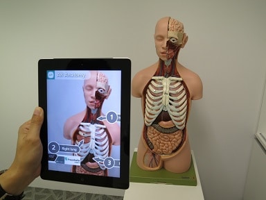
The app Anatomy 4D makes it possible to explore more than two thousand anatomical structures. Undoubtedly, it helps students and medical colleges.
Even with all the advantages, augmented reality in health is still relatively new and is not heavily employed in the area. However, as it proves its value for the quality of patient care, soon AR technologies will be stronger in the medical industry.
Reviewed by Paulo Schor, ophthalmologist, associate professor and director of innovation of the Federal University of São Paulo (Unifesp) and collaborator of the Faculty of Medicine of the Albert Einstein Hospital.
Follow Phelcom blog and learn more about augmented reality in healthcare.