

The retinoscope defines the prescription, but in modern eye care, correlating functional data with high-quality fundus photo-documentation is what truly elevates patient care.
The retinoscope remains one of the most iconic tools for objective refraction. For any eye care professional (ECP), mastering the play of light and shadow in retinoscopy is an art form that ensures refractive precision. It gives us the “how much”—the refractive power.
For decades, that information was enough. Today, however, a patient is more than just a “refractive error”; they are a clinical history in motion. Rapidly progressing astigmatism isn’t just a shift in power—and a standard retinoscopy is a fleeting assessment that leaves no permanent record. How, then, do we track and correlate a precise refraction with the structural health of the eye over time?
Correlating Function and Structure: The New Frontier
In a high-volume practice, efficiency demands that functional evaluation (refraction) and structural assessment (documentation) happen seamlessly. Specialists need to know: Does the patient’s complaint or the retinoscope’s finding match the actual ocular structure?
Retinoscopy may be flawless, but if there is a subtle macular change or suspicious optic nerve cupping, refraction alone doesn’t tell the whole story. Bridging this gap requires a tool that harmonizes functional refraction with structural records, integrating everything into a single, streamlined patient workflow.
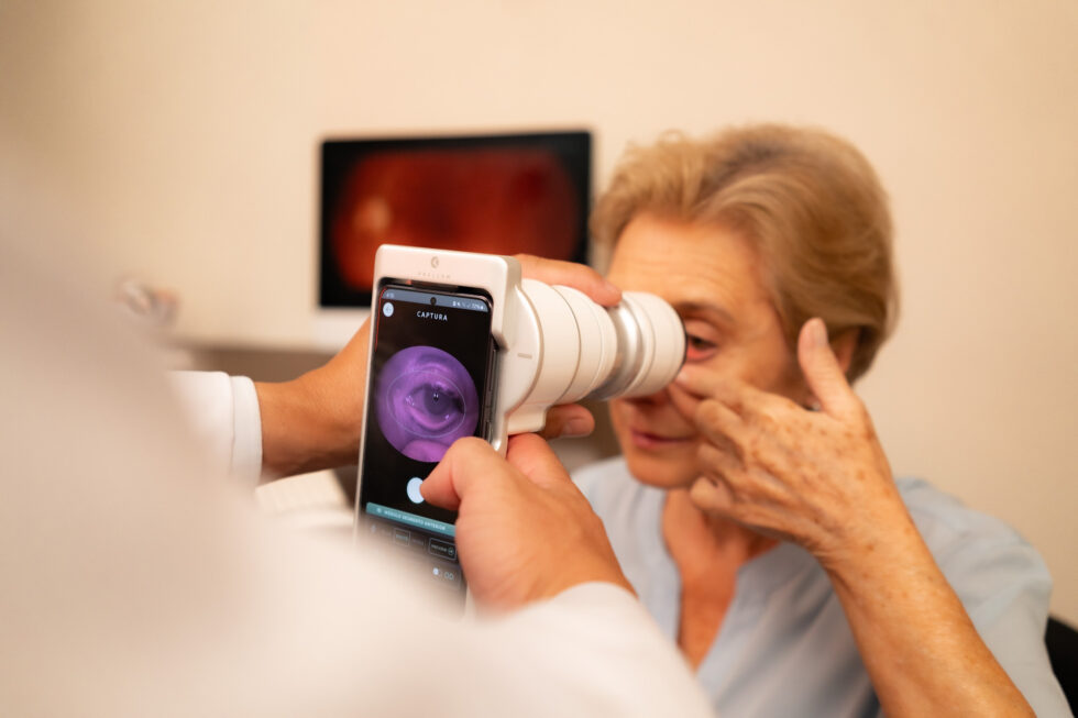
This is the core philosophy behind platforms like the Eyer2. As a portable fundus camera and imaging system, it delivers an immediate connection between refractive data and retinal health.
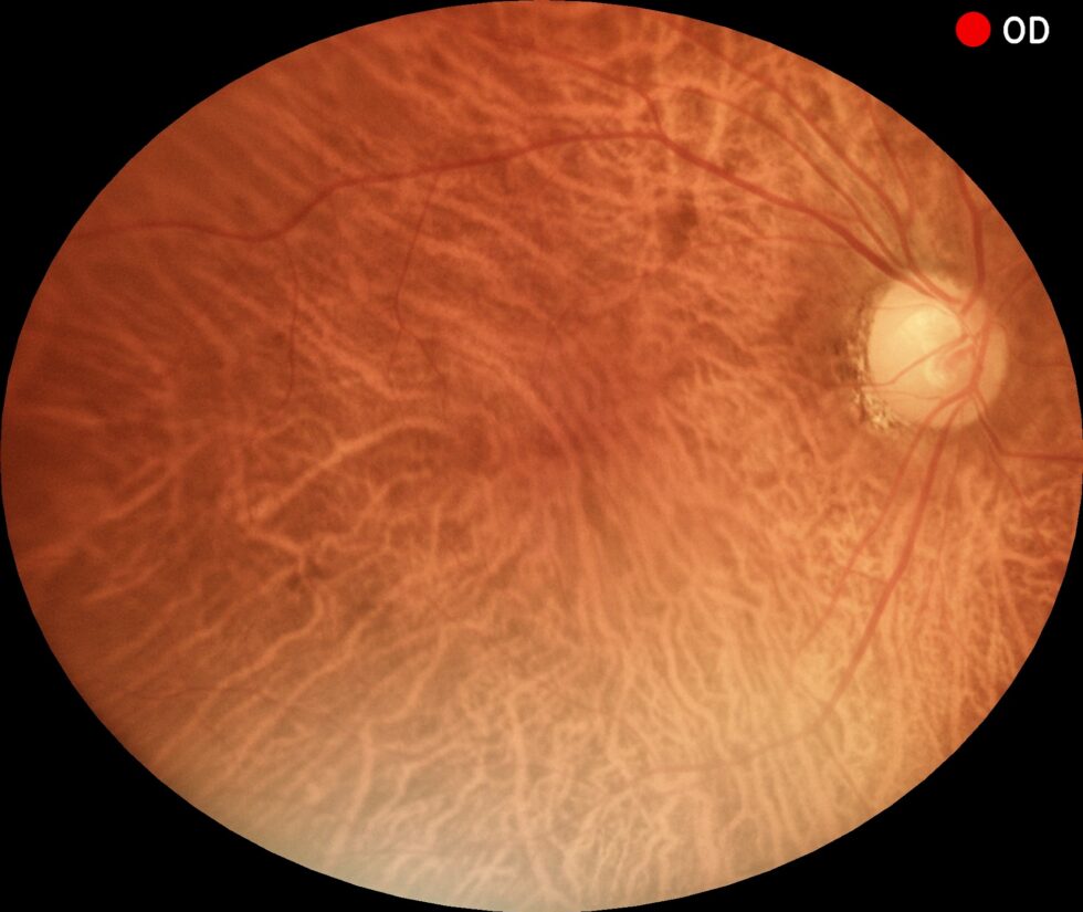
Color fundus photography captured by the Eyer2 portable fundus camera.
Imagine this workflow: The patient undergoes a refractive assessment via retinoscopy. Then, right there in the exam chair, the Eyer2 captures high-definition images of the fundus or ocular surface. What was once just a static data point on a chart is now paired with visual documentation of the retina, macula, and optic nerve.
Instead of juggling two isolated tests—retinoscopy (functional) and fundoscopy (structural)—the specialist now operates within a unified workflow. The precision of the retinoscope gains the context of high-definition documentation. The prescription found through retinoscopy is validated and enhanced by the image captured by the Eyer2.
Meet the Eyer2
The Eyer2 handheld fundus camera supports the diagnosis of over 50 conditions, including glaucoma, cataracts, diabetic retinopathy, age-related macular degeneration (AMD), retinoblastoma, hypertensive retinopathy, retinopathy of prematurity (ROP), and ocular toxoplasmosis.
The device also enables the detection of various ocular surface diseases and conditions, such as blepharitis, eyelash abnormalities, Meibomian Gland Dysfunction (MGD), hordeola, conjunctival and eyelid tumors, advanced cataracts, foreign bodies, burns, corneal lesions, and various types of keratitis (resulting from dry eye, contact lens wear, infections, or ulcers).
Meibography scan captured via the Eyer2 portable retinal camera, evaluating meibomian gland structure for evaporative dry eye.
Key Features & Benefits:
All-in-One Portable Platform: A handheld ocular imaging system capable of performing six different types of exams in a single, non-mydriatic device.
High-Quality Color Fundus Photography: Features a 55° field of view in a single image, making it easy to detect peripheral retinal lesions.
Instant Green-Channel Imaging: Automatically generated immediately following color capture.
Infrared Posterior Segment Imaging: Allows for deeper retinal evaluation with enhanced patient comfort; essential for diagnosing choroidal nevi and assessing evaporative dry eye.
Stereo Disc Photography: Provides 3D visualization of the optic nerve head and cupping.
Panoramic Fundus Imaging: Captures wide-field views up to 120°.
Advanced Analytics: Built-in editing tools and graphics for precise Cup-to-Disc ratio (CDR) analysis.
HD Ocular Surface Documentation: High-definition photo-documentation to track and monitor disease progression.
Cobalt Blue Light: Optimized for the evaluation and documentation of corneal lesions and staining patterns.
Unmatched Mobility: Perfect for multi-clinic practices, remote primary care, and examinations of bedridden or neonatal patients.
Cloud Ecosystem (EyerCloud): Seamless integration with an online platform for comprehensive exam management and secure data storage.
About Phelcom
Phelcom Technologies is an American-Brazilian company combining physics, electronics, and computing to make vision care simpler, more connected, and intelligent.
The company was founded in 2016 by three young researchers: José Augusto Stuchi (computer engineer), Flavio Pascoal Vieira (electronics engineer), and Diego Lencione (physicist). Inspired by Diego’s brother, who struggled with a severe vision condition from childhood, the trio set out to develop a portable retinal camera integrated with a smartphone.
In 2019, Phelcom launched its first product, the Eyer portable retinal camera. Five years later, the company introduced the Eyer2, a comprehensive photo-documentation platform capable of capturing high-quality images of both the posterior and ocular surface segments.
Today, with a 10-year track record, Phelcom’s technology has benefited over two million people across multiple countries, including the United States, Japan, Chile, Colombia, the United Arab Emirates, and Brazil, and has been deployed in more than 200 social outreach initiatives worldwide.


