

 English
  Español
  Português

Diabetes remains one of the world’s fastest-growing chronic diseases and a major public health challenge. In Brazil 12.9% of the population lives with the condition, while in the U.S., the CDC reports over 38 million affected individuals. Left unmanaged, diabetes impairs multiple organ systems.
Among its most concerning complications is diabetic retinopathy (DR), a leading cause of preventable vision loss in adults caused by microvascular alterations in the retina. The primary clinical challenge is that DR often progresses silently, remaining completely asymptomatic during its initial stages.
In this context, fundus photography has solidified its role as a vital tool for screening, early diagnosis, and ongoing ocular monitoring. By providing detailed visualization of the retina, this imaging helps identify structural changes long before vision is compromised.
For endocrinologist and ophthalmologist Dr. Flávia Godinho, monitoring eye health must be integrated into routine diabetic care. “The incorporation of retinal imaging into clinical practice has the potential to change management protocols, increase treatment adherence, and directly impact the prognosis and quality of life for these patients,” she emphasizes.
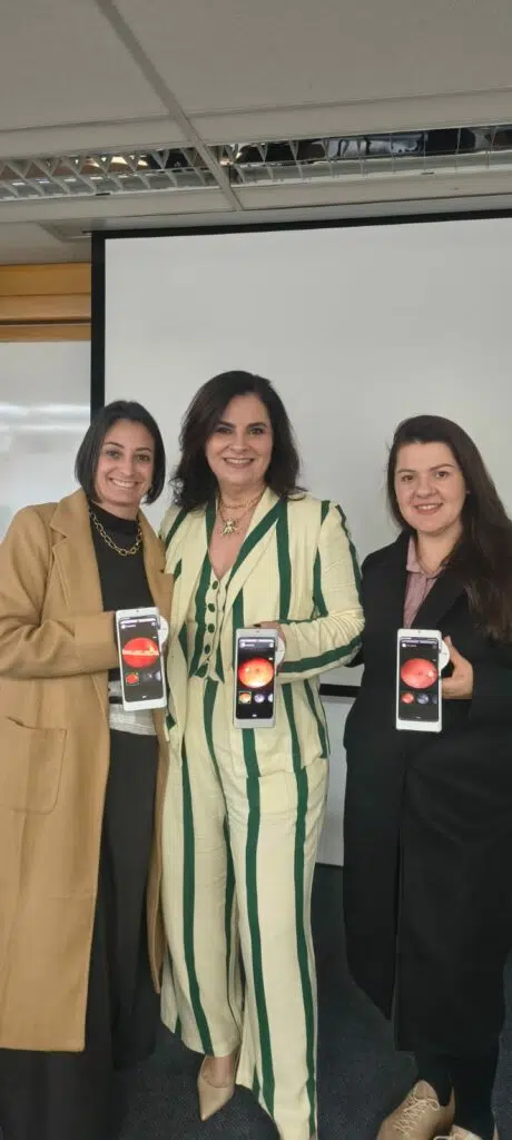
Phelcom commercial analyst Daniele Oliveira, ophthalmologist and endocrinologist Dr. Flávia Godinho, and Phelcom after-sales analyst Maiara Guiliano at the Diabetes 360º event, held May 29–31 in São Paulo, SP.
Beyond blood glucose
One of the lesser-known nuances of diabetes management is that lowering blood glucose as quickly as possible is not always the safest strategy. In patients with pre-existing diabetic retinopathy, a rapid drop in blood sugar levels can trigger “early worsening,” a phenomenon that temporarily exacerbates retinal lesions and increases the risk of severe vision loss.
Dr. Godinho explains that retinal imaging is a fundamental tool for guiding medical management in these scenarios. “We must first understand the status of the retina to determine the appropriate speed and safety of glycemic control,” she states.
She recalls a case that perfectly illustrates this clinical reality: “I once had a patient come into the office saying, ‘Doctor, when my glucose was 400, I could see everything. Now that it is 110 I am practically blind. What happened?’ What had changed was simply the initiation of her diabetes treatment.”
According to Dr. Godinho, safely lowering glucose can sometimes take up to a year. “We do this gradually, monitoring retinal exams and co-managing with the ophthalmologist. If we notice any signs of progression or worsening, we slow the process down and reevaluate the strategy.”
In this dynamic, fundus photography helps endocrinologists balance two equally critical goals: controlling systemic disease and preserving patient vision.
Diabetes and weight-loss injectables
The relationship between diabetes and weight-loss injectables (GLP-1 receptor agonists) has gained widespread attention with the popularization of medications like semaglutide, tirzepatide, and liraglutide. While these drugs deliver significant benefits for glycemic control and weight loss, Dr. Godinho warns that close medical supervision is essential.
“There is no scientific evidence that these drugs directly cause visual alterations or lead to blindness. The problem arises when a diabetic patient experiences a very rapid reduction in glucose levels and already has established retinopathy,” she explains.
This precaution extends beyond injectables to any intervention capable of triggering a steep glucose drop– including insulin initiation or intensification, certain oral antidiabetic medications, highly restrictive diets, or prolonged fasting. “In some cases, the ophthalmologist faces a tremendous struggle to control the progression of diabetic retinopathy once this rapid-reduction process is triggered,” Dr. Godinho notes.
Evaluating the retina before and during treatment can change everything. “Retinal imaging is a simple, non-invasive exam that can completely change how we manage a patient’s treatment,” she points out.
The consequences of vision loss extend far beyond ocular health. “We are talking about independence, quality of life, the ability to work, and performing simple daily activities. When we identify the risk early, we have the opportunity to protect their vision and alter their prognosis for the rest of their life.”
A window to systemic health

During her lecture at the Diabetes 360º event, Dr. Godinho also discussed the broader role of retinal imaging in the endocrinologist’s clinical practice.
Beyond identifying diabetic retinopathy, Dr. Godinho emphasizes that fundus photography offers invaluable insights into overall vascular health, helping detect early signs of systemic diseases before symptoms surface. “Many of these systemic changes appear first in the small vessels. The retina is the only place in the body where we can directly visualize blood vessels and capillaries non-invasively. This allows us to observe microvascular changes related to hypertension, atherosclerosis, inflammation, and other conditions.”
This aspect still receives too little attention in primary and endocrine care. “We frequently evaluate sleep, diet, physical activity, and many other health metrics, but the eyes are often left in the background. However, they can provide critical information about the patient’s overall state of health.”
Because endocrinologists manage populations highly susceptible to age-related conditions, baseline retinal exams can also aid in the early identification of glaucoma and age-related macular degeneration (AMD).
Flávia também lembra que o endocrinologista acompanha uma população frequentemente exposta a doenças associadas ao envelhecimento. Nesses casos, a observação da retina pode auxiliar na identificação precoce de condições como glaucoma, degeneração macular relacionada à idade (DMRI) e outras doenças oculares potencialmente graves.
“By incorporating retinal evaluations into routine monitoring, we can identify problems sooner and refer the patient for appropriate treatment before irreversible vision loss occurs. The eye has a lot to offer to endocrinology,” she believes.
A new tool for the endocrinologist
Dr. Godinho observes that fundus photography remains underexplored outside of ophthalmology, largely because non-eyecare specialists often receive limited training in direct ophthalmoscopy during medical school.
Technologies like the Eyer2 portable fundus camera make integrating this level of care into general or endocrine practices seamless. “The Eyer makes this exam highly accessible for the endocrinologist. Image capture is simple, fast, and can be performed by trained allied health staff. In moments, the doctor has access to the images and can discuss the findings with the patient during the actual consultation,” she highlights.
The Eyer2 captures high-definition images of the ocular surface and posterior segment in just minutes without requiring pupil dilation (mydriasis). Integrated with EyerCloud—an online exam management platform—it enables practitioners to securely store, access, and track images over time. Additionally, EyerMaps artificial intelligence assists in identifying potential retinal abnormalities as a decision-support tool. (Note: EyerMaps is currently available only in the Brazilian market.)
Visualizing these images also strengthens the doctor-patient relationship. “When we show them the image and compare it to a healthy retina, they understand what is happening. This sparks greater engagement with their systemic treatment and compliance with their ophthalmological follow-ups.”

Retinal image displaying diabetic retinopathy captured by the Eyer portable fundus camera.

Retinal image displaying diabetic retinopathy captured by the Eyer portable fundus camera.
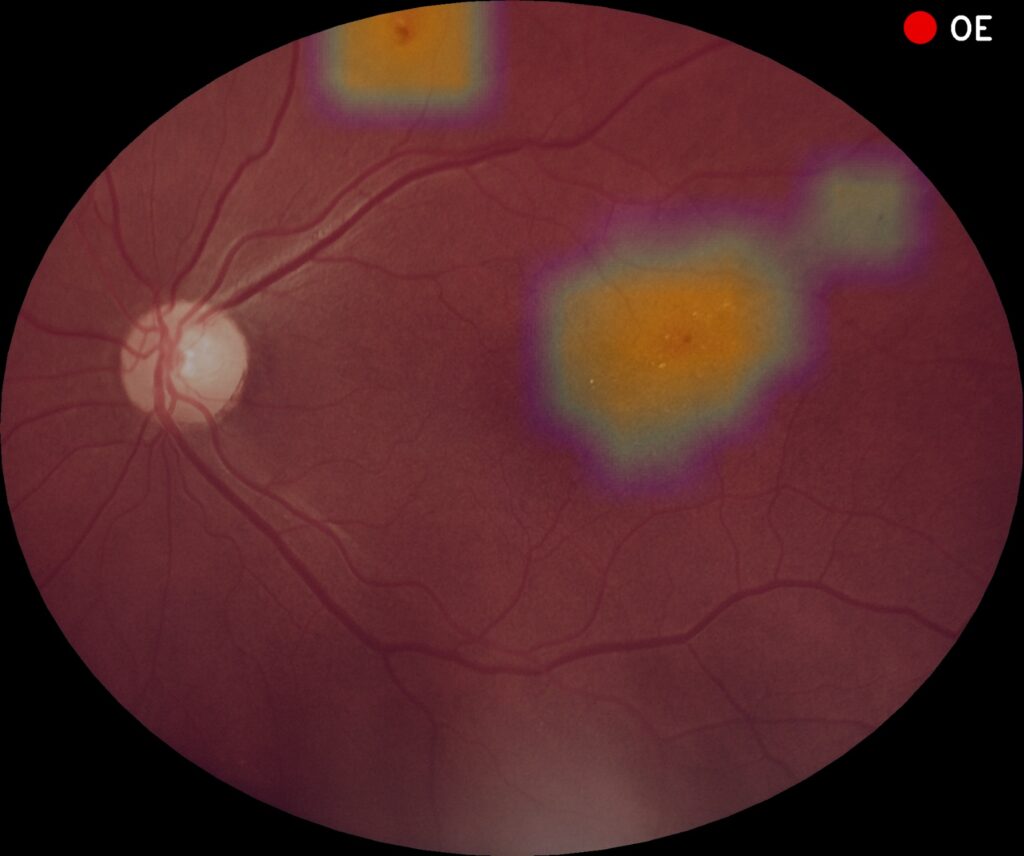
Retinal image captured with the Eyer portable fundus camera. The EyerMaps AI highlights regions with potential abnormalities associated with diabetic retinopathy. (EyerMaps is currently available only in the Brazilian market)
Public health implications
Integrating imaging technologies into primary care and chronic disease management represents a major step forward, particularly given the access barriers to specialized ophthalmological care within public health systems.
According to Dr. Godinho, one of Eyer’s primary advantages is enabling rapid, accessible triage directly at the point of care for patients managing diabetes, hypertension, and other chronic conditions. “Often, a patient waits months, or even over a year, to get a consultation with an ophthalmologist. Meanwhile, the disease continues to progress. Having access to retinal imaging at the point of primary care helps identify exactly who needs a priority referral,” she asserts.
This need is especially critical when managing advanced diabetes, where determining the ideal speed of glycemic control relies heavily on prior retinal evaluation. “The Eyer is a tool that can contribute immensely to the prevention of blindness and to more efficient public health management,” she concludes.


