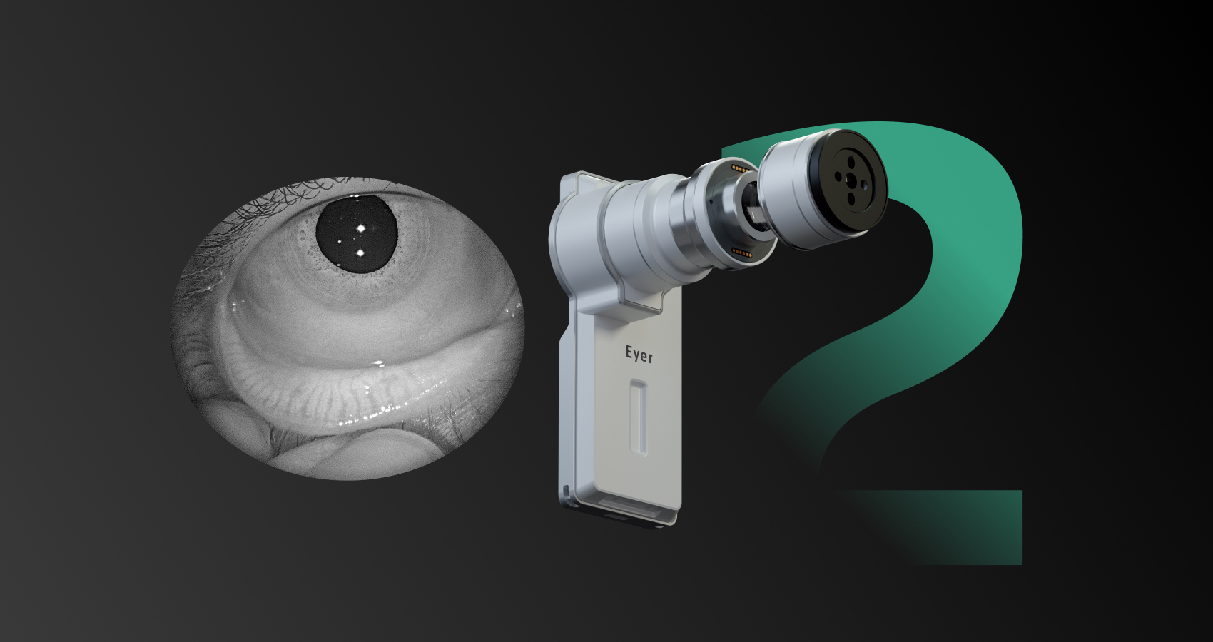
The Meibomian glands, located in the tarsal plates of the eyelids, play a crucial role in producing the lipid layer of the tear film—the outermost layer of the tear film. This layer is essential for stabilizing the tear film, preventing tear evaporation, and maintaining the homeostasis of the ocular surface. Dysfunction of the Meibomian glands (MGD) disrupts tear film stability, leading to ocular surface imbalance.
MGD results from altered meibomian secretion quality, gland dropout, and/or ductal obstruction. One of the most effective tests for assessing Meibomian gland morphology is Meibography, a technique that provides in vivo visualization of these glands.
Propaedeutic evaluation of dry eye: beyond Meibography
Dry eye diagnostic tests include evaluating tear film stability and volume, ocular surface damage with vital dyes such as fluorescein or rose bengal, lid assessments, and Meibomian gland examination.
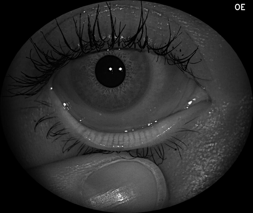
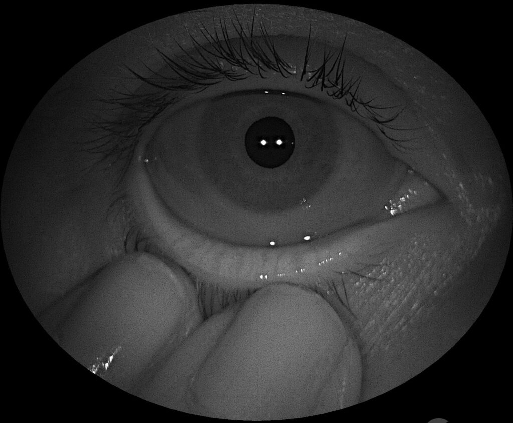
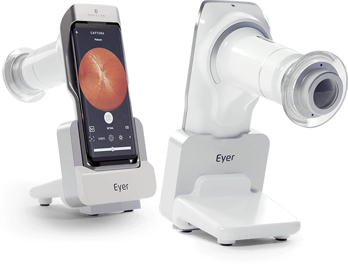
Phelcom has recently revolutionized this field by incorporating infrared imaging into its new Eyer2 portable device, making meibography more practical. The Eyer2’s anterior segment module enables both meibography and examinations for corneal and conjunctival epithelial cell changes using cobalt blue light and fluorescein.
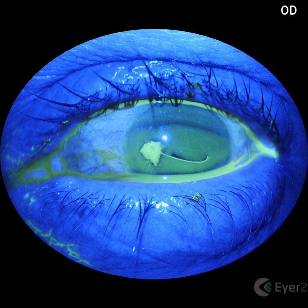
For a comprehensive dry eye diagnosis, other clinical evaluations such as the Schirmer test are recommended to differentiate between aqueous-deficient and evaporative dry eye.
Additional applications of Meibography
Meibography is also valuable in preoperative evaluations for refractive surgery, as dry eye, often linked to MGD, is a common postoperative complication. This exam provides preoperative documentation and gland monitoring, as well as patient education.
Furthermore, meibography can be utilized to assess eyelid impairment due to contact lens wear, allergic conjunctivitis reactions, antiglaucoma treatments, and other clinical conditions.
Eyer2
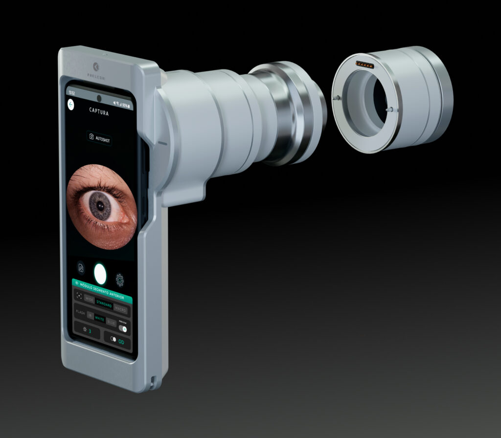
Phelcom proudly introduces the Eyer2, a versatile visual examination platform that delivers high-quality imaging of both the posterior and anterior segments.
This new equipment offers color and red-free fundus imaging with a new 55º field of view, along with tools for anterior segment imaging using various light sources, including cobalt blue light for corneal lesion assessment and infrared light for meibography.
About Phelcom
Phelcom Technologies is a Brazilian medtech company based in São Carlos, in the interior area of São Paulo. The company’s story began in 2016, when three young researchers – a physicist, an electronics engineer and a computer engineer (physics, electronics, computing) – created a portable fundus camera integrated with a smartphone.
The first prototype project was born from Diego Lencione’s interest in visual health, as his brother has a condition that has severely compromised his retina and vision since childhood.
In 2019, Phelcom launched its first product on the Brazilian market: the Eyer portable fundus camera. Today, the technology has reached more than two million people throughout Brazil and in the countries where it is present and has been used in more than 100 community screenings.
Fontes:
Wolffsohn et al. TFOS DEWS II Diagnostic Methodology report 2017
Magno et al. Hot towels: The bedrock of Meibomian gland dysfunction treatment – A review
Arita. Meibography: A Japanese Perspective Reiko. 2018
