

Dr. Eduardo Ferrari Marback, a distinguished ophthalmologist and professor at the Federal University of Bahia (UFBA), has been utilizing the Eyer portable fundus camera since 2019 in his clinic in Salvador, Brazil and during his university lectures. Recently, Dr. Marback incorporated the advanced Eyer2 visual examination platform into his practice, enhancing his ability to capture high-quality images of both the anterior and posterior segments of the eye.
As a specialist in ocular oncology, Dr. Marback employs the Eyer2 to meticulously document and monitor the progression of intraocular and surface tumors, as well as other ocular lesions that require differential diagnosis from eye neoplasms. “Documenting and tracking these changes is crucial for understanding how these lesions behave,” he explains.
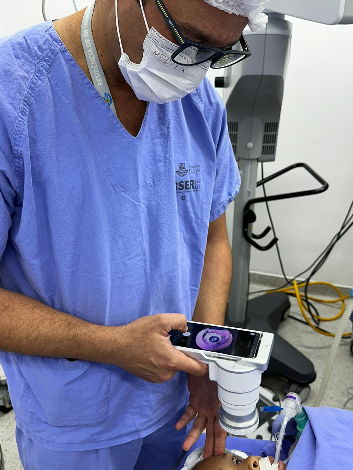
Dr. Eduardo Marback utilizing Eyer2 to examine a patient.
With Eyer2, exam results are seamlessly uploaded to the EyerCloud platform, directly integrating with the patient’s medical records. During follow-up appointments, Dr. Marback can effortlessly review and compare images with patients, providing real-time insights into their condition’s progression.
Eyer2 introduces innovative features like color and red-free fundus imaging with a 55° field of view, along with advanced anterior segment imaging tools. “The expanded field of view, powerful imaging capabilities, and magnetic connection for anterior segment photography allows me to document ocular surface tumors with exceptional clarity,” Dr. Marback notes.
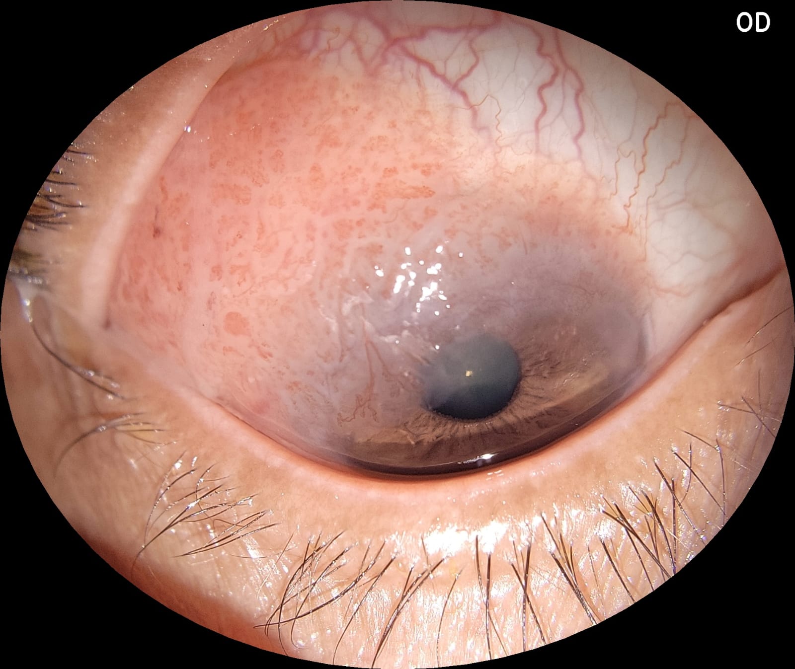
High-resolution image captured with Eyer2, showing a conjunctival squamous neoplasia pre-surgery. Photo: Eduardo Ferrari Marback.
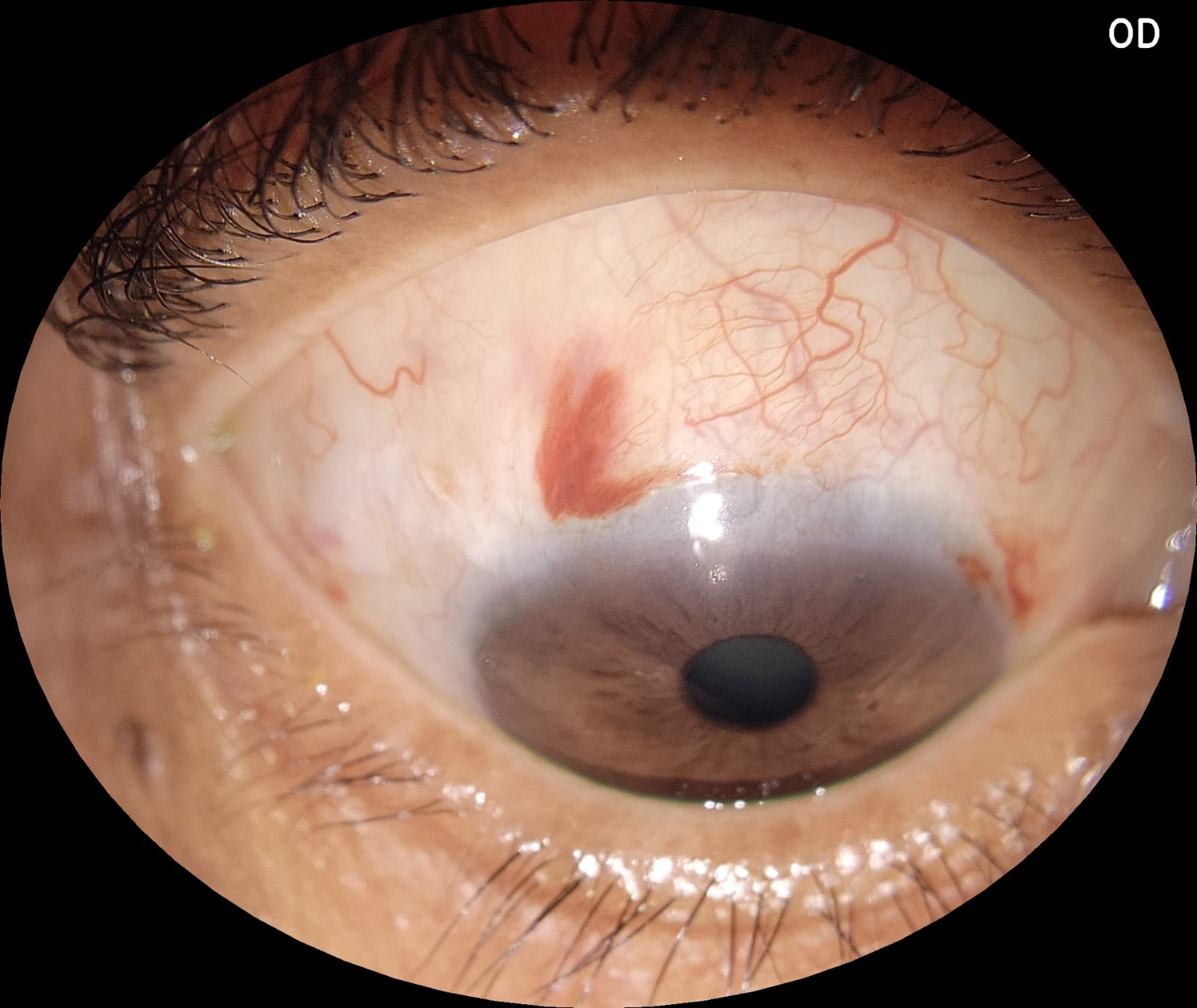
Post-surgical result of conjunctival squamous neoplasia, captured with Eyer2. Photo: Eduardo Ferrari Marback
University
Beyond oncology, Dr. Marback frequently uses Eyer2 in his clinic for diagnosing conditions such as diabetic retinopathy, optic neuropathy, and glaucoma. In emergency situations, the device proves invaluable for thorough documentation which is not only beneficial for patient care, but also crucial for legal documentation especially in trauma cases.
Dr. Marback also leverages Eyer2’s portability in his role as an educator. He incorporates the device into his classroom at UFBA, allowing students to conduct fundus examinations on each other, enhancing their learning through practical, hands-on experience. “The students do the exams among themselves and analyze the structure of their own eyes,” he shares.
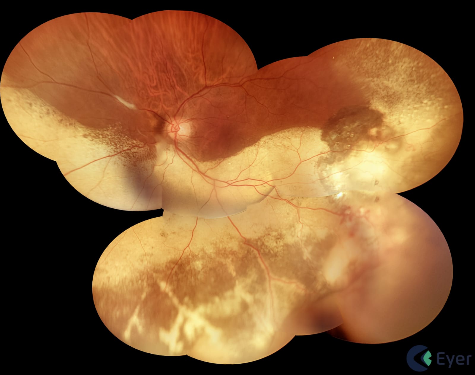
Vasoproliferative retina image captured with Eyer2. Courtesy of Dr. Eduardo Ferrari Marback.
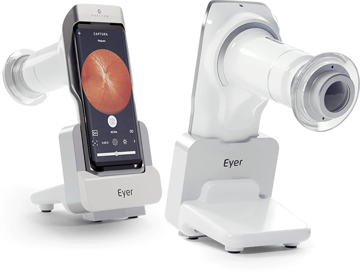
Eyer2
Eyer2‘s portable fundus camera is equipped with cutting-edge features designed to simplify clinical workflow and elevate diagnostic accuracy. Infrared light assists in detecting choroidal changes and performing meibography, while cobalt blue light aids in identifying corneal lesions. This technology allows for the detection of numerous anterior segment conditions, including blepharitis, meibomian gland dysfunction, eyelid tumors, and keratitis.
The Eyer2 platform is a game-changer in clinical practice, offering streamlined examinations, comprehensive diagnostic capabilities, and powerful technological tools that enhance patient care.
Key features include:
- Single-click registration
- Ergonomic design for enhanced comfort during examinations
- Portable imaging platform offering six distinct recording modalities, without the need for pupil dilation
- High-quality 55° color fundus imaging for detecting peripheral retinal lesions;
- Instant red-free imaging following color capture
- Posterior segment imaging with infrared light, crucial for assessing deeper retinal areas without patient discomfort, such as choroidal nevus and evaporative dry eye
- 3D effect of the optic disc
- Panoramic retinal image up to 120°
- High-definition documentation of the ocular surface for disease monitoring;
- Cobalt blue light for corneal lesion assessment
- Portability for use in various clinical settings, remote areas, and for bedridden or neonatal patients
- Seamless integration with EyerMaps, an AI-driven tool that highlights potential retinal anomalies in seconds
- Connectivity with EyerCloud, an online platform for managing examinations.
About Phelcom
Phelcom Technologies is a Brazilian medtech company based in São Carlos, in the interior area of São Paulo. The company’s story began in 2016, when three young researchers – a physicist, an electronics engineer and a computer engineer (physics, electronics, computing) – created a portable fundus camera integrated with a smartphone.
The first prototype project was born from partner Diego Lencione’s interest in visual health, as his brother has a condition that has severely compromised his retina and vision since childhood.
In 2019, Phelcom launched its first product in the Brazilian market: the Eyer portable fundus camera. Today, the technology has reached more than two million people throughout Brazil and in the countries where it is present and has been used in more than 100 community screenings.