A research carried out in two referral centers for covid-19 in Rio de Janeiro (RJ) identified alteration in the retina of patients hospitalized in serious condition. The study occurred in May 2020, in the clinical hospitals Mario Lioni and from Jacarepaguá, and was published in the journal Plos One in December, the same year.
The work was also one of the first to discover retinal damage in critical cases of the disease. It is noteworthy that other Brazilian studies, from the ABC School of Medicine (FAMBC) and the Federal University of São Paulo (Unifesp) , published shortly before, also detected retinal alterations.
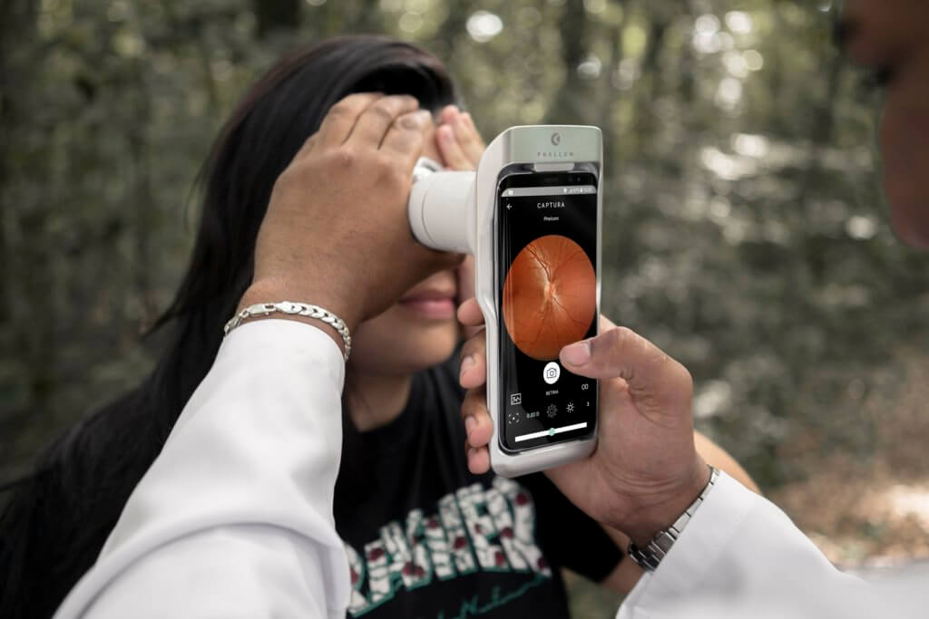
Researchers used the smart device Phelcom Eyer to carry out the retinography in 47 eyes of 25 patients. Coupled to a smartphone, the device carries out high-quality fundus exams, in few minutes and without need of pupil dilation. It connects to an online platform, the Eyer Cloud, which allows remote diagnosis and assures safety to data stored in the cloud.
“Our study showed that, from the patients hospitalized in serious conditions for covid-19 clinical stabilization, 12% presented some finding”, says one of the doctors responsible for the work, Rafael Lani Lozada, ophthalmology resident in Gamboa Hospital (RJ).
A 35-year-old man from the mentioned sample, who had diverse clinical complications during hospitalization, manifested bilateral nerve fiber layer infarctions and micro-hemorrhages in the papillomacular bundle; another man, 56 years old, who needed to undergo full anticoagulation, had a unilateral “flame-shaped” hemorrhage; and a third man, 49 years old and hypertensive, presented discrete and bilateral retinal micro-hemorrhages.
To Lozada, the study shows retinal alterations may occur in severe covid-19 cases. “They were probably secondary to clinical intercurrences or comorbities rather than a direct damage by SARS-CoV-2, since it was not possible to correlate the problem directly to the virus. Thus, the retinal finding may be important and easily accessible markers of therapeutic interventions, as well as sentinels of neurological and systemic diseases during the pandemic”,
The doctor highlights that new studies with more patients are necessary to establish statistic correlations between covid-19 and retinal lesions.
Phelcom Eyer
Louzada states that Phelcom Eyer was essential to the project, since the study, as a base, evaluated hospitalized patients in isolation, with a highly contagious and little studied virus. “Using a clean easy-to-handle portable device made it possible for us to step further in the search for a better comprehension of this disease, that afflicts humanity”, he observes.
The researcher also highlights other advantages on the equipment: short learning curve, quick image backup and a possible thorough analysis from any computer with internet access. “Not to mention that the fundus exams are very beautiful, with a great resolution and the color photos are automatically duplicated with red-free images”, he analyzes.
Beyond the pandemic
Lozada also highlights that the technical ease of retina evaluation with Eyer opens new horizons to triage exams, not only in the present pandemic context, but also for other ophthalmological diseases. “The possibility to obtain retinographies in a portable, easy and practical way, through an accessible image backup, allowing their remote careful study, is fundamental for advances in ophthalmological researches and better diagnostics”, he finishes.

 Português
  Español
  English


