The choroidal nevus is a dark spot that occurs at the eye fundus and is only detectable through routine examinations such as retinal mapping. Usually, treatment only includes an yearly follow-up.
There are also skin nevi, which dermatologists follow-up with dermatoscopy to check for possible changes in their characteristics, such as enlargement. The same follow-up occurs with the spots at the eye fundus, for example.
If they grow, they can evolve into very advanced stages, such as choroidal melanoma, a very rare disease that affects less than 1% of patients diagnosed with the condition. This number is equivalent to five people in a million.
Melanoma (a type of cancer) is asymptomatic at the initial stage. An estimated 85% of cases arise in the uveal tract – iris, ciliary body and choroid. When not identified early, it can metastasize to the liver.
Retinography applies for checking the coroidal nevus size. Learn how this examination can help early diagnosis and disease follow-up.
Choroidal nevus – diagnosis
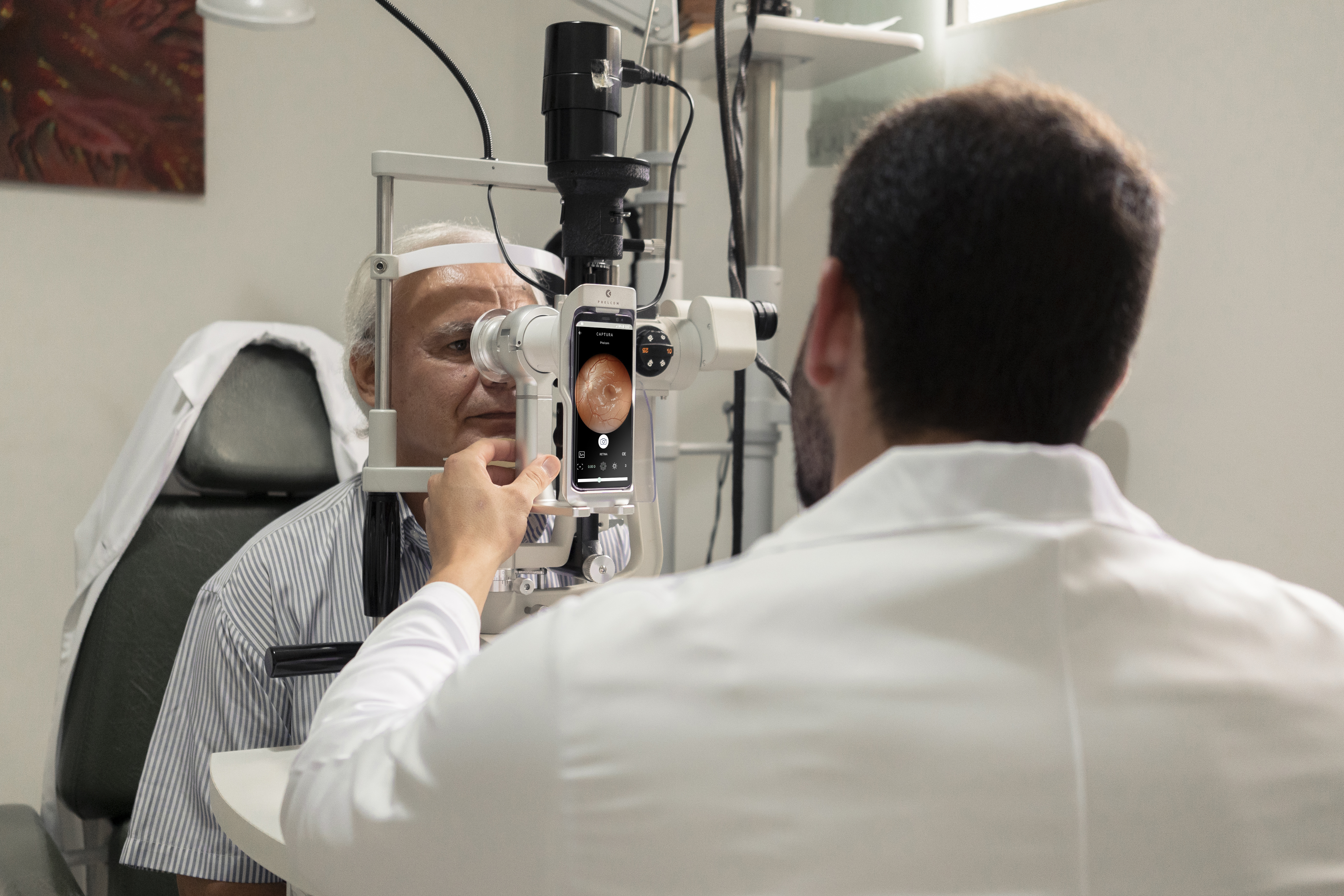
The ophthalmologist can only identify a coroidal nevus in a routine examination, because the disorder is not visible to the naked eye and does not usually present early symptoms.
Retinal mapping is one of them. By observing a nevus, the doctor can carry out further examinations to finish the diagnosis, such as optical coherence tomography (OCT) and retinography.
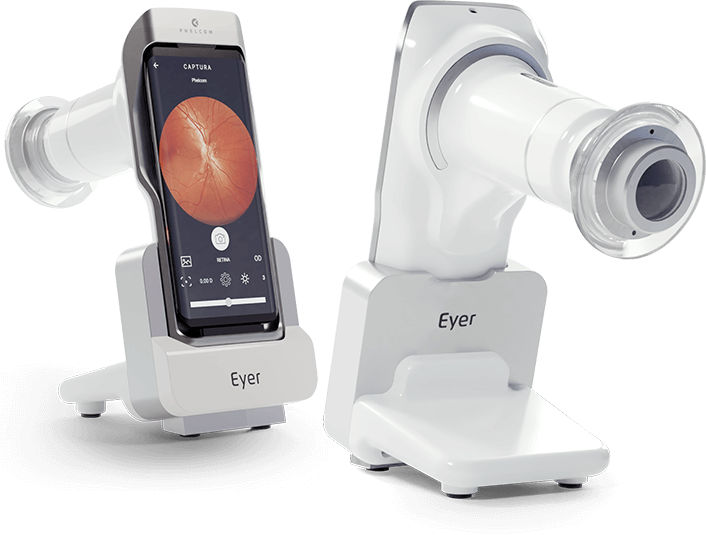
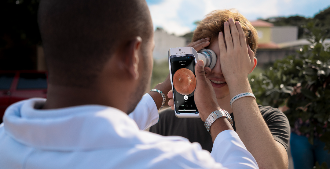
Eye fundus imaging is essential to detect, monitor and check if the spot grows. Currently, there are portable non-mydriatic devices that carry out the examination quickly and store the images on a functional online platform. Thus, it is possible to compare photos over the years. It also allows specialists anywhere in the world to diagnose remotely.
If the nevus grows, the first diagnosis may be undetermined melanocytic lesion, to which the doctor will define a protocol of examinations and follow-up. Observed new nevus increases confirm the choroidal melanoma diagnosis.
Choroidal melanoma – treatment
In fact, choroidal melanoma has no cure, but is treatable and requires lifetime monitoring. Thus, the therapy will be established according to the patient’s vision condition and age, as well as the status, location and extent of the cancer. As with all diseases, an early diagnosis determines a better prognosis.
Brachytherapy is most recommended for small and medium sizes. This surgery has a control rate of approximately 95% and maintains the eye and, in some situations, the ability to see.
An older method was removing the ocular globe. Enucleation may still occur for large tumors with symptoms as intense pain, poor vision and disorganization of internal structures. In some cases, radiotherapy, laser therapy and transpupillary thermotherapy are also indicated.
Reviewed by Paulo Schor, ophthalmologist, associate professor and director of innovation of the Federal University of São Paulo (Unifesp) and collaborator of the Faculty of Medicine of the Albert Einstein Hospital.
Follow Phelcom blog and stay on top of the main news on office management!
Subscribe